Article Text

Abstract
Introduction Preclinical testing of endovascular devices in an in-vivo environment provides a more realistic simulation of critical vessel viscoelastic and physiologic responses. In comparison, in-vitro and cadaveric models, though indispensible, can only assess structural and mechanical responses. Replicating the tortuosity of the carotid siphon in an in vivo model is a challenging endeavor. Models created by complex surgical maneuvers have previously been described1, often requiring the expertise of a skilled surgical team and considerable expense. We describe a novel porcine model of arterial tortuosity with a quantitative comparison of its geometric features with a population representative carotid siphon acquired from a patient dataset.
Materials and Methods Three Yorkshire swine (sex: male, mean weight 46.6 kg) were used for the purposes of the study. The animals were anesthetized and femoral artery access was obtained through a 6F sheath. The microcatheter was successfully navigated to the brachial branch of the axillary artery and a selective digital subtraction angiogram was acquired in both extended and flexed right and left forelimb positions. Three dimensional rotational angiograms (3DRA) were also acquired. The vessel segmentation and centerline was generated using Mimics (Materialise, Leuven, Belgium) and a quantitative measurement of the geometric parameters was obtained as previously described. This was compared with a realistic population representative human carotid siphon model.2
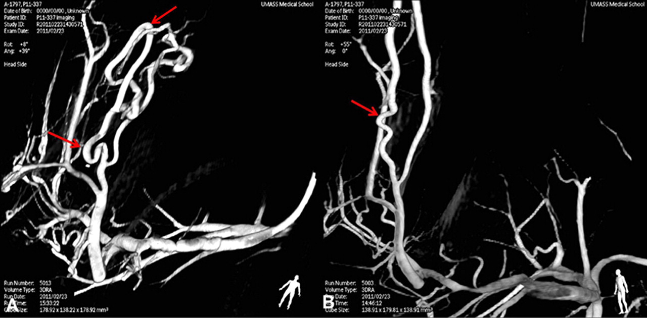
Results The mean average curvature (AC), length and diameter calculated for the arterial segments (average±SE of the mean) in the extended (0.28±0.06 mm−1, 13.99±1.66 mm and 2.60±0.19 mm) and the flexed (0.29±0.02 mm−1, 17.59±0.77 mm and 2.59±0.12 mm) position are similar. In comparison, the values calculated for the population representative carotid siphon are 0.34±0.02 mm−1, 22.60±0.79 mm and 4.15±0.09 mm, respectively. However, positioning the forelimb in the flexed position (Abstract O-023 figure 1A, arrows) increased the number of tortuous bends in comparison to the extended position (Abstract O-023 figure 1B, arrow).
Conclusion We have described a reproducible in vivo model of arterial tortuosity in the brachial artery in the flexed forelimb position in the swine. This model offers a challenging target for the assessment of guidewires, microcatheters and endovascular implants since it closely mimics the curvature experienced at the carotid siphon.
Statistics from Altmetric.com
Footnotes
Disclosures S Carniato: Stryker Neurovascular. M Mehra: None. R King: None. A Wakhloo: Stryker Neurovascular. M Gounis: Stryker Neurovascular.