Article Text

Abstract
Background We retrospectively evaluated the composition of retrieved clots from ischemic stroke patients to study the association between histological composition and stroke etiology
Methods Consecutive patients enrolled in the Stroke Thromboembolism Registry of Imaging and Pathology (STRIP) were included in this study. All patients underwent mechanical thrombectomy and retrieved clots were sent to a central core lab for processing. Histological analysis was performed using martius scarlet blue (MSB) staining, and quantification for red blood cells (RBCs), white blood cells (WBCs), fibrin and platelets was performed using Orbit Image Software. A Wilcoxon test was used for continuous variables and χ2 test for categorical variables.
Results 1350 patients were included in this study. The overall rate of Thrombolysis In Cerebral Infarction (TICI) 2c/3 was 68%. 501 patients received tissue plasminogen activator (tPA) (37%). 267 patients (20%) had a large artery atherosclerosis (LAA) source, 662 (49%) a cardioembolic (CE) source, 301 (22%) were cryptogenic, and the remainder had other identifiable sources including hypercoagulable state or dissection. LAA thrombi had a higher mean RBC density (46±23% vs 42±22%, p=0.01) and a lower platelet density (24±18% vs 27±18%, p=0.03) than CE thrombi. Clots from dissection patients had the highest mean RBC density (50±24%) while clots from patients with a hypercoagulable state had the lowest mean RBC density (26±21%).
Conclusions Our study found statistically significant but clinically insignificant differences between clots of CE and LAA etiologies. Future studies should emphasize molecular, proteomic and immunohistochemical characteristics to determine links between clot composition and etiology.
- thrombectomy
- stroke
Data availability statement
Data pertaining to this study are available upon reasonable request to the corresponding author.
Statistics from Altmetric.com
Introduction
Given the increased utilization of mechanical thrombectomy for the treatment of acute ischemic stroke secondary to large vessel occlusion, there has been growing interest in the analysis of retrieved thrombi.1–7 A number of studies have suggested that histopathological analysis can provide some valuable insights into stroke etiology.8–11 Many of these studies have relied on traditional hematoxylin and eosin (H&E) and martius scarlet blue (MSB) stains to quantify the proportion of clot components including red blood cells (RBCs), fibrin, white blood cells (WBCs) and platelets. Other studies have sought to examine the role that immunohistochemistry and more advanced proteomic analyses can play in differentiating between various clot etiologies.
Understanding that there is a reliable histological signature of retrieved thrombi is important because we can then potentially help to discern between emboli which are more likely to be cardiac in etiology versus those of a large artery atherosclerosis (LAA) etiology, and perhaps influence secondary prevention strategies accordingly. If a large study were to find no such link, then it would suggest that the field should focus on more advanced analyses of clot composition including proteomic signatures, molecular analyses and immunohistochemistry. In order to study the association between stroke etiology and clot histological composition, we studied patients included in the Stroke Thromboembolism Registry of Imaging and Pathology (STRIP). We hypothesized that clots retrieved from patients who had an LAA source would have a higher proportion of platelets and WBCs than those from a cardiac source.
Patients and methods
Patient population
Consecutive patients enrolled in the STRIP Registry from September 2016 to December 2019 were included. The study was institutional review board approved and waiver of consent was granted. Patients were included if they were >18 years of age, had undergone mechanical thrombectomy treatment for acute ischemic stroke, and clot material was retrieved.
Clot processing and histological characterization
Each embolus was immediately fixed in 10% phosphate buffered formalin. Emboli were shipped to a central core laboratory for standard tissue processing and embedded in paraffin. The formalin-fixed paraffin-embedded clot material was cut into 3–5 µm sections. Representative slides from each clot were stained with H&E and MSB. Representative MSB-stained slides were sent for whole slide scanning (Aperio ScansScope AT-Turbo, Leica Biosystems). Histological quantification was performed using Orbit Image Analysis Software (www.Orbit.bio) as per the standard operating procedure.12 Details of the methodology for Orbit image analysis has been previously described.
Data collection
Data regarding patient demographics, clinical presentation, treatment strategies, outcome, imaging findings, and stroke pathogenesis were collected using a data abstraction form. This is provided in the supplement. For the purpose of this study, we collected data on demographics, the use of tissue plasminogen activator (tPA), location of the occluded vessel, number of passes, and final Thrombolysis in Cerebral Infarction (TICI) score. Stroke work-up was performed depending on each institution’s protocol. In general, this consisted of a form of carotid vascular imaging for detection of stenosis (ie, CT angiography of the neck or ultrasound of the neck) as well as echocardiography (transesophageal or transthoracic) and cardiac monitoring. All stroke etiology work-ups were performed by vascular neurologists at the treating institution.
Statistical analysis
The mean (SD) of RBCs, fibrin, platelets and WBCs for each stroke etiology subgroup were calculated. In order to model clot composition as a categorical variable, we also performed analyses based on the dominant component in a clot. If the density of a component was 50% or higher, then the clot was considered ‘rich’ in that component (ie, RBC-rich, platelet-rich, fibrin-rich, etc).
Categorical variables were compared using the χ2 test. Continuous variables were compared using an analysis of variance (ANOVA) test across all groups as well as Student’s t-test for each pair. We also performed a receiver operator characteristic (ROC) analysis to determine if any threshold of RBC, WBC, fibrin and platelet density could be used to differentiate cardioembolic (CE) versus LAA clots. All statistical correlations were assessed using JMP 14.0 (www.jmp.com, Cary, NC).
A machine learning classification algorithms were estimated using the histological characterizations as the features in the algorithm. The classification algorithm was to differentiate CE versus LAA subgroups. The stacked ensemble framework13 was used in the H2o.ai R package.14 The algorithm in the ensemble included regularized logistic regression, gradient boosted machines, deep neural networks, and extremely randomized forests. Fivefold cross-validation estimates of the area under the ROC curve and the area under the precision-recall curve were estimated to evaluate classification performance.
Results
Patient population
A total of 1350 patients were included, with a mean age of 68.5±13.5 years. The overall rate of TICI 2c/3 was 68% (919/1350). A total of 501 patients received tPA (37%) and there was no difference in the proportion of RBCs, fibrin or platelets between clots which were and were not treated with tPA (p=0.89, p=0.98 and p=0.75, respectively). Regarding stroke etiology, 267 patients (20%) had an LAA source, 662 (49%) a CE source, 301 (23%) were cryptogenic, 26 patients (2%) had dissection, and 23 patients (2%) had a known hypercoagulable state. The mean number of passes was 2±2.
Histology results
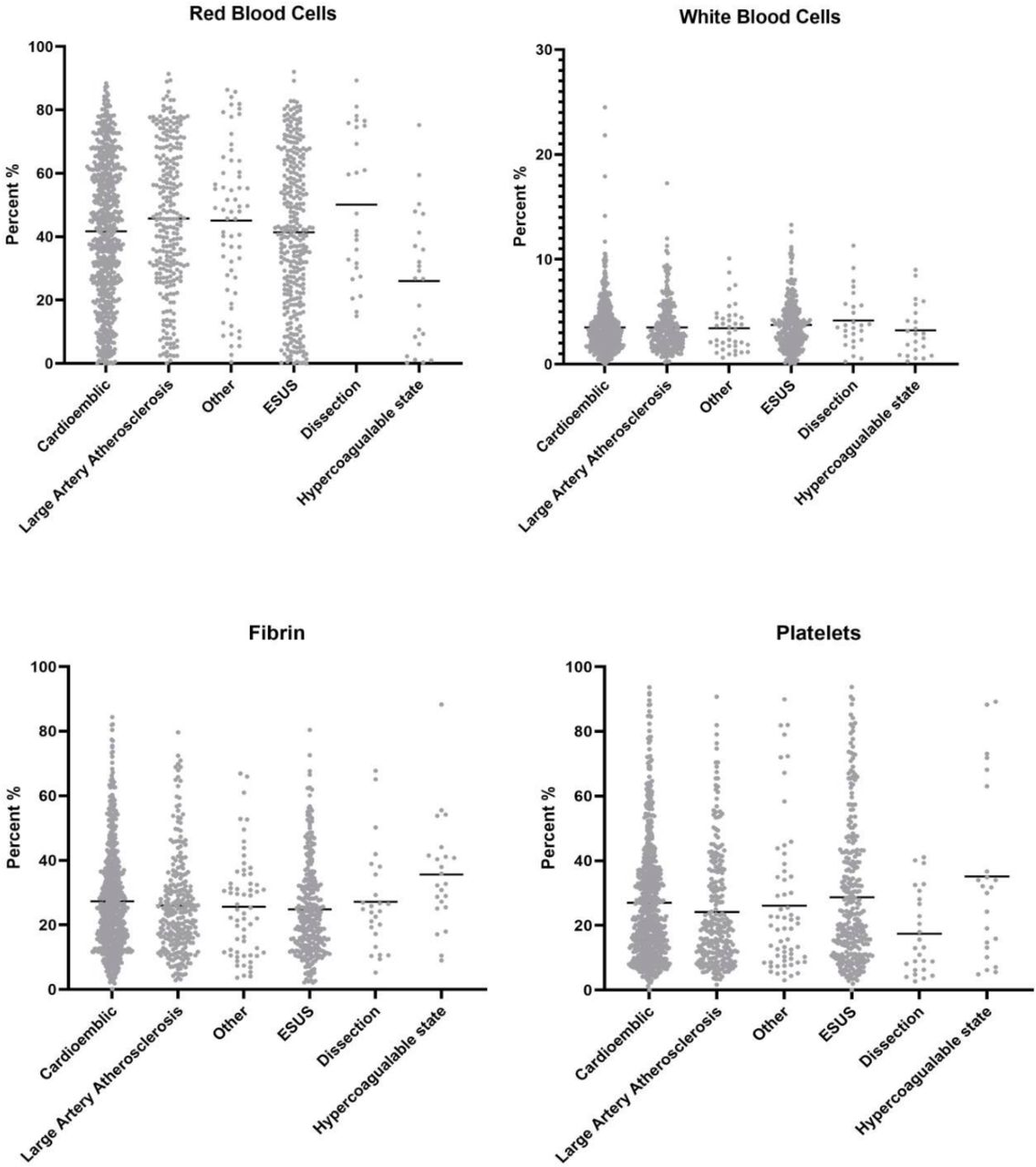
The histological results are summarized in table 1. There were significant differences in RBC density across groups ranging from 26±21% for patients with hypercoagulable state to 50±24% for patients with dissection (p=0.0004). Patients with LAA clots had a higher mean RBC density than those with CE clots (46±23% vs 42±22%, p=0.01). LAA clots also had a significantly higher RBC density than embolic stroke of undetermined source (ESUS) clots (46±23% vs 41±24%, p=0.02). All groups had a significantly higher RBC composition than clots from patients with hypercoagulable states (p<0.001). All other differences in RBC density were insignificant across groups. Figure 1 depicts the distribution of each component across different stroke etiologies.
Scatter plot showing each component of clot for various stroke etiologies.
Clot composition between groups
A total of 530 clots were considered RBC-rich (39%). Hypercoagulable state clots were the least likely to be RBC-rich (13%) while LAA and dissection clots were the most likely to be RBC-rich (44%). There was no difference in the proportion of patients with RBC-rich clots between the LAA and CE groups (44% vs 38%, p=0.11). Hypercoagulable state clots were less likely to be RBC-rich than all other groups (p<0.01).
Mean WBC density was similar across all groups ranging from 3±3% for hypercoagulable state clots to 4±2% for other groups (p=0.223). There were no significant differences in any of the pairwise comparisons.
There were significant differences in mean fibrin density across groups ranging from 25±14% for cryptogenic stroke patients to 36±17% from those with hypercoagulable states (p=0.01). Patients with CE clots had a slightly higher mean fibrin density than those with ESUS (p=0.02). Patients with a hypercoagulable state had a significantly higher fibrin density than all other groups (p<0.05 for each pair-wise comparison). All other differences in fibrin density were insignificant across groups.
A total of 129 clots were considered fibrin-rich (9.6%). Hypercoagulable state clots were the most likely to be fibrin-rich (17%) while ESUS clots were the least likely (7%). There was no difference in the proportion of patients with fibrin-rich clots between the LAA and CE groups (8% vs 11%, p=0.23). There was a significant difference in the proportion of patients with fibrin-rich clots across groups (p=0.28).
There were significant differences in mean platelet density across groups ranging from 18±13% for dissection patients to 35±27% for hypercoagulable state patients (p=0.002). CE patients had a higher platelet density than LAA patients (27±18% vs 24±18%, p=0.03) and a higher platelet density than dissection patients (27±18% vs 18±13%). ESUS patients had a higher platelet density than LAA patients (29±21% vs 24±18%, p=0.005). Hypercoagulable state patients had a higher platelet density than all other groups (p<0.05 for all comparisons).
A total of 169 clots were platelet-rich (13%). The proportion of platelet-rich clots ranged from 10% for LAA clots to 26% for hypercoagulable state clots (p=0.02). There was no difference in the proportion of platelet-rich clots between CE and LAA patients (12% vs 10%, p=0.36).
On ROC analysis, the area under the curve (AUC) for RBC density in differentiating CE from LAA clots was 0.55; the AUC for WBC density was 0.50; the AUC for fibrin density was 0.52; and the AUC for platelet density was 0.55.
Machine learning classification algorithm results
The stacked ensemble for the classification algorithm differentiating CE from LAA had a fivefold cross-validated AUC of 0.55 (area under the precision-recall curve of 0.33).
Other clinical results
On ANOVA analysis, there was no difference in the mean number of passes based on stroke etiology. The mean number of passes was 1.9±1.4 for CE, 2.0±1.5 for LAA, 2.0±1.7 for ESUS source, 2.7±2.3 for hypercoagulable source, and 2.1±1.4 for dissection (p=0.19). First pass TICI 2c/3 for these groups were, respectively, 42.2%, 37.9%, 39.3%, 26.9%, and 43.5% (p=0.18). Overall TICI 2c/3 rates were 70.5%, 65.9%, 66.5%, 61.5%, and 69.6%, respectively (p=0.07). Embolization to previously non-affected territories was significantly higher for dissection patients (13.0%) when compared with CE (2.1%), LAA (3.0%), ESUS (2.7%), and hypercoagulable state (4%) patients (p=0.02).
Discussion
Our study examining clot composition in a large cohort of ischemic stroke patients and its association with stroke etiology demonstrated a number of interesting findings. First, LAA clots had a higher mean RBC density and a lower mean platelet density than CE clots. On ROC analysis, it seems that identification of a reliable threshold with a high AUC for differentiating clots from these two etiologies based on composition analysis alone is not possible. The lack of a reliable histological biomarker on MSB between these two stroke etiologies suggests that conventional histological analyses looking at cellular composition do not provide insights into stroke etiology in cryptogenic cases. One interesting association that we found in our study was that thrombi from patients with a known hypercoagulable state had statistically and clinically significant differences in the mean RBC density and higher fibrin and platelet density than clots from any other known source. Ultimately, we feel these findings are important because they suggest that future investigations into what clues clots can provide for stroke etiology should be focused on more advanced testing such as proteomic composition, structural analyses and immunohistochemical markers rather than conventional histology.
A number of previous studies have examined the histological characteristics of LAA and CE clots to see if they could provide any insight into the source of cryptogenic strokes. Prior studies examining correlations between thrombus composition and stroke pathogenesis have focused specifically on RBCs, WBCs and fibrin/platelet compositions with inconclusive results. A study of 187 patients by Sporns et al found that CE emboli had few RBCs and more fibrin/platelets than non-CE emboli; however, they did not account for fibrin and platelet compositions.11 Meanwhile, a study by Kim et al of 37 patients found that CE clots were actually more likely to have a high RBC composition compared with those related to LAA.15 A similar study by Boeckh-Behrens et al of 145 patients found that CE emboli had higher proportions of fibrin and platelets and fewer RBCs than non-CE emboli.9 Lastly, a prior study from our group found that LAA clots were more likely to be platelet-rich and had a slightly lower RBC density than CE strokes.10 Our study of over 1300 patients showed no findings which allowed us to reliably distinguish CE and LAA clots.
Overall, the findings from our study and the disparate results from multiple prior studies suggest that routine histological staining with H&E or MSB probably will not allow us to differentiate clots of different etiologies and will likely play no role in determining stroke etiology in ESUS. However, some groups have looked into more sophisticated analyses of clot composition to help identify and differentiate stroke etiologies. Recently, Munoz et al have demonstrated the feasibility of mass spectrometry-based proteomic profiling of thrombotic material obtained by embolectomy in ischemic stroke and have help characterize the clot proteome.16 Meanwhile, other groups are examining the role of immunohistochemistry markers staining for proteins such as von Willebrand factor (vWF) and ADAMTS13 to point towards stroke etiology.17 Based on the findings from our study, we feel that histological analyses to determine stroke etiology are likely insufficient and future research should be directed toward more sophisticated analyses focused on immunohistochemistry, proteomics and molecular analyses.
One other feature that we did not specifically examine is the association between clot organization and structure and stroke etiology. While the relative proportion of each cellular component may not provide clues into stroke etiology, the way in which the components are organized might help in differentiation of the embolic source. For example, in a study by Di Meglio et al using scanning electron microscopy, the authors found that ischemic stroke thrombi had an outer shell of densely compacted fibrin, vWF and platelets that made them refractory to thrombolysis.18 Perhaps we may find that clots from CE sources and those from LAA sources are organized differently from one another with different surface components.
Limitations
Our study has limitations. While the MSB stain is more accurate than the H&E stain in identifying major clot components, it still does not specifically identify other potentially key clot components such as vWF and calcification. Therefore, the authors represent this subgroup as ‘platelet and other’ components as we acknowledge that there are potentially other components in addition to platelets in these regions. Immunohistochemical analysis using specific antibodies is the only way to distinguish accurately between platelets and platelet-related factors such as vWF. Also, the determination of stroke pathogenesis and etiology was self-reported at each site, and therefore there may have been some site-to-site variability in the interpretation and implementation of the TOAST criteria. For example, different sites likely had different thresholds to perform transesophageal versus transthoracic echocardiography or ultrasound bubble tests for CE sources. Lastly, there were no data on clinical outcome (ie, modified Rankin Scale) of patients included in the registry.
Conclusions
Our study of over 1300 retrieved emboli in ischemic stroke patients shows no consistent or reliable means to differentiate CE and LAA origin clots as determined by the MSB stain. Further research in this field should focus on more advanced techniques including immunohistochemistry and proteomic research.
Data availability statement
Data pertaining to this study are available upon reasonable request to the corresponding author.
Ethics statements
Footnotes
Twitter @VitorMendesPer1, @AlmekhlafiMa, @FitzSeanT, @PouyaNazari5
WB and MA contributed equally.
Contributors All authors have reviewed and approved the contents of this manuscript. This study is the result of a multicenter registry requiring the collaboration of multiple neurointerventionalists, research fellows and histopathologists and all authors meet the ICJME criteria for authorship.
Funding This work was supported by the National Institutes of Health grant number (R01 NS105853).
Competing interests None declared.
Provenance and peer review Not commissioned; externally peer reviewed.
Linked Articles
- Commentary