Article Text

Abstract
Background Posterior fossa arteriovenous malformations (AVMs) are considered to have a higher risk of poor outcome, as are AVMs with associated aneurysms. We postulated that posterior fossa malformations may be more prone to associated feeder vessel aneurysms, and to aneurysmal source of hemorrhage.
Objective To examine the prevalence and hemorrhagic risk of posterior fossa AVM-associated feeder vessel aneurysms.
Methods A retrospective review of AVMs was performed with attention paid to location and presence of aneurysms. The hemorrhage status and origin of the hemorrhage was also reviewed.
Results 571 AVMs were analyzed. Of 90 posterior fossa AVMs, 34 (37.8%) had aneurysms (85% feeder vessel, 9% intranidal, 15% with both). Of the 481 supratentorial AVMs, 126 (26.2%) harbored aneurysms (65% feeder vessel, 29% intranidal, 6% both). The overall incidence of feeder aneurysms was higher in posterior fossa AVMs, which were evident in 34.4% of infratentorial AVMs compared to 18.5% of supratentorial malformations (p<0.01). The presence of intranidal aneurysms was similar in both groups (9.2% vs 8.8%). Feeder artery aneurysms were much more likely to be the source of hemorrhage in posterior fossa AVMs than in supratentorial AVMs (30% vs 7.6%, p<0.01).
Conclusions Posterior fossa AVMs are more prone to developing associated aneurysms, specifically feeder vessel aneurysms. Feeder vessel aneurysms are more likely to be the source of hemorrhage in the posterior fossa. As such, they may be the most appropriate targets for initial and prompt control by embolization or surgery due to their elevated threat.
- Aneurysm
- Arteriovenous Malformation
- Hemorrhage
- Subarachnoid
- Posterior fossa
Statistics from Altmetric.com
Introduction
Brain or cerebral arteriovenous malformations (AVMs) are vascular lesions that have abnormal direct connections between the arterial and venous systems, without intervening parenchymal-perfusion small vessels. They are relatively rare—estimated to have a prevalence of 0.002–0.5%1 ,2—and associated aneurysms have been reported from 2.7% to 21% of the time.3 ,4 These can further be subclassified as feeder (or pedicle) aneurysms or intranidal aneurysms.5 ,6 This excludes aneurysms on the venous drainage side of the malformation. The aneurysm location has differing implications for treatment and outcome.7 The literature conflicts as to whether intranidal8 or feeder aneurysms carry the higher risk of a hemorrhagic presentation or re-rupture before treatment,4 ,9 although there is generally consensus that these are high-risk features compared with those AVMs without associated aneurysms.2 ,4 ,5 ,8–11
Posterior fossa AVMs have been reported to be an independent predictor of increased morbidity and mortality. The percentage of AVMs presenting with hemorrhage ranges from 30% to 82%.12 Initial presentation with hemorrhage can lead to mortality rates as high as 10–30%.2 ,13 The increased morbidity and mortality of posterior fossa AVMs has also been illustrated,10 ,14–17 even though they represent only 5–15% of all intracranial AVMs.1 ,14 ,15 ,18 This may be due to more frequent presentation with hemorrhage,4 ,7 ,18 or to the more dire consequence of hemorrhage in an enclosed space with highly eloquent functional areas despite often smaller hematoma volumes after rupture.10 We hypothesize that posterior fossa malformations are also more prone to associated aneurysms—specifically feeder vessel aneurysms—and, when present, are more frequently the source of hemorrhage.
Methods
After institutional review board approval, a retrospective review of AVMs treated at our institution from 1995 to January 2015 was evaluated. Demographic data, radiologic characteristics, and clinical parameters were collected, with attention paid to location and presence of associated aneurysms. These associated aneurysms were designated as feeder vessel aneurysms, intranidal aneurysms, or both. The hemorrhage status and origin of the hemorrhage was also reviewed. Two endovascular neurosurgeons determined the site of hemorrhage origination by radiologic review of the presentation CT scan and cerebral angiogram. An extensive literature review was performed for comparison of results with published reports.
Statistical analysis
The demographic data and proportion of AVM patients with feeder aneurysms were compared between a supratentorial and infratentorial location using the χ2 test. ORs were used to evaluate the source of bleeding from a feeder aneurysm in infratentorial AVMs compared with supratentorial AVMs. Statistical analyses were performed using SAS software V.9.4 (SAS Institute, Cary, North Carolina, USA).
Results
Patient characteristics
A total of 571 AVMs that had identifiable locations were analyzed. The age range of the patients was 19 days to 87 years (average 41 years) and the gender was evenly distributed, with 288 women and 283 men. Two hundred and eighty-four (49.6%) of these AVMs initially had a hemorrhagic presentation. One hundred and sixty AVMs (28.0%) had associated aneurysms, 120 (21.0%) of which had associated feeder aneurysms. Of the 571 AVMs, 90 were infratentorial.
Incidence of feeder aneurysms
Of the 90 AVMs located in the posterior fossa (including cerebellar and brainstem locations), 34 (37.8%) had aneurysms: 29 (85%) with at least one feeder vessel aneurysm, 3 (9%) with intranidal aneurysms, and 5 (15%) with both. Of the 481 supratentorial AVMs, 126 (26.2%) harbored aneurysms, 82 (65%) of which were feeder aneurysms, 37 (29%) intranidal, and 7 (6%) both. The overall incidence of aneurysms was appreciably higher in posterior fossa AVMs. This association was most pronounced for feeder aneurysms, which were evident in 34.4% of infratentorial AVMs compared with 18.5% of supratentorial malformations (p<0.01). The incidence of intranidal aneurysms was similar in supratentorial and infratentorial locations (9.2% vs 8.9%; table 1).
Distribution of patients with infratentorial versus supratentorial AVMs, with hemorrhagic presentation between the two groups
Feeder aneurysm rupture status
When evaluating for rupture site, 18 of 60 hemorrhages in the posterior fossa could be attributed to a feeder aneurysm, yielding a 58% rupture incidence for those aneurysms, and accountable for 30% of ruptures among infratentorial AVMs. In comparison, 17 of 224 supratentorial hemorrhages could be clearly attributed to a feeder aneurysm, yielding a 19% aneurysm rupture incidence and accounting for 7.6% of total supratentorial AVM hemorrhages (p<0.01). This amounts to an OR of rupture from feeder aneurysms at the time of presentation of 5.86 (CI 2.4 to 14.2; p=0.001) for infratentorial versus supratentorial hemorrhages.
Illustrative case
A middle-aged patient with no past medical history presented with 1 week of unrelenting headache, which had initially occurred postcoitally. Initial head CT demonstrated a hemorrhage measuring 2 cm×8 mm around the cerebellar vermis and he was admitted to the neurosurgical service (figure 1A). CT angiography (CTA) showed an underlying vermian AVM, and the initial angiogram demonstrated a Spetzler–Martin grade II, Spetzler–Ponce grade A malformation measuring 2 cm and fed by the superior cerebellar artery (SCA) and posterior inferior cerebellar artery (PICA) (figure 1B). It drained into the straight sinus, as well as the transverse sinus and torcular herephili. There were associated feeder and intranidal aneurysms of the SCA (figure 1C) and PICA. Embolization of all feeder aneurysms was performed using n-butyl cyanoacrylate (n-BCA) (Codman Neuro, Raynham, Massachusetts, USA) in one session (figure 1D). Postembolization angiographic images demonstrated occlusion of all aneurysms. Two weeks later, suboccipital craniotomy was performed for resection of the residual malformation with no resultant complications or symptoms (figure 1E).
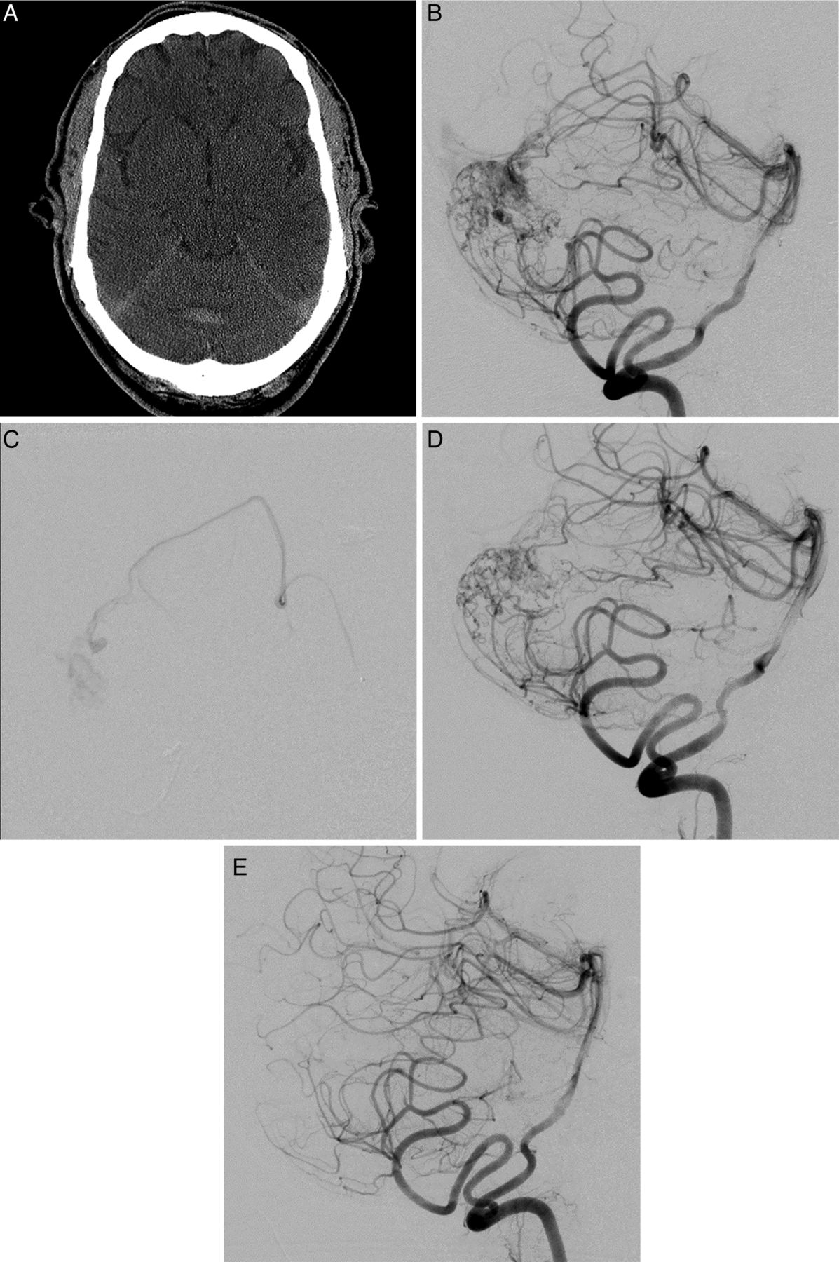
(A) Initial non-contrast head CT and CT angiography showing vermian cerebellar hemorrhage and right paravermian arteriovenous malformations (AVM). (B) Initial angiogram image, lateral, demonstrating a paravermian Spetzler–Ponce grade A AVM with associated posterior inferior cerebellar artery (PICA) and superior cerebellar artery (SCA) aneurysms. (C) Selective catheterization of right SCA pre-embolization. (D) Post-embolization angiogram showing absence of associated aneurysms with residual AVM nidus. (E) Postoperative angiogram, lateral, demonstrating complete resection of the AVM.
Discussion
Comparison of data in the literature on the topic of AVM-associated aneurysms can be difficult due to varying classification schemes and differing denominators for reported prevalence. As shown in table 2, the occurrence of associated aneurysms is listed as the percentage of total AVMs (ie, supratentorial and infratentorial) in some papers, while others only evaluated infratentorial malformations. Some only cataloged ruptured presentations while others reported all AVMs in an institution. The aneurysms themselves are described alternately as ‘feeder’, ‘proximal’, or ‘prenidal’ aneurysms to separate them from ‘intranidal’/‘nidal’ or ‘remote’/‘unassociated’ aneurysms. For instance, Platz et al19 classifies AVM-associated aneurysms as feeding (type 1), nidal (type 2), or unassociated (type 3), but further divides type 1 into 1a and b, where 1a is closer to the circle of Willis than the malformation itself while the opposite is true with type 1b. In contrast, Yu et al20 used a four-tier classification system where I was unassociated, II was flow-related but at the proximal portion of a feeding artery, III was ‘dissecting type’ on a feeding artery, and IV was intranidal. Some papers do not subclassify feeder, remote, or intranidal aneurysms at all. However, the overall incidence of aneurysms associated with AVMs from our literature review is 2.6–47%. In the posterior fossa specifically, the incidence is 20.8–47% (table 2).
Literature review presenting the incidence of associated aneurysms with AVMs with respect to location and hemorrhagic presentation
Our results support previous implications that feeder aneurysms are more common infratentorially.21–24 The reason for this increased incidence and predilection for hemorrhage is unclear, although the differential impact of AVM hemodynamics in smaller posterior fossa vessels may be relevant. Increased wall shear stress (WSS) and, to some degree, smaller vessel size has previously been demonstrated as a risk factor for supratentorial feeder vessels to be associated with aneurysms;25–27 thus, smaller vessel feeders such as the PICA and SCA, as encountered in the posterior fossa, may be more prone to developing increased WSS in the setting of high AVM flows and, consequently, aneurysms.21 ,26
This study is drawn from one of the largest databases of AVMs reported. Only three other groups have reported on a volume of patients larger than this one, ranging from 600–662, some by joining data from multiple centers.6 ,8 ,23 In correlation with the majority of the literature, we demonstrate no statistically significant gender predilection for hemorrhage or associated feeder aneurysms, although articles such as those by Platz et al19 and Thompson et al23 did find a difference, with increased associated aneurysms and hemorrhagic presentation, respectively, in women. In our study, slightly more men presented with hemorrhage, although more women had associated aneurysms. However, we demonstrate, in a large single-center sample, an increased risk of hemorrhagic source specifically from AVM-associated aneurysms in the posterior fossa in comparison with supratentorial locations, with an OR of 5.86.
AVMs have been represented in the literature most recently due to controversy over the best treatment regimen, which has been heavily debated, especially for lower grade lesions. The ARUBA trial made sweeping recommendations to leave all of these malformations to conservative medical management only. There has been significant discussion as to whether low-grade lesions should, instead, be addressed using surgical resection as the primary modality. Many authors and experts advocate a multimodality approach to many of these malformations,16 ,22 ,28 especially high-grade lesions. The literature suggests that posterior fossa AVMs represent an especially dangerous subset of lesions, with associated aneurysms an especially high-risk feature. We suggest, as have other authors,29 ,30 that immediate elimination of the feeder aneurysm should be a priority, often by endovascular means, if it cannot be addressed during the surgical resection easily.
In this study, the designation of the site of origin of the hemorrhage was somewhat subjective as this could not be performed blind to the location of the AVM. However, every effort was made to report the source definitively only if the location was disparate enough to be clear and often reinforced by the location of n-BCA liquid embolisate after embolization on CT. The hemorrhage rates reported to be attributable to the feeder aneurysms are, if anything, likely to be underestimated. Only those that could be definitively attributed to the aneurysm due to some distance or an appropriate trajectory were documented as such, whereas those that could have been either from the aneurysm or the AVM itself—or an intranidal aneurysm—were not. Other study limitations include the single-center nature of the data; as a tertiary referral center, our data may not be generalizable as the true incidence of aneurysms in AVMs. However, referral bias is unlikely to be AVM location-specific, thus our comparison between supratentorial and infratentorial locations maintains validity. Given the retrospective nature of the data, we cannot make definitive statements as to the prospective risk of hemorrhage attributable to associated aneurysms, but we have identified the increased likelihood that posterior fossa AVM feeder vessel aneurysms are significantly more likely to be the source of hemorrhage in ruptured AVMs of this location.
Conclusion
Posterior fossa AVMs are more prone to developing associated aneurysms, specifically feeder vessel aneurysms. Compared with supratentorial AVMs, feeder vessel aneurysms are also more likely to be the source of hemorrhage in the posterior fossa. These data support targeting posterior fossa feeder aneurysms for initial and prompt control by embolization or surgery.
Acknowledgments
Part of this manuscript was presented at the 12th Annual Meeting of the Society of NeuroInterventional Surgery in San Francisco, California, USA in July 2015.
References
Footnotes
Contributors JO: design of the work, including the acquisition, analysis, and interpretation of data, drafting the paper, revising it critically for important intellectual content, and contribution to the final approval of the version to be published. SA-H: design of the work, drafting the work, revising it critically for intellectual content, and contribution to the final approval of the version to be published. YH: acquisition of data, analysis, and final approval of the version to be published. XD: statistical analysis. ZH: design of the work and final approval of the version to be published. VA: design of the work, interpretation of the results, drafting of the paper and final approval. FC: revising the manuscript and final approval of results. AA: design of the work, drafting the work, revising it critically for important intellectual content, and final approval of the version to be published.
Funding This study is partially supported by the Dr Ralph and Marian Falk Medical Research Trust, fund # 629138.
Competing interests AA and VA are consultants for Cordis-Codman.
Ethics approval Ethics approval was obtained from the University of Illinois at Chicago Institutional Review Board.
Provenance and peer review Not commissioned; externally peer reviewed.
Data sharing statement All relevant data have been presented in this paper. The entire data are protected and available from the principal investigator. More detailed clinical and demographic data are unidentified and locked in the office of the corresponding author and are available on request.