Article Text

Abstract
Introduction Following satisfactory benchtop testing of a new liquid embolic agent, animal implant studies were performed.
Materials and method Elastase aneurysms were created in the right common carotid artery of New Zealand rabbits under approved institutional guidelines. Using direct fluoroscopic control and commercially available microcatheters, the device was introduced into the aneurysms. At 2 months, 12 months and 24 months, follow-up angiography was performed and analyzed. The animals were sacrificed, the brachiocephalic arteries were explanted and fixed, and the histologic appearance of the treated aneurysms was evaluated.
Results The Neucrylate polymerized into an open pore elastic sponge. The open pores permitted fibrous tissue ingrowth. By 2 months, all of the aneurysm necks had been covered by fibrous tissue and a neointima. Two of the aneurysms originally inadequately filled allowed opportunity for retreatment. The reactive change within the aneurysms demonstrated fibroblastic proliferation, collagen and some giant cells but no vascular necrosis. Results at 2 months, 12 months and 24 months were for all practical purposes similar.
Conclusion The lack of necrosis, the mild inflammatory response and the permanence of the implant are interesting in a cyanoacrylate based embolic agent, especially in light of the experience with lower chain homologs and other liquid embolic agents.
Statistics from Altmetric.com
Introduction
Endovascular coil therapy of cerebral berry aneurysm has, in many centers, become the standard of care.1–3 Unfortunately, many aneurysms of complex shape are not amenable to coil packing, and given the recurrence and failure rate of coil treatments,4–13 an additional treatment tool could be valuable to the neurointerventional surgeon. But before any human trials are undertaken, the intra-arterial behavior of this device should be studied in an animal model.
Based on the results of benchtop (previously reported) and unpublished animal feasibility studies, we decided to begin intravascular implant evaluation of this new endovascular liquid embolic device, Neucrylate AN. Neucrylate AN is a cyanoacrylate based liquid embolic system whose properties have been specifically crafted and modified to fill cerebral berry aneurysms.
We have implanted this device into rabbit elastase induced aneurysms and now report the endovascular behavior, gross explanted appearance of the aneurysms and brachiocephalic vessels, and the histologic results of that study.
Materials and methods
Aneurysm creation
Under general endotracheal anesthesia (ketamine 35 mg/kg, xylazine 5 mg/kg, glycopyrrolate 0.03 mg/kg, buprenex 0.04 mg/kg, enrofloxacin 10 mg kg), administered and monitored by a veterinary physician, following all institutional animal use guidelines and approval of the Institutional Animal Care and Use Committee, aneurysms were created in the proximal right common carotid artery of 2.8–5 kg albino New Zealand rabbits using the elastase digestion technique.14 15
The rabbits were recovered, and for 2–8 weeks were fed a normal laboratory diet.
Aneurysm treatment
The animals were brought back to the operating room, anesthetized, a cutdown was created in the right common femoral artery and a 5 F sheath was introduced into the artery. Through that sheath, using fluoroscopic guidance, a 4.0 mm semicompliant balloon microcatheter (usually the MIS occlusion balloon catheter, but also the Hyperglide balloon microcatheter; Micro Therapeutics Inc, now ev3 Neurovascular, Irvine, California, USA) was directed across the neck of the aneurysm keeping it within both the proximal and distal arterial lumen to bridge the aneurysm. Also through the sheath, a second microcatheter with a tip diameter of 0.022 inches (such as the Excelsior SL10, Boston Scientific Neurovascular, Natick, Massachusetts, USA), was placed into the aneurysm sac—its tip in the mid-portion of the aneurysm.
Then, using real time fluoroscopic control, Neucrylate AN, a liquid radio-opaque device (a proprietary compound of plasticizers, additives and 1 hexyl n cyanoacrylate; VALOR Medical Inc, San Diego, California, USA) was introduced into the aneurysm lumen, attempting to place it adjacent to the balloon. The adequate and effective quantity was determined by viewing the real time fluoroscopic image of the aneurysm as the treatment progressed (figure 1).

Left frontal angiogram, rabbit 034. Contrast agent has been injected through the guiding catheter. The oval is a US dime coin used for comparative measurements, and since it is on the skin, some overestimation of the size of the aneurysms is expected. (A) The aneurysm and the relationships to the other brachiocephalic vessels are shown. Note the oblique nature of the neck and the differing size of the parent vessel immediately before and beyond the aneurysm neck. (B) At 1 year, the radiodensity of the Neucrylate remains densely opaque and has not changed in size or appearance from the immediate pretreatment angiogram. Now, however, some radiolucent material (shown to be fibrous tissue in the histologic specimen) separates the dense Neucrylate from the contrast agent. A slight but definite aneurysm recurrence is evident. Compare with figure 2B, C.
Vessel explant
Again following institutional rules, five animals were sacrificed 2 months following implantation, three animals were sacrificed at 12 months and three animals were sacrificed at 24 months. Three additional rabbits died or were sacrificed for other reasons, as shown in table 1. Untreated but aneurysm induced rabbits that died from other systemic causes provided some limited control analyses. As many of the aneurysms were treated before maturation (because of logistical and travel problems), there were opportunities for retreatment because of aneurysm continued growth.16
The experiment in general and gross post mortem findings
At time intervals proposed in the original protocol (2 months, 1 year and 2 years), the rabbits were brought back to the operating room, anesthetized and a follow-up angiogram was performed. They were then sacrificed, and after sacrifice the vessels were fixed in situ with neutral buffered formalin perfused intra-arterially under physiologic pressures. After 30 min, the brachiocephalic vessels were explanted, examined for gross changes—especially for necrosis and for periadventitial inflammation or scarring—and were placed in neutral buffered formalin.
After complete fixation, the tissues were dehydrated, embedded in paraffin, sectioned, stained and then a minimum of four sections of each specimen were examined microscopically. As standard staining procedures, in particular the xylene used in most steps, dissolve cyanoacrylate, the preparation was carried out by hand, according to the process described by Galil and colleagues17 and by Lundie and colleagues.18
The microscopic sections were evaluated for degree and type of inflammatory change, whether there had been shrinkage of device away from the aneurysm wall, whether there was ingrowth of fibrous tissue into the device, any compaction or change in size of the device, the presence of wall necrosis and covering of the aneurysm neck either with fibrous tissue or with neointima. The degree of inflammatory change was graded using the ordinal scale proposed by Dai et al19 (table 2).
Aneurysm character, size and histologic findings
Results
Rabbit mortality
Although rabbits are known to be sensitive animals to anesthesia and surgical manipulation, we were surprised by the high mortality rate found during these experiments. We could find nothing in the recovery phase or in the subsequent laboratory care to explain our high death rate from the procedures. A re-evaluation of the induction protocol was performed (without effect) and we began treating all animals prophylactically with a proton pump inhibitor. Thereafter, no additional gastric ruptures occurred.
Endovascular behavior
There was no significant change in the behavior of the device in the animal aneurysms compared with the benchtop aneurysm replica tests previously reported. The device passed easily through microcatheters, and even in the face of the hyperdynamic arteries found in the rabbit, could be visualized easily (figure 1). The vessel excursion combined with the limitations of the available fluoroscopic equipment did preclude our use of combining mechanical ventilation with roadmap technique, which has been described as a solution to this motion problem.20 21 This lack made precise deposition of the device against the balloon difficult. Consequently, many of the aneurysms were incompletely filled.
One of the older MIS balloons ruptured during infusion of the device, which allowed passage of Neucrylate into the distal branches. This untoward embolization led to cerebral infarction and death of one animal.
In all but one animal the microcatheters and balloons were removed without difficulty and without adherence of Neucrylate to either. The one exception, an early animal, a miscommunication between operators allowed one of us to pull the entire polymerized device from an aneurysm. The nearly spherical device mass was brought down into an iliac artery, removed from the catheter and then the entire treatment procedure was repeated with satisfactory results.
Gross appearance
The treated aneurysms and surrounding brachiocephalic vessels, including the aortic arch, were removed from the animals without difficulty; there was no visible or palpable inflammatory response surrounding any of the aneurysms. There was no evidence of necrosis.
In general, the aneurysms were relatively small and had broad oblique necks (tables 2 and 3). Because of the anatomic relationships of elastase aneurysms—close to the opposite carotid and close to the ipsilateral vertebral artery—aneurysm neck relationships were complex. Further complexity was caused by the parent vessel just proximal to the aneurysm neck (the innominate artery) being larger than the subclavian artery at the distal portion of the neck.
Histology
Figure 2 demonstrates the responses seen in a typical treated aneurysm, changes that were remarkably consistent and similar in each of the specimens.

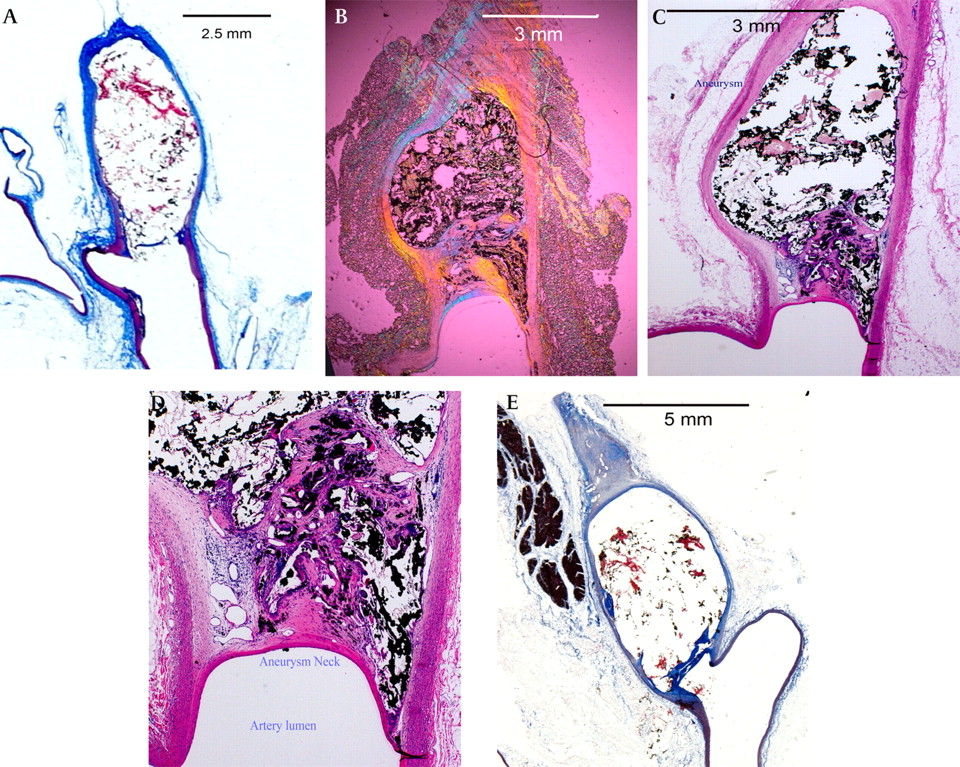
Histologic preparations of typical aneurysms. (A) Rabbit 024 sacrificed 2 months following treatment. The histologic trichrome preparation has essentially dissolved all of the device but the black particles of gold remain. The pink material appears to be collagen that has grown in through the interstices. The fibrous tissue covering the neck of the aneurysm is thin compared with the later more mature 12 and 24 month specimens. (B). Rabbit 034 sacrificed 12 months following treatment (compare with figure 1B). This dark field illumination before histologic preparation shows the extent of the device within the aneurysm. The open pore character is visible. (C). Rabbit 034, haematoxylin–eosin preparation. Compare with (B). There is almost no inflammatory change within the walls of the aneurysm. Dense fibrous tissue covers the neck. (D) Rabbit 034, close-up of neck of the aneurysm. On the left, neovascularity is evident. Toward the right, a few giant cells are visible, but primarily the tissue covering the neck is dense fibrous tissue, probably fibroblasts and collagen fibers. A single cell covering separates the fibrous tissue from the flowing blood. Although this cellular layer has the appearance of intima, our repeated immunohistochemistry attempts failed in every specimen, so we cannot confirm that these cells are true endothelium. (E) Rabbit 046 sacrificed 24 months following treatment. This low power trichrome stained view demonstrates that the majority of the Neucrylate has been dissolved but the dense fibrous tissue at the neck remains.
There was no significant change in the amount or location of inflammation of the specimens at the three sacrifice times. The primary changes consisted of dense collagen, fibroblastic spindle cell infiltration and occasional neovascularity. The neck was covered by neointima (multiple attempts at immunohistochemistry were unsuccessful in determining whether the neointima was actually composed of endothelial cells).
Polymerized Neucrylate AN has a rubbery tough physical character and so it was difficult for the histology technologist to microtome the paraffin embedded tissues, and on many of the cuts the device did not exactly register with the artery wall. Worse, despite our hand processing the specimens and using the described preparation technique,17 18 much of the device dissolved during final mounting, which requires a xylene based plastic. Sadly, the europium fluorescence visualization process described by Calvo et al22 was not available to us.
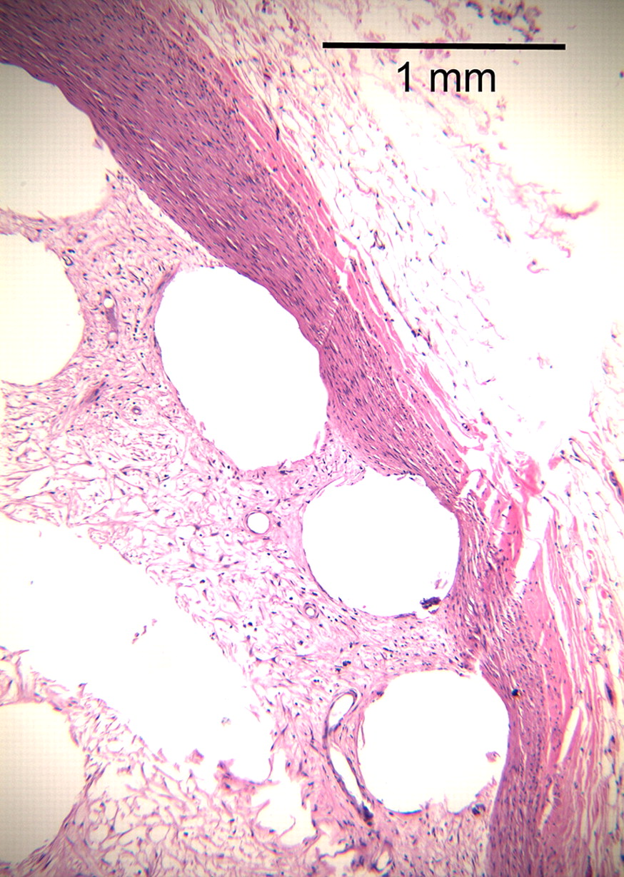
In figure 2A, an unstained specimen viewed with polarizing light, the actual extent of the device and its close apposition to the aneurysm walls can be appreciated. Polarized light was the only method we found that did not disturb the relationships of Neucrylate to the wall, and allowed us to evaluate the actual appearance and extent of the device within the aneurysm, and to search for clefts. In no specimen did we see separation of the device from the aneurysm wall. Importantly, in the unprocessed images, the open pore character of the polymerized device was best appreciated (see also figure 4).
In all stained specimens (figure 2B), mild chronic inflammatory changes consisting of plasma cells and lymphocytes occupied portions of the wall but were more concentrated at the junction of the new neck tissue with the artery wall. These inflammatory changes did not extend outside the wall into the adventitia or into the periadventitial fat. Occasionally, multinuclear giant cells occupied portions of the domes but were more frequently seen in the dense collagen-like fibrous tissue that had formed near the necks (see especially figure 2C,D). The fibrous tissue at the neck consisted mostly of spindle shaped cells, probably fibroblasts. Although the preparation dissolved much of the cyanoacrylate, some residual pink amorphous Neucrylate was visualized in each specimen but the true extent of Neucrylate was determined only on the unstained specimens viewed with polarized light.
In the stained specimens, the black gold particles were conspicuous and, because of their geometry, it could be inferred that fibrous tissue had grown in and through the open sponge of the polymerized device (see also figure 4). Fibrous tissue was seen throughout the central portions of all stained aneurysms.
Importantly, in all rabbits, dense fibroblastic response occurred at the aneurysm neck, and a smooth, endothelial-like neointima had formed to cover the neck. In no specimen were there excrescences or focal cavities remaining which might disturb blood flow: the blood/device interface was uniformly smooth.
Many of the aneurysms had continued to grow despite adequate filling, especially in the region of the neck. Several parent arteries expanded as well (see table 2).
In summary, each cyanoacrylate treated aneurysm contained solid but flexible sponge-like material, material that in its interstices had allowed the ingrowth of usually dense elongated spindle-shaped cellular material whose microscopic appearance was consistent with fibroblasts and collagen. Importantly, each neck was filled with dense fibrous tissue whose surface was covered with smooth, flattened cells having the appearance of endothelial cells.
Discussion
Some thoughts about the model
If intra-arterial device testing is difficult, testing intra-aneurysmal treatment devices is particularly problematic because the traditional model, the German and Black surgically constructed sidewall aneurysm,23 does not replicate the complex flow dynamics found in the human.
In 1998, Miskolczi et al developed a rabbit aneurysm model that provided a close to ideal testing milieu for endovascular, particularly intra-arterial, devices.14 White New Zealand rabbits have a coagulation system similar to humans, their brachiocephalic vessels are of similar size as the intracranial vessels in humans and, most importantly, the aneurysms that develop following elastase digestion are subjected to the high shear stresses and impact pressures found in human berry aneurysms. Finally, the aneurysms frequently continue to enlarge if treated before maturation, as did many of ours, and can provide an opportunity for retreatment.16
An extensive literature has accumulated since Miskolczi's original paper—a literature describing device behaviors and a wall response to the introduction of foreign devices24–28 (figure 4). There is a relative scarcity of histologic data for human aneurysms treated with platinum coils, but in large aneurysms it appears that the intraluminal thrombus caused by and contained within the coils remains chronically unorganized.28 More importantly, the evidence suggesting a 15–30% recurrence rate shows that bare platinum coils lack significant stimulus to organize a significant dense fibroblastic response (see figure 3). Fibrosis should be desirable, as it would avoid the results of coil compaction, and presumably lead to a longer term efficacy of treatment.
Section through the wall of a platinum coil treated elastase aneurysm. Courtesy of David Kallmes and colleagues. The open circles are of the ghosts of the previously removed coils. Note the loose granulation-like tissue between the coil ghosts. Compare this with figure 2D.

An explanted Neucrylate bolus from an endovascularly treated silicone aneurysm. The aneurysm had been filled by a physician during a training session. The open pore nature of the sponge is evident. The pink material is the Neucrylate, the light yellow are gold particles and the orange regions are aggregates of red blood cells.
The only disadvantages to the rabbit model is that the entry site—the femoral artery—is relatively small and, consequently, in one smaller rabbit, we destroyed the artery during sheath insertion. Further, the brachiocephalic vessels are hyperdynamic, making it more difficult to use a subtraction technique—more about that below.
The endovascular technique
In this model, we have introduced and evaluated a liquid device that was intended to fill aneurysms of complex shape and secondarily to provide a smooth bypass for the blood away from the aneurysm. Using all current endovascular procedures, techniques, microcatheters and guidewires, a balloon microcatheter was placed across the neck of the aneurysm in the parent vessel as the surgeon would, for example, do to introduce a stent. Then a microcatheter was placed into the aneurysm itself, without difficulty, and the device was infused using real time fluoroscopic control. There was no difference in the behavior of the device, despite the hyperdynamic movement of the arteries compared with the benchtop aneurysm models.
The gross findings
That no periadventitial inflammatory change or actual extravasation was found is reassuring, especially when compared with the known long term angionecrosis caused by other cyanoacrylate devices.
In light of the high rate of device migration and parent vessel occlusions reported in the CAMEO trial which evaluated ethylene vinyl alcohol copolymer,29 and in later trials that showed a parent vessel occlusion rate by Onyx 500 of some 12%,30 31 we especially searched for any abnormality in the region of device deposition—and found none. We searched distally only with the fluoroscope and did not find distal embolization, although a more careful microdissection or microradiography would have been more accurate. In one animal, because of our technical error, we allowed some device to pass proximally into the innominate and then into the left carotid artery. It caused no vascular stenosis or occlusion on follow-up.
The histologic responses
Despite our using the technique described by Galil et al17 and Lundie et al,18 a majority of the Neucrylate was either distorted or removed during histologic processing, and only ghosts of the Neucrylate that had been introduced into the aneurysm remained. Artifacts were also introduced by the particulate gold, and by the difficulty in cutting through the cyanoacrylate by even new microtome knives. We were unable to re-create the europium fluorescence technique described by Calvo and colleagues.22
We looked for separation—clefts—along the artery wall, as shrinkage of some liquid devices (the vinyl alcohol copolymers) allow blood to enter aneurysms following treatment.4 No separation from the wall or change in size of the device was found, nor was there any evidence of device compaction (comparison made by measuring the device size at the post-treatment angiogram and comparing it with the angiogram performed at 12 and 24 months).
That the implant caused only a mild arterial wall inflammatory response is interesting, especially in light of other reports. Sadato et al found that rabbit vessels embolized with another cyanoacrylate, the n butyl (4 carbon) homolog reacted with an extensive foreign body response,32 and Mazal et al found that brain arteriovenous malformations treated with the same material developed acute inflammation to the point of angionecrosis, a finding that persisted for years.33
The negatives
This model can be criticized on several levels. Firstly, we must acknowledge that there is no perfect replica of a human aneurysm, with walls of varying thinness and fragility. Secondly, our lack of quality fluoroscopic image amplification and subtraction technique often made it technically impossible to fill the aneurysms completely. Thirdly, aneurysms treated in this study were generally small, and they usually had wide necks.
Another problem with our experiments was the continuing growth of some aneurysms, a result of treatment before maturation, a maturation that usually occurs by 4 weeks.16 These operational and logistic errors were ours, and were not the fault of the host laboratory.
This treatment before maturation (and possibly some technique errors in containing the elastase) not surprisingly caused continuing aneurysmal enlargement in many of our specimens and although at first glance such instability might be considered a disadvantage of this preparation, continued growth did in fact allow for retreatment, simulating human aneurysm failed coil treatments. But there is another way to look at the continued enlargement of the aneurysm. It is possible that the enlargement may have occurred because of some inadequacy of the device itself and not the model preparation. That possibility must be evaluated during the human safety study.
And finally
Modest inflammation, the first stage in healing, is the desired response when the practitioner wishes a permanent arterial occlusion. On the other hand, it is important that the inflammatory change does not progress to the point of wall necrosis, lest rupture occur, especially in human thin wall aneurysms, particularly those containing a Murphey's teat. In all stained specimens, mild chronic inflammatory changes consisting of plasma cells and lymphocytes developed within the aneurysm wall but were more concentrated at the junction of the new neck tissue with the aneurysm wall. These inflammatory changes did not extend outside the wall itself into the adventitia or the periadventitial fat. Multinuclear giant cells were generally concentrated in the dense fibrous tissue that had formed near the necks (see especially figure 2C,D). The fibrous tissue at the neck consisted mostly of spindle shaped cells, probably fibroblasts. Although the preparation dissolved much of the cyanoacrylate, some residual pink amorphous Neucrylate was visualized in each specimen but the true extent of Neucrylate was determined only on the unstained specimens viewed with polarized light.
Importantly, in every rabbit, dense fibroblastic response occurred at the aneurysm neck, and a smooth, endothelial-like neointima covered each neck. There were no excrescences or focal cavities remaining following the treatment that might disturb blood flow.
There was surprisingly little histologic difference in the 2 month, 12 month and 24 month specimens, likely indicating stability of the device.
But why does this device allow fibroblastic ingrowth whereas coils do not? The answer may lie in the physical character of the polymerized mass. Figure 4 is an explanted polymerized Neucrylate mass created in a silicone aneurysm replica. The open sponge-like nature of the device is evident. This physical character plus the lack of device toxicity likely accounts for the ability of fibrous tissue to grow into the Neucrylate.
Most importantly, we were impressed by the ability of the endothelial-like cells to grow over the aneurysm neck, and by the ingrowth of fibrous tissue through the interstices of the spongy cyanoacrylate mass. These two characteristics may provide smooth blood flow and long term effectiveness of the Neucrylate device when used in humans.
Conclusion
The rabbit elastase digestion model produces a high shear and relatively stable aneurysm model, allowing us to implant and study a new device using basic endovascular catheter techniques. Given the large literature that has accumulated showing both the histologic behavior of the model and the behavior of various coil implants, we have been able to compare the histologic response of Neucrylate to the prior reports. We found no compaction, a desirable modest chronic inflammatory response, dense fibrous tissue at the neck and a smooth, endothelial-like cover at the blood/device interface. There was no technical difficulty in performing these treatments, and endovascular surgeons who have experience in basic interventional techniques should be able to use this device after the specific training we have described in our prior paper.
Key messages
The development of endovascular treatment of cerebral berry aneurysm has increased in safety and efficacy significantly during the past 20 years, but endovascular treatment remains difficult, and its failure rate and utility can be improved upon. We hoped to add to the neurointerventional surgeon's armamentarium by developing a new liquid endovascular device. Neucrylate AN, a proprietary compound of hexyl cyanoacrylate, has passed toxicology testing and is now entering the pathway to achieve human use. In this second of three papers, we describe the gross and histologic results of implant studies in rabbit elastase induced aneurysms. The results demonstrate device filling the entire aneurysm, a mild inflammatory response and no evidence of resorption, even after 2 years.
Acknowledgments
The authors sincerely thank Ms Jacqueline Rodriguez for her untiring efforts to make satisfactory histologic slides from this difficult material.
References
Supplementary materials
JNIS editor Rob Tarr talks to Chuck Kerber in the JNIS podcast
Footnotes
Competing interests Charles Kerber MD is a shareholder and chief medical officer in Valor Medical Inc. Ramin Sean Pakbaz is a shareholder in Valor Medical Inc. There are no other competing interests.
Ethics approval This study was conducted with the approval of the Institutional Animal Care and Use Committee.
Provenance and peer review Not commissioned; externally peer reviewed.