Article Text

Abstract
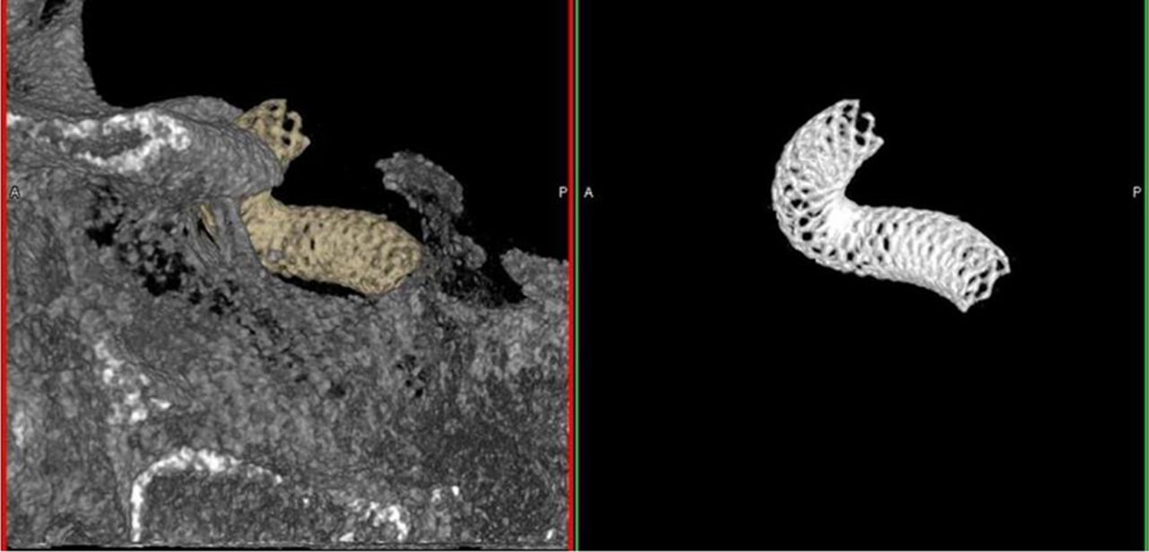
Purpose In addition to stent-assisted coiling, endovascular treatment of intracranial aneurysms using flow diverters, like the Pipeline™ Embolization Device (PED), has become a viable treatment option. Visualization of the device after deployment or during routine follow-up imaging is important to assess expansion and vessel wall apposition. Currently, PEDs are visually assessed from 3D C-arm CT images because of their limited visibility under 2D fluoroscopy. However, tedious post-processing is required to segment the device from the surrounding bony anatomy to better visualise the deployment. The main objective of this study is to report preliminary results of prototype software that automatically detects and segments PEDs from C-arm CT images.
Materials and methods C-arm CT images from 15 patients with internal carotid artery (ICA) aneurysms that underwent endovascular treatment with PEDs were evaluated using prototype software (Flow Diverter Detection Prototype, Siemens AG, Forchheim, Germany). PEDs were deployed in the right cavernous ICA (n = 7), right supraclinoid ICA (n = 2), left cavernous ICA (n = 3), and left supraclinoid ICA (n = 2). C-arm CT images (Syngo Inspace 3D 5s DSA, Siemens AG, Forchheim, Germany) were obtained immediately after deployment or during follow-up angiography as part of the routine imaging for device assessment. The software was applied to the native mask reconstructed around the PED using a sharp kernel with a matrix size of 512 × 512 × 512 voxels and a voxel size of 0.11–0.16 mm3. The software requires the operator to use one click to automatically segment the PED.The segmentation results were analyzed qualitatively. Quality of segmentation was rated based on the following: 0 – software failed to detect PED, 1 – software detected PED with additional bony contamination that required additional cropping, 2 – software automatically detected PED without any need for additional post-processing.
Results Software automatically segmented the PED in all fifteen datasets. Six datasets were given a rating of 2. Nine datasets were given a rating of 1, indicating small parts of bone were still included in the segmentation. This did not limit the visualization of the device and can be cropped out with minor post-processing. The average time needed to automatically segment the PED was 20.39 ± 3.5 seconds.
Conclusions The prototype software was able to identify and segment the PED in all cases. Hence, automatic detection of PED from C-arm CT images is feasible. This tool can provide fast visualization of the flow diverters in 3D to assess device expansion and vessel wall apposition.

Disclosures M. Jagani: 5; C; Siemens Medical Solutions USA, Inc. P. Chinnadurai: 5; C; Siemens Medical Solutions USA, Inc. G. Chintalapani: 5; C; Siemens Medical Solutions USA, Inc.. H. Shaltoni: None. H. Morsi: None. M. Mawad: None.