Article Text

Abstract
Introduction CTA is used to triage acute stroke patients for potential thrombectomy. Single, multiphase1 or dynamic2 CTA may demonstrate areas of parenchymal hypoattenuation on source images(CTASI). These areas may represent either delayed arrival of contrast or could be ischemic/infarcted tissue. The area can be variable depending on the arrival of contrast on the time-tissue attenuation curve (TAC). Areas of hypoattenuation distal to an occlusion are perceived because of differences in contrast compared to normally-perfused areas and can be objectively measured. Patients with significant hypoattenuation may be potentially excluded from thrombectomy.
We evaluated conspicuity of hypoattenuation on dynamic multiphase CTASI. Specifically, we sought to determine if there is a specific time point on the TAC curve which optimizes conspicuity.
Materials and methods We retrospectively identified 45 consecutive large vessel strokes between 2012–14 who had dynamic CTA and CTP.
We identified ischemic areas from areas of low CBV. An ROI was drawn on the corresponding CTASI and in normally-perfused tissue.
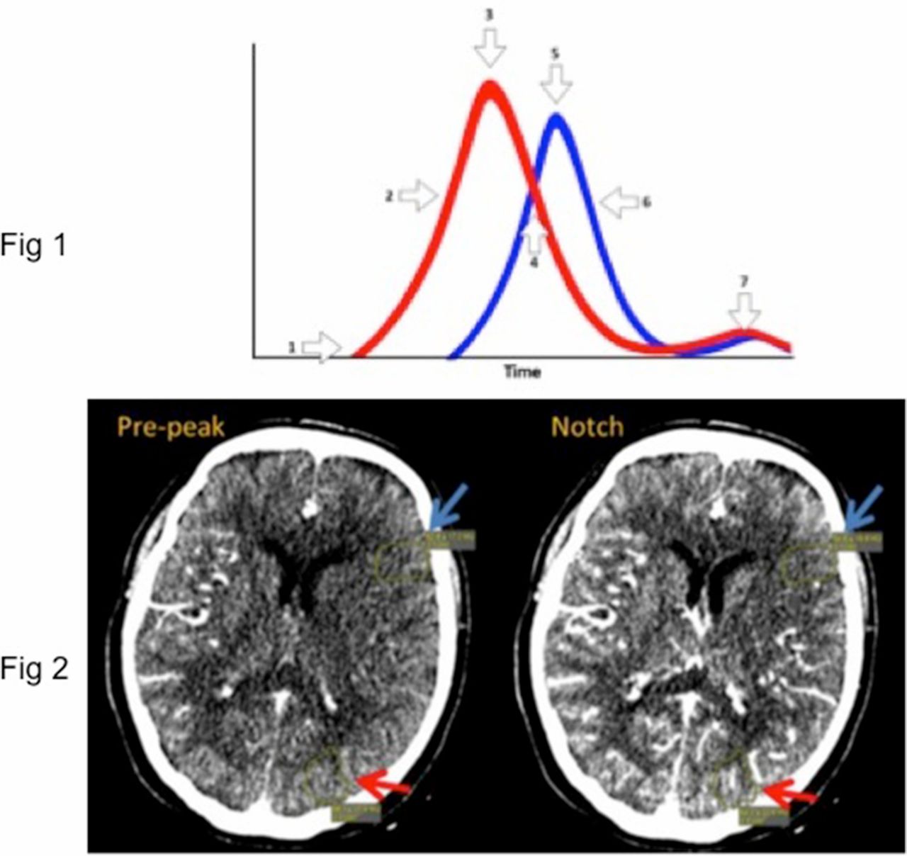
We evaluated CTASI in 7/18 points along the TAC (Figure 1). We calculated absolute and relative change in attenuation between potentially ischemic and normally-perfused tissue.

TAC curve; red = arterial input, blue = venous. 1: Baseline Arterial, 2: Prepeak, 3: Peak, 4: Notch, 5: Peak venous, 6: Venous downslope, 7: Venous eq.
Differences in conspicuity were evaluated using a Friedman test with Bonferroni correction.
Results There were 22 males/23 females, age 69, ±16, median NIHSS 10.6. All occlusions were in the anterior circulation; 64% on the left.
There was a significant effect of phase on the TAC for both absolute and relative conspicuity of ischemic (Figure 2) vs normally-perfused areas (P < 0.00001).

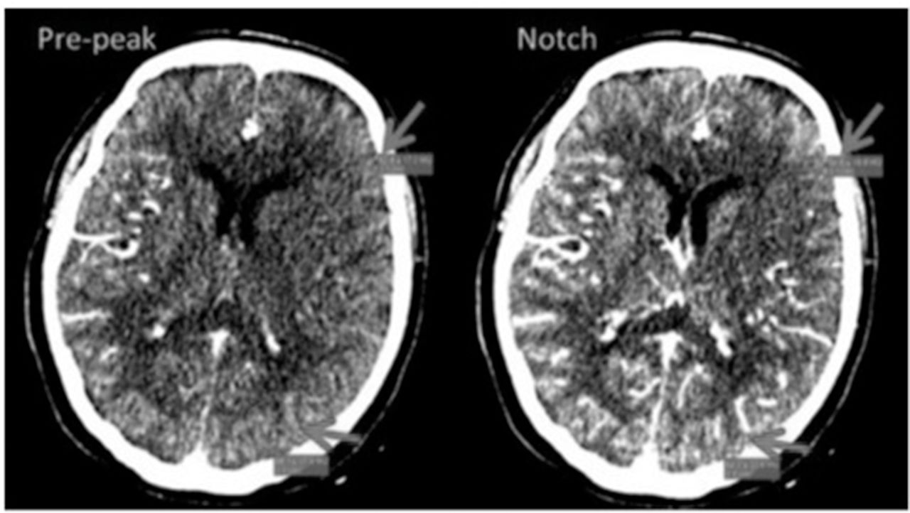
Identical slices at 2 different (pre-peak & notch) phases of CTASI. The conspicuity of the ischemic area in the left frontal lobe (blue arrows) compared to normally-perfused tissue in the left parietal lobe (red arrows) is greater on the notch phase
The median absolute and relative conspicuity of ischemic:normally-perfused tissue was greatest at the peak arterial (8.6 HU, 1.15), notch (9.4 HU, 1.17) and peak venous phases (7 HU, 1.13) vs other portions of the TAC (2.5–3.3 HU, 1.04–1.09).
Conclusion The conspicuity of ischemic areas distal to a large artery occlusion in acute stroke is dependent on the phase of contrast arrival on time-resolved dynamic CTASI. The greatest conspicuity of areas of hypoattenuation between ischemic and normally perfused tissue is in the middle of the TAC.
References 1 Radiology 2015 May;275(2):510–20.
2 Stroke 2014 Sep;45(9):2683–28.
Disclosures C. Lum: None. P. Dave: None. R. Thornhill: None. S. Chakraborty: None. D. Dowlatshahi: None.
This is an Open Access article distributed in accordance with the Creative Commons Attribution Non Commercial (CC BY-NC 4.0) license, which permits others to distribute, remix, adapt, build upon this work noncommercially, and license their derivative works on different terms, provided the original work is properly cited and the use is non-commercial. See: http://creativecommons.org/licenses/by-nc/4.0/