Article Text

Abstract
The use of flow diverters to treat intracerebral aneurysms is highly successful with a low rate of morbidity of mortality. One of the primary concerns following endovascular aneurysm treatment is recurrence, and close imaging followup is often required. High resolution MRI is being employed in an increasingly wide variety of pathologies, but its use as a surveillance tool following flow diversion has not been extensively explored. We present three cases where high resolution MRI was performed following flow diversion for intracranial aneurysms and discuss its utility in this patient population.
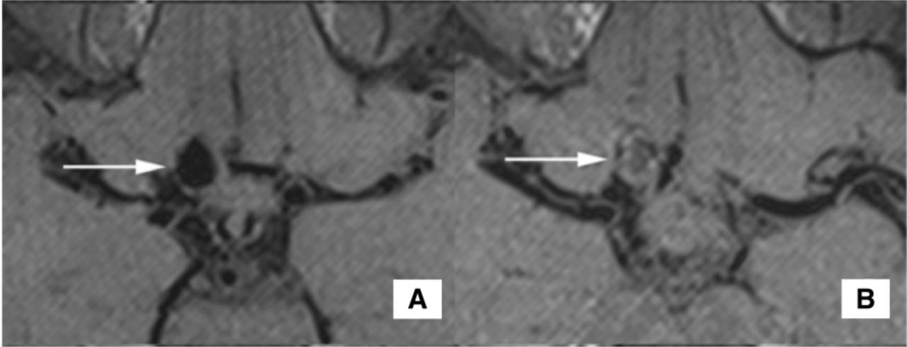
A) Pre-flow diversion 3D T1w Black Blood pre-contrast image; B) Post-flow diversion 3D T1w Black Blood pre-contrast image
Disclosures J. Guan: None. S. McNally: None. A. de Havenon: None. P. Tuassky: 2; C; Covidien. S. Kim: None. M. Park: None.
This is an Open Access article distributed in accordance with the Creative Commons Attribution Non Commercial (CC BY-NC 4.0) license, which permits others to distribute, remix, adapt, build upon this work noncommercially, and license their derivative works on different terms, provided the original work is properly cited and the use is non-commercial. See: http://creativecommons.org/licenses/by-nc/4.0/