Article Text

Abstract
Purpose Experience with the endovascular treatment of middle cerebral artery (MCA) aneurysms by flow diverter devices (FDD) is still limited. This study examines the results and complications of FDD for small aneurysms at this location.
Methods From February 2010 to December 2013, 14 patients (10 women; mean age 59 years) with 15 small MCA aneurysms were treated with FDD. All procedures were performed with the Pipeline embolization device (PED).
Results Complete occlusion was obtained in 12/15 aneurysms (80%) and partial occlusion in 3 (20%). Among 13 aneurysms with a side branch, this was patent at the angiographic control in 4 cases, showed decreased filling in 6, and was occluded in 3 (with neurological deficits in 2). All PEDs were patent at follow-up. Post-procedural ischemic complications occurred in 4 (27%) procedures with permanent neurological deficit (modified Rankin score 2) in 3 (21%). No early or delayed aneurysm rupture, no subarachnoid or intraparenchymal hemorrhage and no deaths occurred.
Conclusions Endovascular treatment with FDD is a relatively safe treatment for small MCA aneurysms resulting in a high occlusion rate. The findings of this study suggest that complete occlusion after endovascular treatment with FDD can be delayed (>6 months). Ischemic complications may occur as early or delayed, particularly at clopidogrel interruption.
- Flow Diverter
- Aneurysm
- Angiography
- Intervention
Statistics from Altmetric.com
Introduction
Aneurysms of the middle cerebral artery (MCA) are usually located at the bifurcation; they are often wide-necked and may have an unfavorable anatomical arrangement with a side branch from the sac.1 They are therefore mainly treated by surgical clipping, allowing remodeling and exclusion of the sac.2
Endovascular treatment of MCA aneurysms may be realized by several techniques3 including coiling,4 stent-assisted coiling,5 Y-stenting,6 balloon-assisted coiling,7 and new endosaccular devices (eg, WEB, pCONus).8 ,9 These last techniques have been developed to improve the occlusion rate and decrease the risk of recanalization.
Flow diverter devices (FDD) are new-generation stents placed in the parent artery at the level of the neck to disrupt the flow into the aneurysm.10 We report a series of 14 patients with 15 MCA aneurysms treated by the Pipeline embolization device (PED; Covidien, Mansfield, Massachusetts, USA). The aim of the study is to examine the results and complications of FDD for aneurysms at this location.
Materials and methods
Study design
This study was designed to define the role of endovascular management with FDD in unruptured MCA aneurysms.
Patient recruitment began in February 2010 in two neuroradiological services and was closed in December 2013. Inclusion criteria were no previous neurosurgical treatment, patient consent to the endovascular procedure, and aneurysms difficult to treat with other techniques (neurosurgical and endovascular) because of the anatomical configuration (wide neck and side branch).
During the period of recruitment 14 patients with 15 MCA aneurysms were treated with FDD. All patients had complete clinical and radiological pre- and post-procedural data. The radiological definition was obtained in all cases by CT angiography (CTA), with native images and two-dimensional multiplanar reconstructions, and by digital subtraction angiography (DSA).
Aneurysm location, size, neck, dome to neck ratio, and side branches were identified. The endpoints were angiographic evidence of complete aneurysm occlusion, recanalization rate, occlusion or stenosis of the parent artery and side branches, and clinical and radiological evidence of brain ischemia.
Patient characteristics
The 14 patients comprised 10 women and four men of median age 59 years (range 39–71 years) (table 1). All patients had unruptured incidental aneurysms with no previous subarachnoid or cerebral hemorrhage.
Patient and aneurysm characteristics
The FDD was the first and only treatment in 10 patients (72%) and in four patients (28%) the device was implanted because of recanalization of a previous coil and additional coiling was not indicated due to aneurysm morphology.
One patient was treated with two PEDs at the same aneurysm in two different procedures; because of shortening of the first device which was evident at 6 months, a second device was placed distally. In two patients with another aneurysm at a different location, this was previously treated with endovascular coiling. Another patient with two contiguous aneurysms of the MCA bifurcation was treated by a single device.
Aneurysm characteristics
Among the 15 aneurysms, 13 (87%) were located at the M1–M2 bifurcation and two (13%) at the M1 segment (table 1); 14 (93%) were saccular and one (7%) was fusiform.
The aneurysm size ranged from 4 to 10 mm (median diameter 7 mm). The neck size ranged from 2.5 to 7.5 mm (mean 4.5 mm); eight aneurysms (57%) had a neck of <5 mm and six (43%) had a neck of ≥5 mm. The neck to sac ratio ranged from 0.3 to 1 (mean 0.7) and side branches were found in 13 of the 15 aneurysms (87%).
Endovascular treatment and medication
All patients were pretreated with 75 mg daily clopidogrel for 5 days and 150 mg aspirin. Thrombocyte inhibition tests were not performed routinely before treatment in all patients, but only in those with a history of thrombophilia (ie, deep venous thrombosis, previous lung embolism). All patients had a continuous intravenous infusion of heparin during the procedure and a bolus of 1000 IU every hour during the procedure to maintain an activated clotting time of >250–300 s.
Clopidogrel (75 mg) and aspirin (100 mg) were administered daily until aneurysm occlusion was confirmed by DSA, after which only aspirin was continued indefinitely. Corticosteroids were not administered.
All endovascular treatments were performed by two interventional neuroradiologists with experience of >10 years in the management of cerebral aneurysms.
The procedure was performed under general anesthesia through catheterization of the right common femoral artery using a 8 F vascular sheath and a triaxial system (8F Cordis Multipurpose or Neuronmax Penumbra, Reflex 072 Reverse Medical or Navien Covidien, Marksman Covidien). The PED was then placed at the level of the aneurysm. The correct apposition to the vessel wall was assessed with DSA and non-subtracted angiographic images. The procedure was considered successful if the PED completely covered the aneurysm.
Intra-aneurysmal contrast stasis was observed in 12 patients (85%) immediately after the end of the procedure.
Follow-up schedule
All patients underwent clinical examination and CTA 1, 3, 6, and 12 months after the procedure, and every 6 months until complete aneurysm occlusion was verified.
Complete aneurysm occlusion was confirmed in all cases by DSA. The actual follow-up ranged from 6 to 48 months.
Results
Treatment characteristics
Thirteen patients (93%) had one device and one patient had two devices (table 2). In addition, one patient had one device for the treatment of two contiguous aneurysms.
Treatment characteristics
No early or delayed aneurysm rupture, no subarachnoid or intraparenchymal hemorrhage and no deaths occurred.
There were no intra-procedural technical complications. Four patients (27%) had post-procedural ischemic complications. The neurological deficits included dysphasia in three patients and hemiparesis in two. They occurred early (<72 h) in one case and later in three (at 2 weeks, 1 and 6 months, respectively). The deficits, although moderate, were definitive in three patients (21%). Two of these four patients had abnormal post-procedural angiographic studies (thrombosis at the site of the overlapping of two devices in one case and occlusion of the right temporal branch and perforators covered by the FDD in the other). In two other patients the post-procedural angiography was normal.
All four patients with ischemic complications had aneurysms of the M1–M2 bifurcation and all had a side branch. No difference in aneurysm size was found.
Angiographic outcome
Complete occlusion was obtained in 12 of the 15 aneurysms (80%; table 3). Partial occlusion was observed in three (20%) at 12, 18, and 24 months, respectively. According to the aneurysm location, complete occlusion was obtained in 11 of 13 aneurysms at the M1–M2 bifurcation (84.5%; figures 1⇓⇓–4) and in one of two of the M1 segment aneurysms (figure 5).
Angiographic outcome

(A) A 63-year-old man with a M1–M2 bifurcation aneurysm treated with coils. (B) Six-month follow-up angiography shows recanalization of the aneurysm sac. (C) Non-subtracted digital subtraction angiography: Pipeline embolization device (PED) deployment. (D) Twelve-month follow-up angiography after PED placement shows complete exclusion of the aneurysm.

(A) A 58-year-old woman with a M1–M2 bifurcation aneurysm (digital subtraction angiography (DSA)). (B) Non-subtracted DSA: Pipeline embolization device (PED) deployment: immediate contrast stasis. (C) Non-subtracted DSA: PED placement at the end of the procedure. (D) Twelve-month follow-up angiography shows complete occlusion of the sac.

(A) A 67-year-old woman with a M1–M2 bifurcation aneurysm treated with coils. (B) Six-month follow-up angiography shows recanalization of the aneurysm sac. (C and D) Non-subtracted digital subtraction angiography: Pipeline embolization device (PED) deployment. (D) Immediate angiogram following PED deployment. (E) Non-subtracted and (F) subtracted 12-month follow-up angiography shows complete exclusion of the aneurysm.
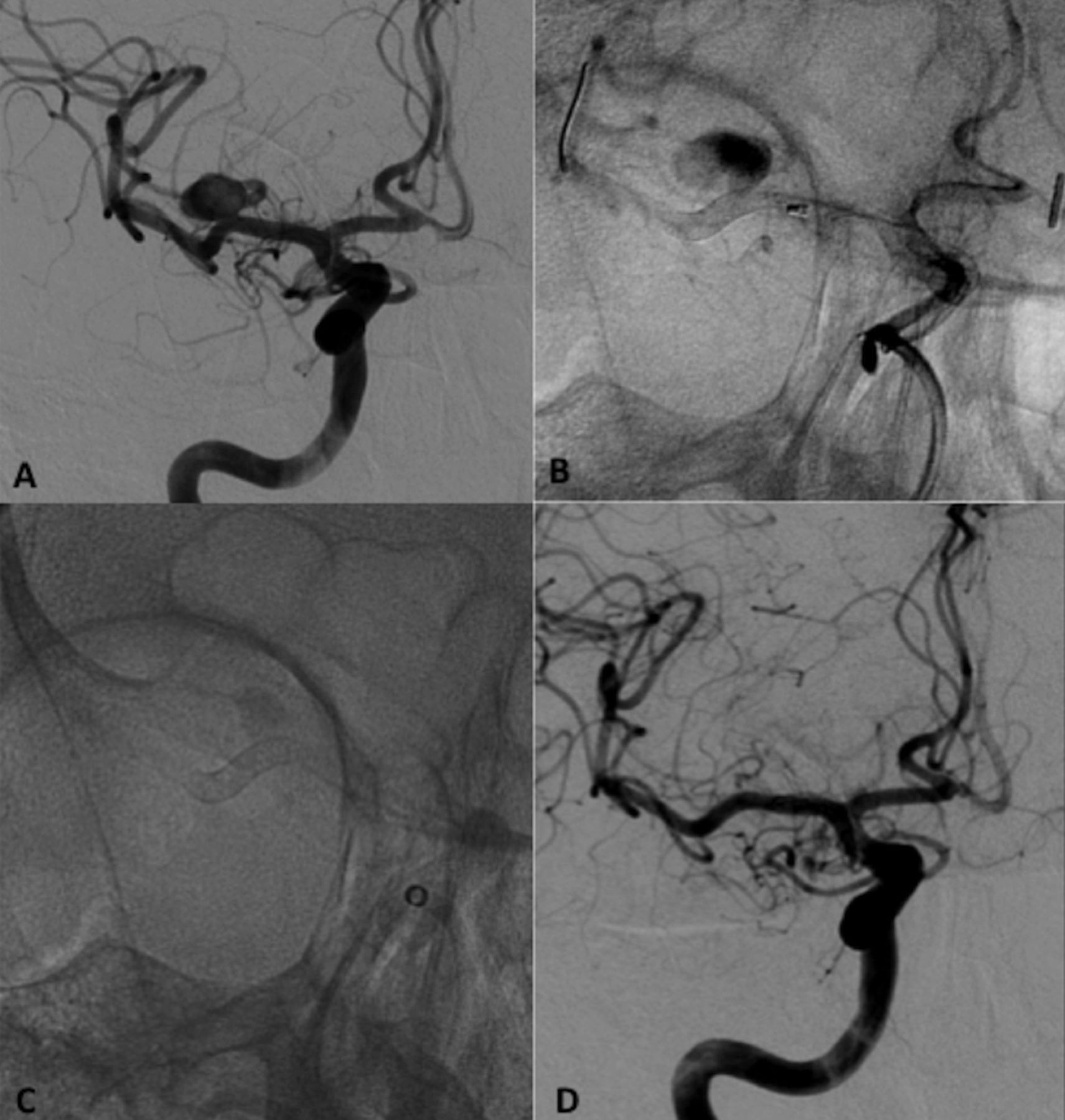
(A) A 68-year-old woman with a M1–M2 bifurcation middle cerebral artery aneurysm (digital subtraction angiography (DSA)). (B) Non-subtracted DSA: Pipeline embolization device (PED) deployment. (C) Non-subtracted DSA shows contrast stasis in the sac at the end of the procedure. (D) Six-month follow-up angiography shows complete occlusion of the sac; the side branch covered by the PED is reduced in filling.

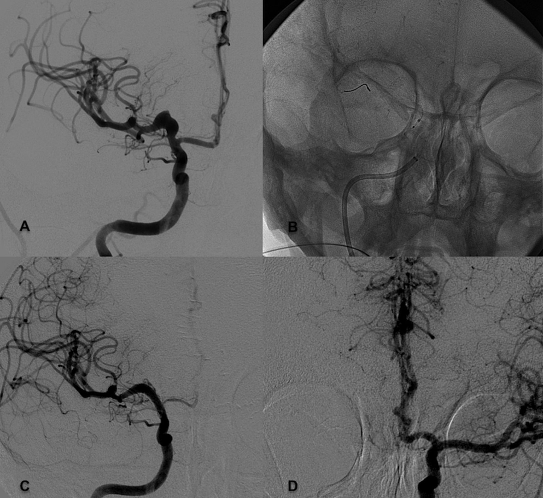
(A) A 53-year-old woman with a fusiform right M1 middle cerebral artery aneurysm (digital subtraction angiography (DSA)). (B) Non-subtracted DSA: Pipeline embolization device deployment. (C and D) Three-month DSA: complete occlusion of the aneurysm; right A1 covered by flow diverter device is reduced in filling while both A2 are supplied by the anterior communicating artery.
The three aneurysms with partial occlusion were stable during follow-up; CTA showed a decrease in the size of the sac. No further treatment was undertaken because of difficulty in both coiling and clipping. The further outcome of these three cases (complete occlusion or regrowth) will be defined in a longer follow-up.
Complete occlusion (12 patients) occurred within 3 months in two cases (17%), at 6 months in four (33%), at 12 months in five cases (42%), and at 24 months in one (8%).
The side branches were patent at the angiographic control in four of 13 cases, showed a decrease in filling in six and were occluded in three (with no symptoms in one and neurological deficits in two).
All PEDs were patent at follow-up.
Correlation of occlusion time with aneurysm characteristics (table 4) showed no difference in occlusion time according to aneurysm size, neck and neck/sac ratio, probably because of the absence of large and giant aneurysms in this series.
Correlation of occlusion time with aneurysm characteristics (15 aneurysms)
Discussion
The use of FDD has been reported in the recent literature mainly to treat internal carotid artery aneurysms proximal to the circle of Willis;11–20 in these and other studies3 ,20–22 a few cases of MCA aneurysms were included. Only two series23 ,24 and one case report25 have focused on the treatment of MCA aneurysms by FDD.
The primary aims of treatment with FDD are disrupting the intra-aneurysmal flow and favoring intra-aneurysmal thrombosis while preserving patency of the adjacent small vessels. It also offers good support for the development of the neointima.10 Preclinical studies have shown that the process is not immediate and requires variable time.10 The occlusion time is determined by several factors including aneurysm size and morphology, presence of a side branch, and choice of technique (ie, ‘oversizing’ the device to prevent occlusion of small branches and perforators).
Several studies including aneurysms at all locations have shown a high rate of occlusion after treatment with FDD (76–85%) with acceptable morbidity (3.7–7.3%).12–14 ,17 ,18 However, a significantly higher rate of complete angiographic obliteration compared with other standard endovascular techniques was mainly obtained for internal carotid artery aneurysms,12 whereas MCA aneurysms showed partial or late occlusion (>18 months).
Five recent studies have reported on the treatment of MCA aneurysms with FDD.19–21 ,23 ,24 The data from these series and our own, which include 81 patients with 85 MCA aneurysms, are summarized in table 5. The location was mainly at the M1–M2 bifurcation (66 aneurysms, 78%) and more rarely at M1 (13%) or distal (9%) segments. The aneurysm morphology was mainly saccular (80%), with 14 (19.5%) fusiform and three (3.5%) blister. The size was mainly small. Among the 66 patients for whom the clinical presentation was cited, only nine (14%) had subarachnoid hemorrhage whereas, in 57 (86%), the aneurysm was incidental. Sixteen patients (20%) had undergone a previous endovascular procedure other than FDD resulting in incomplete occlusion or recanalization.
Data on reported cases of MCA aneurysms treated by flow diverter devices (FDD)
The type of FDD used for the treatment was mainly the PED (74 aneurysms, 87%), while three cases (3.5%) were treated by the Silk embolization device (Balt Extrusion, Montmorency, France) and eight (9.5%) with Surpass (Stryker Neurovascular, Fremont, California, USA).
The rate of complete occlusion ranged from 60%19 ,21 to 84%.23 In the overall series of 60 patients, when the occlusion rate was cited (excluding one with parent vessel occlusion25) the occlusion rate was 77% (46 cases). In our series we achieved an aneurysm occlusion rate of 80%.
No death occurred in the five series analyzed. Only three of 81 patients (3.7%) had intra-procedural complications and nine (11%) had late complications. These included one arterial perforation,21 one complete PED thrombosis,21 one symptomatic occlusion of a side branch,20 a peri-procedural subarachnoid hemorrhage of unknown origin,23 and morbidities related to antiplatelet therapy.23 ,24 In our series, post-procedural ischemic complications occurred in four out of 14 patients (27%), including slight dysphasia in three cases and slight hemiparesis in two.
The causes of the ischemic complications are both technical and medical. Technical causes include the use of more than one overlapping device in the MCA and occlusion of the perforator arteries covered by the FDD. Oversizing of the device with a reduced flow diversion effect should preserve patency of the perforators but may delay aneurysm occlusion. Another therapeutic option is the use of new devices with double-ended non-flow diverter or non-double layer parts. In this way, the flow diverter part should be limited at the neck and perforator arteries should be covered by the non-flow diverter segment.
Medical causes of the ischemic complications are mainly correlated to platelet function and clopidogrel resistance. Several recent studies have stressed the correlation between platelet function and postoperative ischemic complications after neurointerventional procedures.26–30 These studies suggest that patients who experience thromboembolic complications could have been clopidogrel hyporesponders. This may explain the ischemic events in two our cases. The identification of these cases in pre-procedural testing of platelet function may allow adjustments to the clopidogrel dose, thus reducing the risk of ischemic events.
The patient with a M1 fusiform aneurysm in our series showed an unexpected early occlusion (at 3 months), probably related to the absence of perforators (figure 5). A similar experience was reported by Yu et al.16 FDD seems to be the best treatment modality for fusiform aneurysms. However, this needs to be verified in a larger series.
MCA aneurysms with a larger neck and those with side branches show a longer occlusion time than internal carotid artery aneurysms. However, FDD may be a good option for unruptured aneurysms.
In partially occluded aneurysms treated by FDD, the risk of rupture is very low. In these cases we therefore recommend neuroradiological follow-up with CTA or contrast-enhanced MR angiography. Retreatment is advised only when aneurysm regrowth occurs.
Conclusion
Endovascular treatment with FDD is a relatively safe treatment for unruptured MCA aneurysms, resulting in a high rate of occlusion. The results of our study suggest that complete occlusion after endovascular treatment with FDD can be delayed (>6 months). Ischemic complications may be both early and delayed, particularly at clopidogrel interruption; however, occlusion of the side branches can occur with or without symptoms.
Further series with a larger patient population may define the best endovascular management for small MCA aneurysms.
References
Footnotes
Contributors FB designed the data collection tools, monitored data collection, cleaned and analyzed the data, and drafted and revised the paper; he is the guarantor. LD analyzed the data and drafted and revised the paper. GL monitored data collection, analyzed the data and drafted and revised the paper. CS monitored data collection for the whole study and revised the draft paper. GB designed the data collection tools, monitored data collection, and revised the draft paper. MM designed data the collection tools and drafted and revised the draft paper. FT analyzed the data and drafted and revised the paper. FC monitored data collection for the whole study and revised the draft paper. FM analyzed the data and drafted and revised the paper.
Competing interests FB serves as proctor for Covidien with a modest remuneration.
Patient consent Obtained.
Ethics approval The study was approved by the Ethics Committe of our Institution and was performed in accordance with the ethical standards laid down in the 1964 Declaration of Helsinki and its later amendments.
Provenance and peer review Not commissioned; externally peer reviewed.
Data sharing statement Partecipants gave informed consent for data sharing.