Article Text

Abstract
Introduction/purpose The treatment of symptomatic chronic subdural hematomas is challenging. Patients are often elderly, on anticoagulation, and have multiple medical comorbidities. Less invasive surgical techniques such as the subdural evacuating port system (SEPS) have been supported in this high risk surgical patient population. However, there is a high rate of recurrence of chronic subdural hematomas requiring repeat intervention. Recurrence is thought to occur due to bleeding from the friable vascularized membranes that form in chronic subdural hematomas. Case reports suggest that embolization of the middle meningeal artery can reduce the arterial supply to the membranes and reduce recurrence rates.
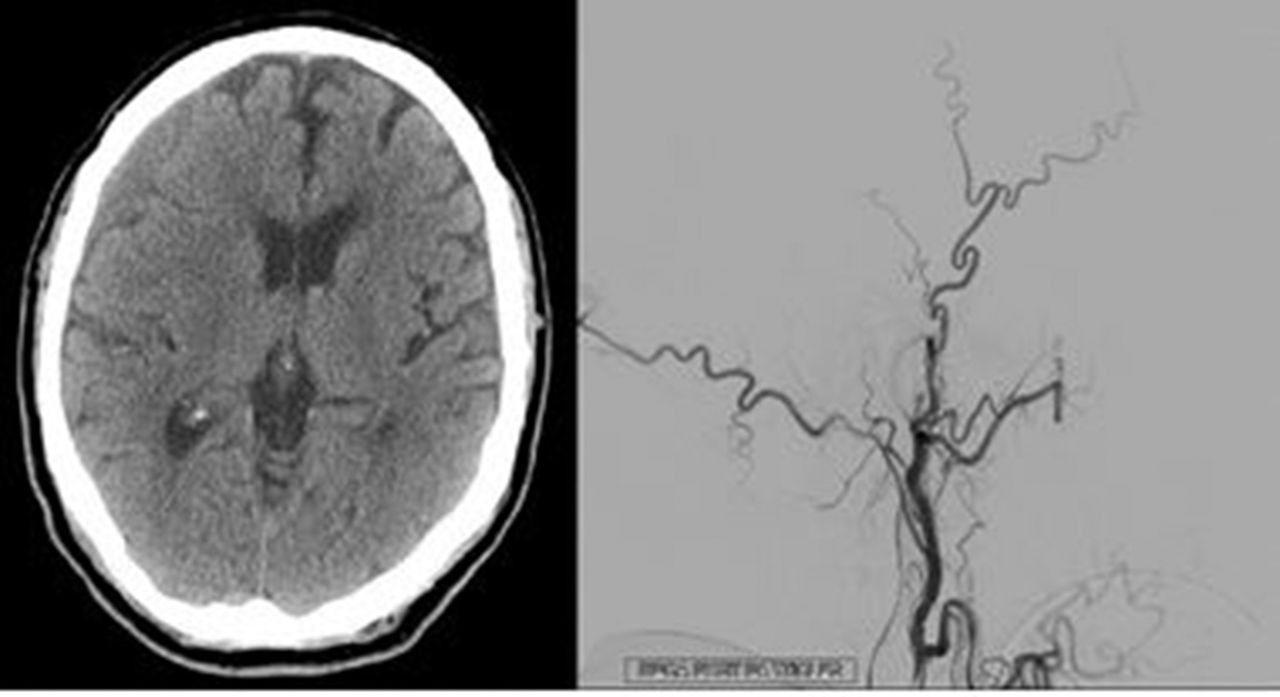
A) CT head showing right-sided chronic subdural hematoma; B) Lateral cerebral angiogram of right external cartid artery showing PVA microparticle embolization of the right middle meningeal artery
Materials and methods We present a case of a 74-year-old male on aspirin with a history of recurrent symptomatic chronic right-sided subdural hematoma treated successfully with a SEPS and right middle meningeal artery embolization with poly vinyl alcohol (PVA) microparticles. The patient initially presented to the emergency department with headaches, difficulty walking, and left sided hemiparesis. CT Head showed a large chronic right-sided subdural hematoma measuring 2.7 cm thick with 1 cm of leftward shift. Patient underwent placement of a right-sided SEPS and the subdural hematoma decreased in size to 1.0 cm with 2 mm of leftward shift. The patient had resolution of headaches and neurological symptoms and was discharged home. Three months later, the patient returned to the emergency department with headache and left hand numbness. CT Head showed an acute on chronic right-sided subdural hematoma measuring 1.4 cm with 3 mm of leftward shift. Patient underwent right-sided SEPS placement. Repeat CT Head showed reduction in the subdural hematoma to 1.2 cm. The SEPS was removed and the patient had resolution of neurological symptoms. The patient then had a diagnostic cerebral angiogram with PVA microparticle embolization of the right middle meningeal artery. A SEPS was placed at the time of the angiogram to further reduce the size of the subdural hematoma.
Results Repeat CT Head after SEPS and middle meningeal artery embolization showed decrease in size of the subdural hematoma. Follow-up CT Head showed stability of the subdural hematoma and patient had no further neurological symptoms. Patient was discharged home.
Conclusion Middle meningeal artery embolization is a useful endovascular technique for reducing the arterial supply to the membranes in chronic subdural hematomas. Middle meningeal artery embolization can reduce the recurrence rate of subdural hematomas.
Disclosures A. Strickland: None. B. Bohnstedt: None.