Article Text

Abstract
Background Revascularization after endovascular therapy for acute ischemic stroke is measured by the Thrombolysis In Cerebral Infarction (TICI) scale, yet variability exists in scale definitions. We examined the degree of reperfusion with the expanded TICI (eTICI) scale and association with outcomes in the HERMES collaboration of recent endovascular trials.
Methods The HERMES Imaging Core, blind to all other data, evaluated angiography after endovascular therapy in HERMES. A battery of TICI scores (mTICI, TICI, TICI2C) was used to define reperfusion of the initial target occlusion defined by non-invasive imaging and conventional angiography.
Results Angiography of 801 subjects was available, including 797 defined by non-invasive imaging (154 internal carotid artery (ICA), 583 M1, 60 M2) and 748 by conventional angiography (195 ICA, 459 M1, 94 M2). Among 729 subjects in whom the reperfusion grade could be established, using eTICI (3=100%, 2C=90–99%, 2b67=67–89%, 2b50=50–66%) of the conventional angiography target occlusion, there were 63 eTICI 3 (9%), 166 eTICI 2c (23%), 218 eTICI 2b67 (30%), 103 eTICI 2b50 (14%), 100 eTICI 2a (14%), 19 eTICI 1 (3%), and 60 eTICI 0 (8%). Modified Rankin Scale shift analyses from baseline to 90 days showed that increasing TICI grades were linked with better outcomes, with significant distinctions between TICI 0/1 versus 2a (p=0.028), 2a versus 2b50 (p=0.017), and 2b50 versus 2b67 (p=0.014).
Conclusions The benefit of endovascular therapy in HERMES was strongly associated with increasing degrees of reperfusion defined by eTICI. The eTICI metric identified meaningful distinctions in clinical outcomes and may be used in future studies and routine practice.
- stroke
- angiography
Statistics from Altmetric.com
Endovascular therapy is a highly effective treatment for acute ischemic stroke caused by large artery occlusion in the anterior cerebral circulation, significantly increasing the likelihood of recovery to independence.1 Reperfusion of the ischemic territory downstream from an arterial occlusion is the therapeutic mechanism responsible for benefit. The extent of reperfusion, however, may vary across individual cases depending on numerous factors, including the degree of collateral circulation.2–5 Most commonly, reperfusion is evaluated on angiography performed immediately after recanalization or when reopening of the arterial occlusion is achieved.
Several iterations of the Thrombolysis In Cerebral Infarction (TICI) score, adapted from the Thrombolysis In Myocardial Infarction (TIMI) coronary circulation scoring system to the cerebral circulation, have been used to quantify reperfusion.6 Successive refinements of the original TICI scale were implemented to enhance standardization for different sites of arterial occlusion and to optimally discern subtle distinctions in the amount of blood flow restored after thrombectomy. Multiple studies have examined the inter-rater reliability of TICI grades and future such analyses are warranted. Variability exists, however, in definitions, nomenclature, use, and reporting. Consensus recommendations for angiographic revascularization standards developed in 2013 defined successful reperfusion by a modified TICI (mTICI) score signifying filling of 50% or more of the downstream territory.7
Reperfusion of the ischemic territory downstream from an arterial occlusion in stroke is distinct from recanalization, defined as restoring patency in the occluded arterial segment.8 9 Reperfusion specifically refers to re-establishing blood flow via normal arterial routes, in contrast to indirect collateral perfusion. The extent of reperfusion is quantified by the percentage of the downstream territory and is therefore dependent on defining the specific site of initial arterial occlusion. Such measurement of reperfusion is most often conducted on biplane angiography where the three-dimensional nature of the arterial territory must be inferred. Grading the extent of reperfusion or assigning a TICI score is typically conducted by the local operator or treating physician at the end of the procedure. Achieving a favorable TICI grade is considered a quality metric for endovascular stroke therapy. Rating is influenced by local rater experience, and central core laboratory adjudication is commonly more conservative compared with local ratings. Since the original description of TICI 15 years ago, several intermediate grades of reperfusion have been introduced. The entire range of grade 2 TICI reperfusion, extending from a minimum of any distal branch filling to almost complete downstream perfusion, has been subdivided and confusingly labeled with inconsistent terminology.
Availability of data from reperfusion trials now allows critical review of TICI reperfusion grade definitions. Standardization of TICI grading is essential for clinical practice and for future trials. We examined the relationship of reperfusion grades and clinical outcomes using individual patient data in the Highly Effective Reperfusion Evaluation in Multiple Endovascular Stroke (HERMES) trials that comprise the majority of randomized controlled trials undertaken with modern endovascular treatments.10 Data were systematically analyzed by an experienced core laboratory to delineate TICI reperfusion and substantiate prior scale distinctions with the associated clinical outcomes after stroke treatment.
Methods
Study design
HERMES included seven distinct endovascular therapy trials that established the efficacy of mechanical thrombectomy for acute ischemic stroke. Detailed methodology of each trial has been previously reported.11–13 Ethics approval was obtained from the local institutional review board and written informed consent was obtained from patients. In HERMES, the entire imaging and angiography datasets of the seven participating trials were centrally pooled in the Neurovascular Imaging Research Core. Anonymized images of each enrolled subject were indexed and relabeled by a randomly assigned HERMES subject identification number to mask any possible association with the original randomized controlled trial. In each of the original trials, only the subjects randomized to endovascular therapy underwent angiography. As a result, the HERMES angiography dataset reflects solely those subjects assigned to endovascular therapy.
Angiography core laboratory evaluation
The HERMES angiography dataset was analyzed by an independent core laboratory with extensive experience in adjudication of imaging and angiography from numerous multicenter stroke trials and registries. The angiography images were provided to the core laboratory without any additional information other than the site of the initial target occlusion determined by non-invasive imaging in the original trial. As technical differences in imaging technique or dynamic changes in occlusion due to recanalization or distal thrombus migration may occur between baseline non-invasive imaging and conventional angiography, reperfusion was graded based on location of the conventional angiography procedure start.
The HERMES angiography core laboratory performed a quality assessment of the angiography data, denoting availability, adequacy, and limiting factors associated with each subject’s data. As TICI reperfusion in the downstream territory is critically contingent on the specific location of the arterial occlusion, it was imperative that a diagnostic run or contrast injection of the occlusion was available at the procedure start to determine the conventional angiography target occlusion location. Similarly, a final run or diagnostic injection of the same arterial territory was required to determine TICI reperfusion. In addition, the angiography core laboratory noted when limited data were available, such as the lack of biplane angiography or failure to acquire adequate runs that precluded evaluation of reperfusion. A battery of various TICI scores in this study population (table 1) was used to define reperfusion of the initial target occlusion on non-invasive imaging and conventional angiography. This 7-point compilation of TICI grades, termed the expanded TICI (eTICI), reflects all previously reported thresholds used to define reperfusion after endovascular stroke therapy. In brief, eTICI grade 0 is equivalent to no reperfusion or 0% filling of the downstream territory; eTICI 1 reflects thrombus reduction without any reperfusion of distal arteries; eTICI 2a is reperfusion in less than half or 1–49% of the territory; eTICI 2b50 is 50–66% reperfusion, exceeding the modified TICI (mTICI) 2B threshold but below the original TICI 2B cut-off point; eTICI 2b67 is 67–89% reperfusion, exceeding TICI but below TICI 2C; eTICI 2c is equivalent to TICI 2C or 90–99% reperfusion; and eTICI 3 is complete or 100% reperfusion, tantamount to TICI 3. Multiple studies have already examined the inter-rater reliability of all the components of the eTICI.14 15 In order to define the inter-rater reliability of the distinction between eTICI 2b50 (50–66% reperfusion, mTICI 2B) and eTICI 2b67 (67–89% reperfusion, TICI 2B), a cohort of 52 subjects was rated independently by two readers.
Study population
Statistical analyses
Descriptive methods were used to characterize baseline angiographic features, including distributions across categories. For each HERMES subject, the blinded evaluation of the conventional angiography target occlusion location was noted and compared with the initial target occlusion location for each case. Inter-rater reliability was analyzed using a correlation coefficient and Cohen’s kappa statistic. The distribution of eTICI scores for the conventional angiography target occlusion was described and analyzed with respect to the key stroke outcome variable of the modified Rankin score (mRS) at 90 days. Graphical analyses were also used to describe the distribution of mRS at day 90 stroke clinical outcomes for each eTICI grade. mRS distribution analyses at day 90 were used to compare clinical outcomes between neighboring eTICI thresholds, with statistical comparisons by Wilcoxon’s rank-sum test for non-parametric analysis. Finally, receiver operating characteristic (ROC) curves were constructed to illustrate the discriminative value of various TICI classification schemes on the mRS outcomes. The ROC results were described by the area under the curve (AUC) statistic and associated p values computed by the method of DeLong. For all analyses, a two-tailed p value <0.05 was considered statistically significant, without adjustment for multiple testing. Analyses were performed using SAS version 9.3 (SAS Institute, Cary, North Carolina, USA) and R version 3.2 (R Foundation for Statistical Computing, Vienna, Austria).
Results
Arterial occlusions
Angiography was available in a total of 801 subjects in HERMES, reflecting 801/871 (92%) of patients assigned to the endovascular treatment arms of the seven participating trials. The target occlusion on non-invasive imaging was located at the internal carotid artery (ICA) in 154 (19%), proximal middle cerebral artery (MCA) or M1 in 583 (73%), and M2 arterial segment in 50 (6%), and on conventional angiography at the ICA in 195 (24%), proximal MCA or M1 in 459 (57%), and M2 arterial segment in 94 (12%).
Extent of reperfusion on eTICI
The extent of reperfusion was distributed across the entire range of the eTICI scale, including the spectrum from absolutely no reperfusion to complete restoration of flow in the territory downstream of the conventional angiography target occlusion. Among the 801 subjects noted above, reperfusion grade could not be established due to imaging limitations in 72, giving 729 evaluable eTICI scores. No reperfusion or eTICI 0 was noted in 60 (8%) and only reduction in thrombus without filling of distal arterial branches (eTICI 1) in 19 (3%). Reperfusion in less than half the territory (eTICI 2a) was noted in 100 (14%). Reperfusion in 50–66% of the territory (eTICI 2b50), equivalent to mTICI 2B yet less than TICI 2B, was noted in 103 (14%). Restoration of flow in 67–89% of the territory (eTICI 2b67), above the TICI 2B threshold yet less than TICI 2C, occurred in 218 (30%). Extensive reperfusion in 90–99% (eTICI 2c), equivalent to TICI 2C, was noted in 166 (23%). Finally, complete or full reperfusion of eTICI 3, equivalent to TICI 3, was found in only 63 (9%).
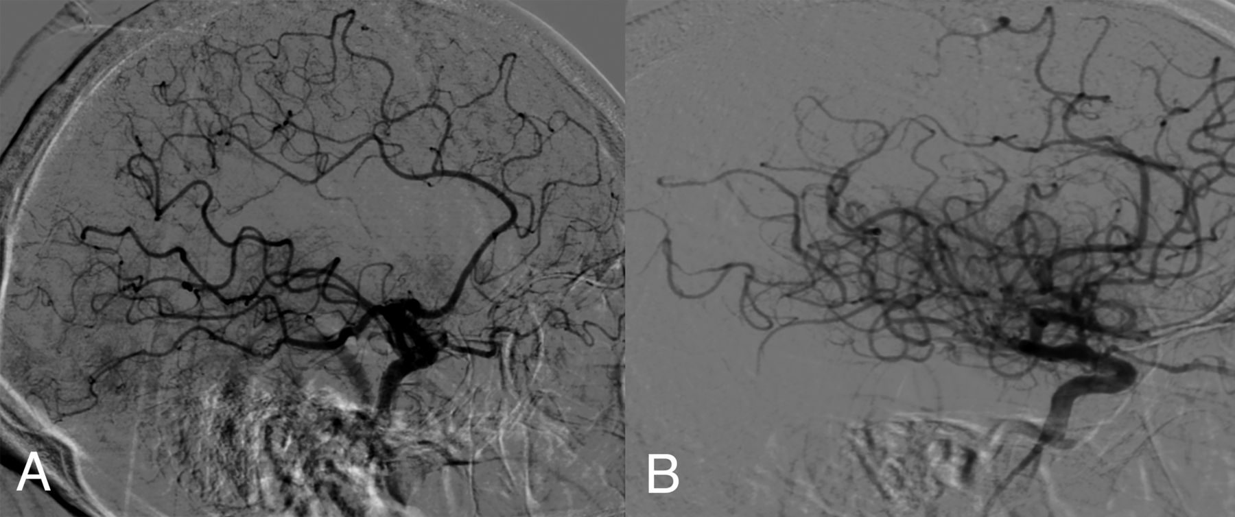
The distinctions between individual eTICI grades and the resultant discriminant ability of these perfusion categories was critically dependent on the availability of adequate angiographic views. Diagnostic confidence or potential limitations in the separation of eTICI categories was documented in 25/729 (3%) of subjects. The most common difficulty (n=12) occurred at the intermediate eTICI grades that demarcated eTICI 2b50 from eTICI 2b67, demarcating reperfusion thresholds around the 67% level (figure 1). Interestingly, the subtle distinction of eTICI 2c and 3, discriminating the TICI 2C (90–99%) category from full or TICI 3 (100%) categories (figure 2) occurred in only 8/729 (1%) with difficulties between eTICI 2b67 and eTICI 2c in the remaining five cases. Within the eTICI 2c or TICI 2C category, overt distal emboli manifest as vessel cut-offs or menisci were visualized in 67/166 (40%).

Angiography of final reperfusion of the middle cerebral artery territory showing (A) eTICI 2b50 (50–66%) versus (B) eTICI 2b67 (67–89%).

Angiography of final reperfusion of the middle cerebral artery territory showing (A) eTICI 2c (90–99%) versus (B) eTICI 3 (100%).
Inter-rater reliability for the distinction between eTICI 2b50 and 2b67 (mTICI 2B vs TICI 2B) showed an agreement of 92% (48/52), with a Cohen’s kappa statistic of κ=0.83, p<0.001.
Relationship between eTICI reperfusion and clinical outcomes
More extensive eTICI reperfusion was associated with better outcomes. Graphical depictions of this relationship for subsets of ICA and M1 MCA occlusions are illustrated in figure 3. There was an unequivocal graded pattern of an increased proportion of subjects with no or minimal disability (mRS 0–1) hierarchically linked with higher eTICI grades. Similarly, the proportion of severe disability or death (mRS 5–6) was less with higher eTICI grades. Interestingly, even intermediate levels of disability (mRS 2–4) exhibited a clear relationship between decreasing disability with more extensive reperfusion. It should be noted, however, that across almost all eTICI reperfusion grades there was still a broad distribution of mRS 90-day clinical outcomes.

Graphical depiction of modified Rankin Scale outcomes at 90 days based on eTICI grades for internal carotid artery (ICA) occlusions (above) and M1 middle cerebral artery (MCA) occlusions (below).
Direct comparison of the distribution in clinical outcomes of individual neighboring eTICI grades revealed specific differences between reperfusion categories (table 2). The relatively small number of subjects with extremely limited (eTICI 0– 1) or complete (eTICI 3) reperfusion may have limited such distinctions between eTICI categories at the extreme ends of the scale, whereas differences between intermediate categories of reperfusion were more apparent. The distribution of clinical outcomes was clearly different between eTICI grades 0/1 versus 2a, 2a versus 2b50, and 2b50 versus 2b67 that demarcate the extent of reperfusion at any versus 0%, 50%, and the 67% thresholds, respectively.
Distinctions between eTICI categories with respect to clinical outcomes for all cases
Multivariable modeling of mRS shift on eTICI with covariate adjustment demonstrates that eTICI is an independent predictor of outcome in the presence of covariate adjustment and, more importantly, that adjacent categories 2a/2b50/2b67 are important distinctions (table 3).
Multivariable model of eTICI categories and clinical outcomes
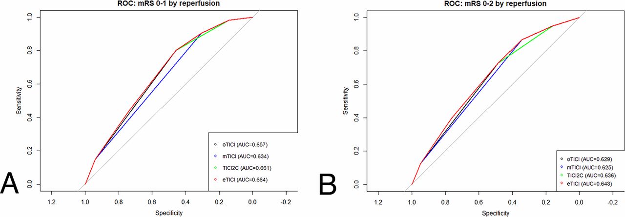
ROC curve analyses (figure 4) showed similar performance of TICI (AUC 0.657), TICI 2C (AUC 0.661), and eTICI (AUC 0.664) in discriminating better clinical outcomes. All three of these were statistically superior to the mTICI AUC (0.634) (p=0.013 for eTICI vs mTICI in particular).

ROC curves showing the predictive ability of various TICI grades with respect to clinical outcomes using (A) modified Rankin Scale (mRS) 0–1 and (B) mRS 0–2.
Discussion
Our study shows that grades of better reperfusion are incrementally associated with better clinical outcomes. This study is novel as HERMES is a large and ideal dataset to examine TICI grading as it relates to clinical outcomes, pooling many of the large landmark trials in endovascular therapy. The use of all previously described TICI variants demonstrates the utility of using an expanded or eTICI scale that encompasses these previous scale definitions. We also demonstrated that these eTICI grades are linked with subsequent clinical outcomes after stroke therapy, providing a cogent rationale for future adoption and large-scale use of eTICI. However, while more extensive reperfusion is associated with better clinical outcomes, a wide range of outcomes was evident even at the extremes of poor and full reperfusion, indicating that additional factors remain important determinants of outcome. Previous reports have demonstrated better outcomes with more extensive reperfusion, yet application of all TICI grades and their relative impact on outcomes from HERMES provides important data for the literature.16 17
We confirmed a predominance of M1 occlusions with considerably fewer ICA or M2 occlusions, as reported in individual trials and in previous pooled analyses. We noted that the target occlusion defined on conventional angiography at procedure start differed from that on baseline non-invasive imaging. This may result from interval recanalization or thrombus migration, or alternatively be ascribed to technical differences in acquisition of non-invasive versus invasive imaging techniques. Based on the conventional angiography target occlusion site determined by our HERMES core laboratory, we corroborate the reperfusion rates reported in the original trial reports, exhibiting substantial predominance of flow restoration to much of the ischemic territory. Importantly, however, the proportion of complete or full reperfusion was substantially less than originally described. There were key distinctions in reperfusion categories when specific thresholds were used to subdivide the ischemic territory. Established consensus recommendations for angiographic standards in endovascular therapy have defined successful reperfusion as exceeding 50% of the territory.7 We have demonstrated, however, that within the >50% reperfusion category, a considerable number of cases fell into previously defined finer subdivisions of 50–66%, 67–89%, and 90–99%, and that these refinements identify meaningful differences in clinical outcomes.
Our use of all previously defined TICI categories, redefined as eTICI grades, showed that such categories identify gradation of outcomes associated with the extent of reperfusion. Comparison of adjacent or ordinal reperfusion grades on the eTICI scale showed that clinical outcomes vary between each grade, with more pronounced differences within intermediate eTICI categories. Prior studies have provided preliminary evidence that, compared with less granular TICI scales, the more fine-grained TICI scales have added prognostic value and clinical utility.18–22 The previous studies have been relatively small and with limited geographic scope, limiting their precision and generalizability. We therefore undertook the current study to validate and quantify the superiority of the eTICI in the large multinational HERMES dataset. We have extended previous reports concerning inter-rater reliability of eTICI cut-off points by demonstrating excellent reliability for distinguishing eTICI 2b50 and 2b67 (mTICI 2B vs TICI 2B).14 15 This distinction between eTICI 2b50 and 2b67 may be difficult to discern, unlike or more so than other category distinctions (eg, 2a vs 2b50; 2b67 vs 2c; 2c vs 3). Furthermore, these other category distinctions have been addressed in prior publications, hence it was important to demonstrate inter-rater reliability in this subset. Our results also indicate that this eTICI category distinction is the most significant as it relates to different subsequent clinical outcomes.
Although this analysis establishes that the 7-point eTICI scale predicts increasingly better clinical outcomes, it is important to note that, even at the extreme ends of the scale, a wide distribution of outcomes may be evident. For example, not all subjects with eTICI 0/1 reperfusion have poor clinical outcomes and, conversely, there are subsets of patients with eTICI 2c or 3 reperfusion with outcomes of severe disability or death. Such examples suggest that angiographic outcomes alone have limited utility for clinical outcome prediction. The AUC values for predicting good clinical outcomes were higher for eTICI 2b67 or 2c (>66% or >90%, respectively) compared with eTICI 3 (>50%), yet such AUC values of 0.66 are relatively modest at best. Factors beyond reperfusion, including the underlying pathophysiology such as extent of established tissue injury, or collateral circulation, clinical variables, or subsequent events, such as recurrent stroke or hemorrhage, likely influence clinical outcomes. It also remains unclear whether the cause of limited reperfusion, due to distal emboli or increased downstream resistance, have different impact in clinical outcomes, even when reperfusion is almost complete as in eTICI 2c or TICI 2C flow.
Limitations of our systematic angiographic evaluation in HERMES include the availability of adequate images, already restricted to subjects in the endovascular arm of these randomized controlled trials. There are numerous aspects of angiography that we may not have been able to elucidate in this overall or primary paper on eTICI reperfusion in HERMES. Many of such limitations and data considerations have been addressed in the vast experience of the IMS I–III trials.4 23–25 Evaluation of reperfusion may have been limited by incomplete depiction of the conventional angiography target occlusion or final diagnostic runs with biplane angiography. Aside from potentially missing data, variations in technique or local practice may have precluded adequate visualization of key angiography data. The degree of reperfusion and clinical outcomes may vary by location of occlusion site and availability of data may be limited even for ICA versus M1 MCA occlusions (figure 3). For ICA occlusions, the role of the ipsilateral anterior cerebral artery (ACA) and collateral flow may be handled differently with respect to subsequent reperfusion grading.24 25 In our analyses, most of the ICA occlusions did not have any pre-treatment digital subtraction angiography information on collaterals. As a result, we defined eTICI for ICA lesions without accounting for potential collateral flow; we scored all based on the ICA lesion being responsible for the ipsilateral ACA and MCA territories. It is agreed that this scenario could be further addressed by a new scoring system in the future. We did not account for the time required to achieve final eTICI reperfusion or consider local factors that may have influenced the duration of the procedure.26 Such challenges in blinded readings may not capture subtle aspects apparent at the time of intervention. Importantly, it remains unclear how the angiography was used in real time to decide how far to continue with revascularization. The endpoints of successful reperfusion or the need to continue may have varied widely from operator to operator. Furthermore, lack of additional clinical demographics and medical history (eg, atrial fibrillation, atherosclerosis, other risk factors) beyond those noted in the multivariable model in table 3 limit our analyses. In addition, wider application of any rating scale requires ongoing examination.
Conclusions
The benefit of endovascular therapy in HERMES was strongly associated with increasing degrees of eTICI reperfusion. The eTICI scale shows important distinctions in the degree of reperfusion with respect to clinical outcomes, underscoring the need to implement standard methodology for reporting of angiography in stroke treatment in trials and routine practice. eTICI provides granularity in distinguishing the extent of reperfusion that is clinically meaningful. Defining successful reperfusion should be linked with good clinical outcomes, making it unlikely that a single threshold of eTICI reperfusion will work in all cases. Our analyses suggest that, if a dichotomous threshold were to be used for the definition of successful reperfusion, then eTICI 2b67, equivalent to TICI 2B, is optimal.
References
Footnotes
Contributors DSL prepared the first draft of the report based on an analysis plan agreed by the HERMES Executive who also contributed to study interpretation. SB performed the statistical analyses. DSL coordinated the central imaging repository. All authors participated in patient enrollment, data collection, critically reviewed the report and approved the final version.
Funding The authors have not declared a specific grant for this research from any funding agency in the public, commercial or not-for-profit sectors.
Competing interests DSL reports having received grant funding from NINDS and consulting fees as an imaging core laboratory from Stryker and Medtronic. RJ reports consulting with Medtronic. CBLMM reports having received grant funding from the Dutch Heart Foundation and European Commission and an unrestricted grant from Stryker. AvdL reports consulting fees from Stryker and grant funding from the Dutch Heart Foundation, AngioCare BV, Medtronic/Covidien/EV3, MEDAC Gmbh/LAMEPRO, Penumbra, Top Medical/Concentric, and Stryker, received by the Erasmus University Medical Center. LSR proctors for Stryker and Medtronic. PW discloses institutional research grant support within the last 2 years from Microvention Terumo. He declares the following relevant professional relationships: Chair of the European Society of Minimally Invasive Neurotherapeutics Guidelines Committee, sits on the Policy Working Group for Thrombectomy of NHS England and represents the Royal College of Radiologists on the UK Intercollegiate Stroke Working party (none of these are associated with financial reimbursement). He reports the following modest consultancy work: member of Stryker’s Global Hemorrhagic Stroke Advisory Board and educational consultancy work for Microvention Terumo. He has no other interests to declare. MG reports being the principal investigator of an unrestricted research grant to the University of Calgary for the HERMES collaboration by Medtronic. He also reports consulting services with Medtronic, Stryker, Microvention, Cerenovus and a licensing agreement with GE Healthcare re systems of acute stroke diagnosis.
Patient consent Not required.
Ethics approval Ethics approval was obtained from the local institutional review board and written informed consent was obtained from patients.
Provenance and peer review Not commissioned; externally peer reviewed.