Article Text

Abstract
Objective To report the feasibility of a combined imaging and treatment strategy for acute ischemic stroke (AIS) utilizing a hybrid CT-Angiography suite.
Methods A descriptive case report covering the technical aspects of whole-brain vascular and perfusion imaging with a single intra-aortic injection of 7 ml of iodinated contrast for suspected large vessel occlusion (LVO).
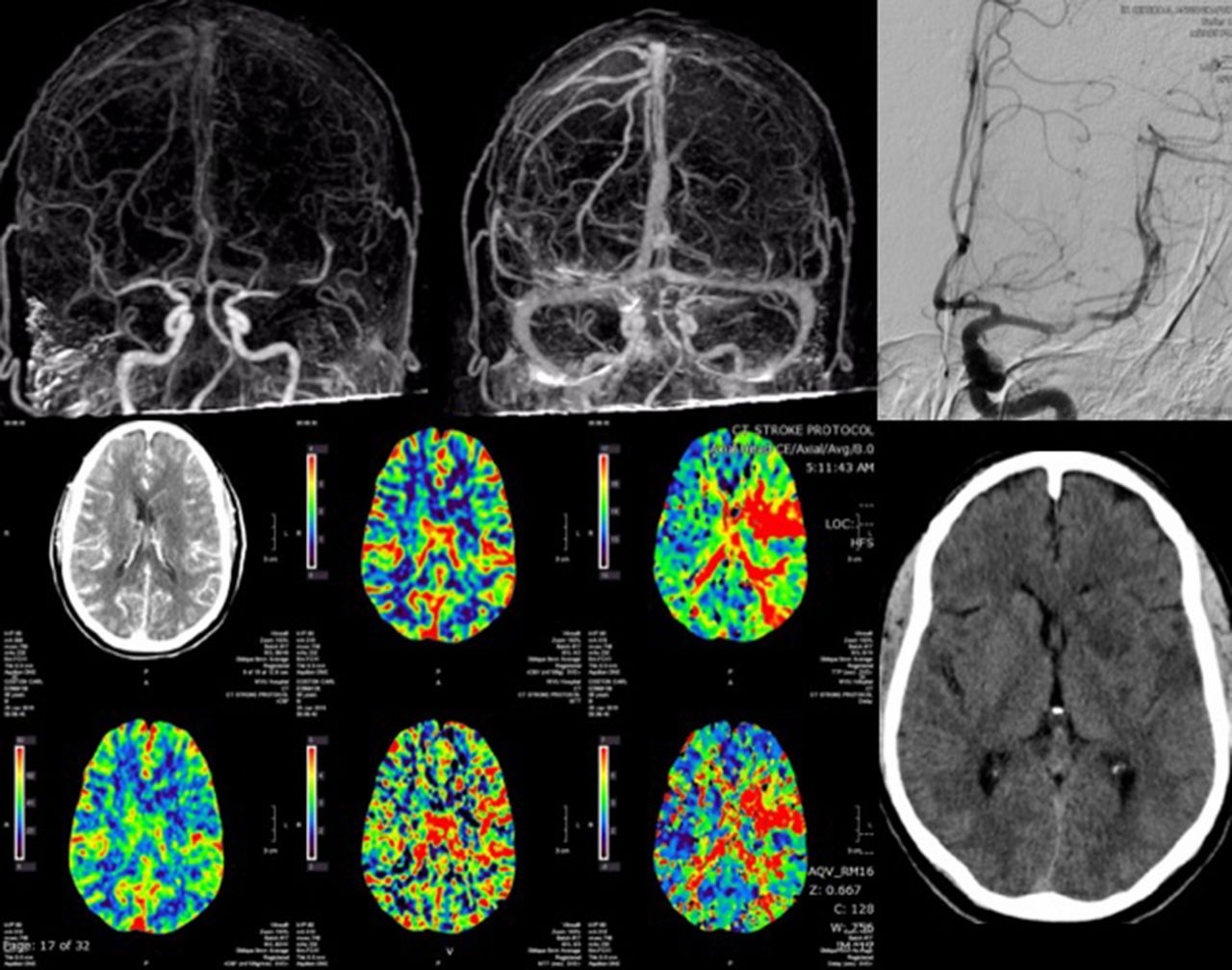
Results A middle-aged man was transferred after 20-hours with a suspected LVO on imaging. The NIHSS was 12 with right sided hemiplegia and slurred speech. A 5F pigtail catheter was placed in the aortic arch in the hybrid interventional (AlphenixTM) and CT (GenesisTM Aquilion One) suite (Canon Medical Systems, Tustin, CA). A 2-second, intra-aortic injection was performed with 20 ml of 30% contrast-saline mixture at 10 ml/s. Simultaneous triggering of the continuous CT volumetric acquisition was performed with a peak of 80 KVP at a constant 320 mAmp. Volumetric angiography reconstruction of the data set was performed at 0.3s interval (320 images, 0.5 mm thickness with 16cm of coverage). The 12-second single acquisition generated a non-contrast CT of the brain, multiphase angiography at 3 frames/second and whole-brain perfusion imaging (figure 1). The non-contrast CT showed patchy infarcts. The angiography showed a sub-occlusive left MCA thrombus with good collaterals. The perfusion imaging showed matched defects corresponding to the infarcts but no significant penumbral tissue. A standard selective left internal carotid artery catheter angiography confirmed the sub-occlusive thrombus and good collaterals. Repeat on table clinical exam had improved from admission. Given the sub-occlusive thrombus, good collaterals and no at-risk ischemic tissue on perfusion – thrombectomy was not performed.
Conclusion Rapid assessment of suspected LVO with simultaneous treatment is feasible in select stroke patients, e.g. transfers, using a hybrid CT-Angiography suite and direct intra-aortic diluted contrast injection. An intra-arterial injection gives superior signal-to-noise ratio and temporal resolution than intravenous injection. Immediate thrombectomy following the acquisition in the same room can shorten door to recanalization times.
Disclosures A. Rai: 2; C; Stryker, Microvention, Cerenovus. A. Tarabishy: None. S. Boo: 2; C; STRYKER.