Article Text

Abstract
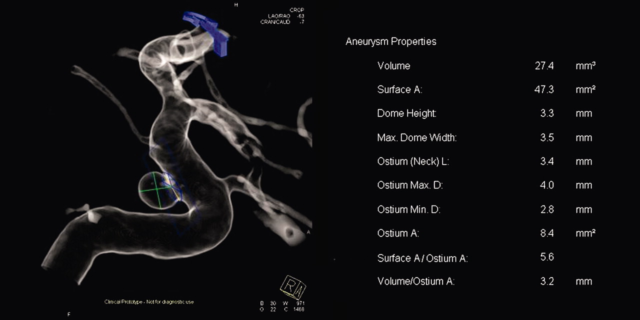
Introduction/purpose Endovascular treatment planning for intracranial aneurysms requires accurate quantification of aneurysm geometry. Today, aneurysm geometry is typically measured manually on an angiographic workstation based on three-dimensional (3D) Digital Subtraction Angiography (DSA) images. Since accuracy and reproducibility of manual measurements are user dependent, computer-aided approaches have been recently proposed. We evaluated prototype software for intracranial aneurysm geometry quantification and compared generated measurements to manual measurements from two experienced raters.
Materials and Methods We examined 25 saccular unruputured single intracranial aneurysms (sidewall = 17, bifurcation = 8) treated via endovascular coiling at our institution between August 3, 2011 and February 23, 2012. Based on 3D DSA images, Manual measurements of the neck length (N), dome height (H), dome diameter (D) were obtained by two independent experienced raters. The aneurysm volume (V) was computed manually via an image punching and voxel counting approach. Subsequently, prototype cranial aneurysm analysis software (Siemens AG, Forchheim, Germany) was applied to the 3D DSA images to measure the same quantities. The software requires the user to manually mark the aneurysm dome via one point click and the parent artery via two point clicks, proximal and distal to the aneurysm.
Results The software to average rater errors were compared to inter-rater variability. No statistically significant differences were found between the two error distributions for N, H, D (p=0.371, p=0.567 and p=0.195, respectively). The average software-to-rater errors were N=0.55±1.03 mm, H=0.49±0.58 mm and D=0.45±0.39 mm. The largest errors in N (4.23 mm) and H (2.42 mm) occurred in cases where the 3D image could not adequately separate the aneurysm dome from the parent vessel. For V, data from only one rater were available. The average relative error was 7.76%±6.68% with maximum of 20.9%.
Conclusions Results indicate good agreement between measurements obtained by the software and two independent manual sets of measurements. In cases where 3D imaging did not adequately separate the aneurysm dome from the neighboring blood vessels, the aneurysm neck length, dome height, and volume measurement errors were the largest. Manual corrections may be necessary in such cases. Further studies on a larger dataset of various aneurysm geometries are needed.
Competing interests Y Murayama: Siemens AG. A Mohamed: Siemens AG. H Takao: None. T Ishibashi: None. I Yuki: None.