Article Text

Abstract
Purpose Unruptured intracranial aneurysms (IA) are potentially life threatening conditions. Rupture risk based on specific data regarding interval growth or destabilization is lacking. We studied the feasibility of 4D-Flow MRI to analyze 3D flow patterns (vorticity) and aneurysm wall shear stress (WSS) in large and giant IAs.
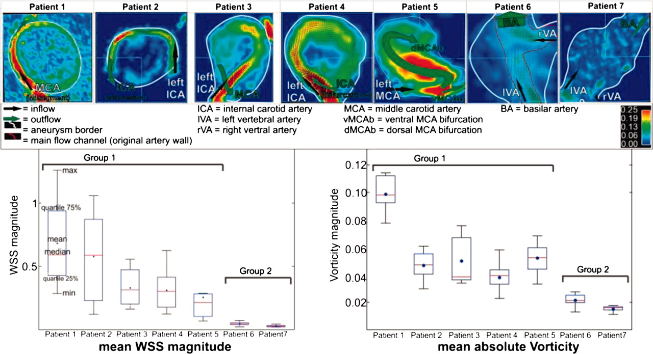
Materials and Methods Seven patients (4F:3M, age=3.6±15.4 years) with large IAs (mean diameter=2.6±0.9 cm) were studied. IAs were located near the ICA bifurcation (n=5) or basilar artery (n=2) with saccular, spherical, or fusiform morphology. 4D flow MRI (3T TRIO & 1.5T Avanto, Siemens, Germany, voxel =0.99–1.8×0.78–1.46×1.2–1.4 mm, TE/TR=2.9–3.3/5.5–6 ms, temporal-res=44–48 ms, 3-directional velocity encoding with venc =70–80 cm/s) data were analyzed using Matlab-based tools and 3D blood flow visualization software. The WSS pattern along the aneurysm wall was calculated by cubic spline interpolation of the velocity gradient along the aneurysm contour as described previously. The vorticity defined as Vort=abs(ζx,ζy,ζz) (with ζx=δw/δy –δv/δz, ζy=δu/δz–δw/δx, ζz=δv/δx– δu/δy and u,v,w being the velocity vector components2) was calculated within the plane transecting the aneurysm.
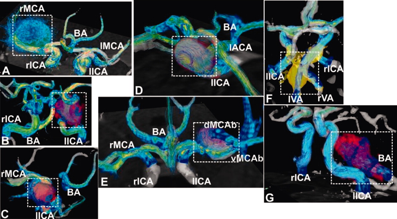
Results 3D spatial and 3-directional velocity encoding allowed for the 3D visualization of complex intracranial flow patterns. Flow patterns in the aneurysms could be classified in two morphological groups. Narrow high-flow channels in combination with large central low flow regions were identified in five saccular/spherical IAs. Slow flow with less defined flow channels were noted in two fusiform IAs. The distribution of WSS was significantly more heterogeneous and increased and vorticity in the plane through the IA center was significantly higher for saccular/spherical vs fusiform IAs (WSS=0.63N/m2±0.33 N/m2 vs 0.038 N/m2±0.016; Vorticity =0.073±0.028 vs 0.018±0.005; p<0.01).
Conclusion Our feasibility study shows the potential of 4D flow MRI to identify differences in flow characteristics such as vorticity and WSS patterns in two IA morphology groups which may improve risk stratification and treatment planning.
Competing interests None.