Article Text

Abstract
Introduction/purpose The geometry and organization of intranidal vessels that underlie flow within cerebral arteriovenous malformations (AVM) have not been clearly elucidated. Here, we examine the relationship between intranidal artery characteristics and AVM flow rate.
Materials and methods Records of patients with cerebral AVMs evaluated at our institution between 2007–2013 were retrospectively reviewed. Patients were included if a surgical specimen of the nidus was available and if flows were obtained before treatment using quantitative magnetic resonance angiography. Specimens were mounted on slides and stained with hematoxylin and eosin as well as with elastic special stain. Intranidal arteries were identified and the diameter and cross-sectional area of each artery were measured from digitized images of the slides. The total area of the slide studied and magnification used was the same for each patient. Total AVM flow was estimated as aggregate flow within primary arterial feeders or flow in single draining veins. AVM volume was determined from digital subtraction angiography. The relationship between vessel diameter, vessel cross-sectional area, AVM volume, and AVM flow was assessed.
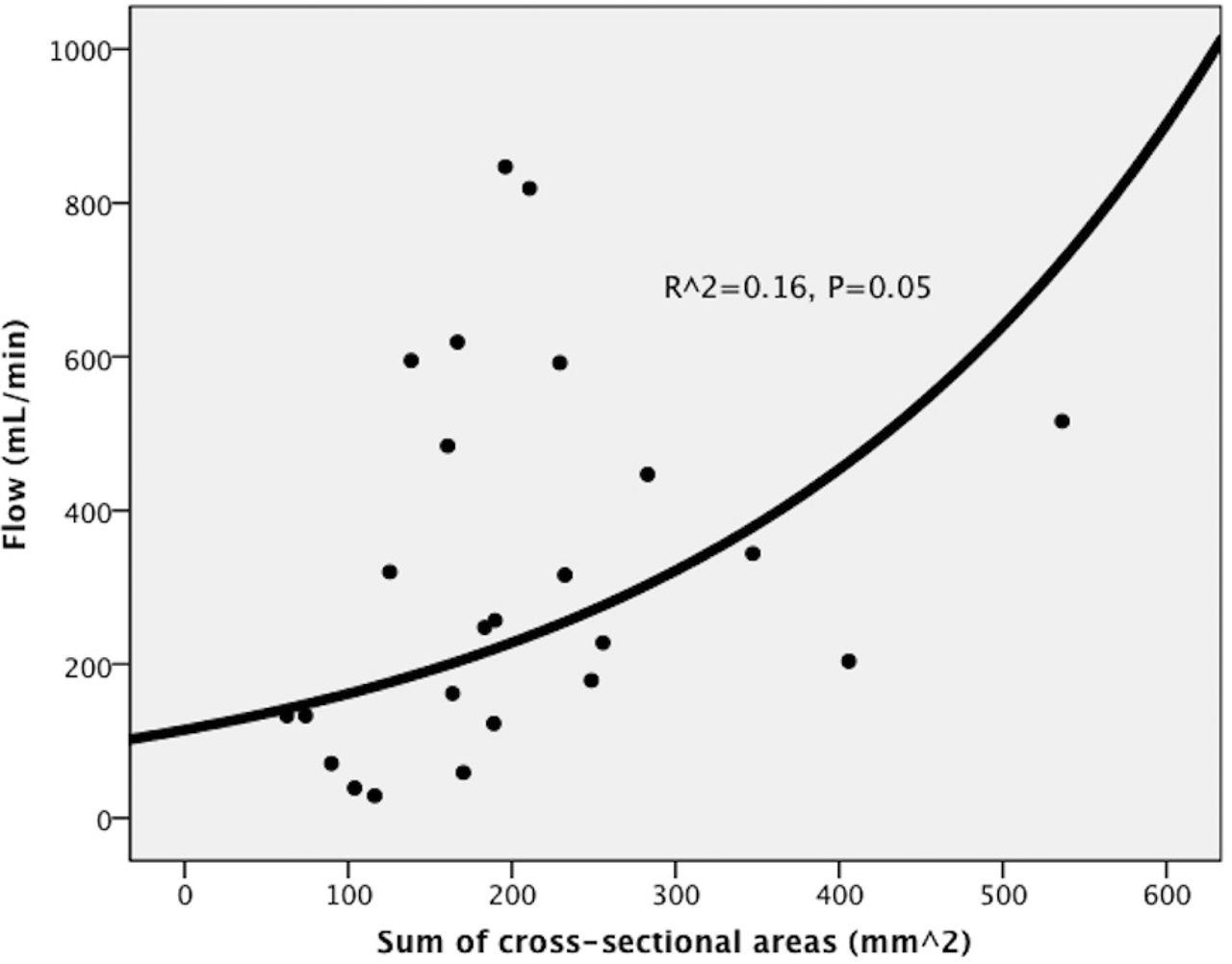
Results 29 patients were included. Cohort characteristics are summarized in Table 1. Mean total number of arteries per specimen was 133. Mean total AVM flow was 340 ± 276 mL/min. Mean vessel diameter ranged from 0.18–2.37 mm and mean vessel cross-sectional area ranged from 0.09–9.46 mm2. Linear regression analysis showed that total flow is significantly associated with larger AVM volume (R2 = 0.28, P = 0.007), but not with the number of arteries per section of the specimen (P = 0.20) or mean vessel diameter (P = 0.92). Exponential regression analysis demonstrated that AVM flow is significantly correlated to the sum of the cross-sectional areas within each specimen (R2 = 0.16, P = 0.05) (Figure 1).
Patient and AVM characteristics
Conclusion Total AVM flow is significantly related to the sum of the cross-sectional areas of all arteries within each nidus, rather than to the total number of arteries or mean nidal vessel diameter. This finding suggests that the sum of the cross-sectional areas of intranidal arteries likely determines the permeability of the cerebral AVM nidus.

Total AVM flow versus sum of the cross-sectional areas of all arteries within each nidus
Disclosures S. Shakur: None. T. Valyi-Nagy: None. S. Amin-Hanjani: None. L. Ya'qoub: None. V. Aletich: None. F. Charbel: None. A. Alaraj: None.