Article Text

Abstract
Introduction/purpose Sigmoid sinus diverticulum (SSD) is one of the potentially treatable causes of pulsatile tinnitus (PT). How SSD causes PT is unknown, but it is thought to be secondary to aberrant blood flow in the diverticulum or parent sinus. We performed velocity field mapping using MR 4 D Flow (MRV) and computational fluid dynamics (CFD) in cerebral venous sinuses and internal jugular veins (IJV). We aim to determine if a distinct blood flow pattern may be responsible for PT in SSD.
Materials and methods Patients suspected of venous etiology of PT underwent MRI at 3 T, using contrast-enhanced MRA (timed to venous phase), MRV and CFD. SSD was confirmed on MRA. Flow pathlines were evaluated. In patients with confirmed SSD, additional CFD modelling was performed with the SSD excluded from the models.
Results Nineteen patients with suspected venous etiology of PT and 10 controls were evaluated. Six (31.5%) had SSD and five of these had transverse sinus stenosis upstream from the SSD. These five patients also demonstrated a unique pattern of flow not seen in the controls characterized by:
1. High velocity flow jet in an up-stream stenosis in the transverse sinus directed at the SSD opening,
2. Flow jet into the SSD along the long axis of the SSD, either anteriorly or laterally directed,
3. Vortex of flow in the SSD,
4. Prominent vortex component of flow in the sigmoid sinus downstream from SSD,
5. Vortex of flow in the jugular bulb
Three of the patients had simulated post-coil treatment models developed excluding the SSD from the models. CFD showed no flow in SSD and decreased vortex component of flow in the sigmoid sinus downstream from the SSD.
Conclusion PT caused by SSD may be caused by a unique flow pattern in the SSD and sinuses as visualized on both MRV and CFD.
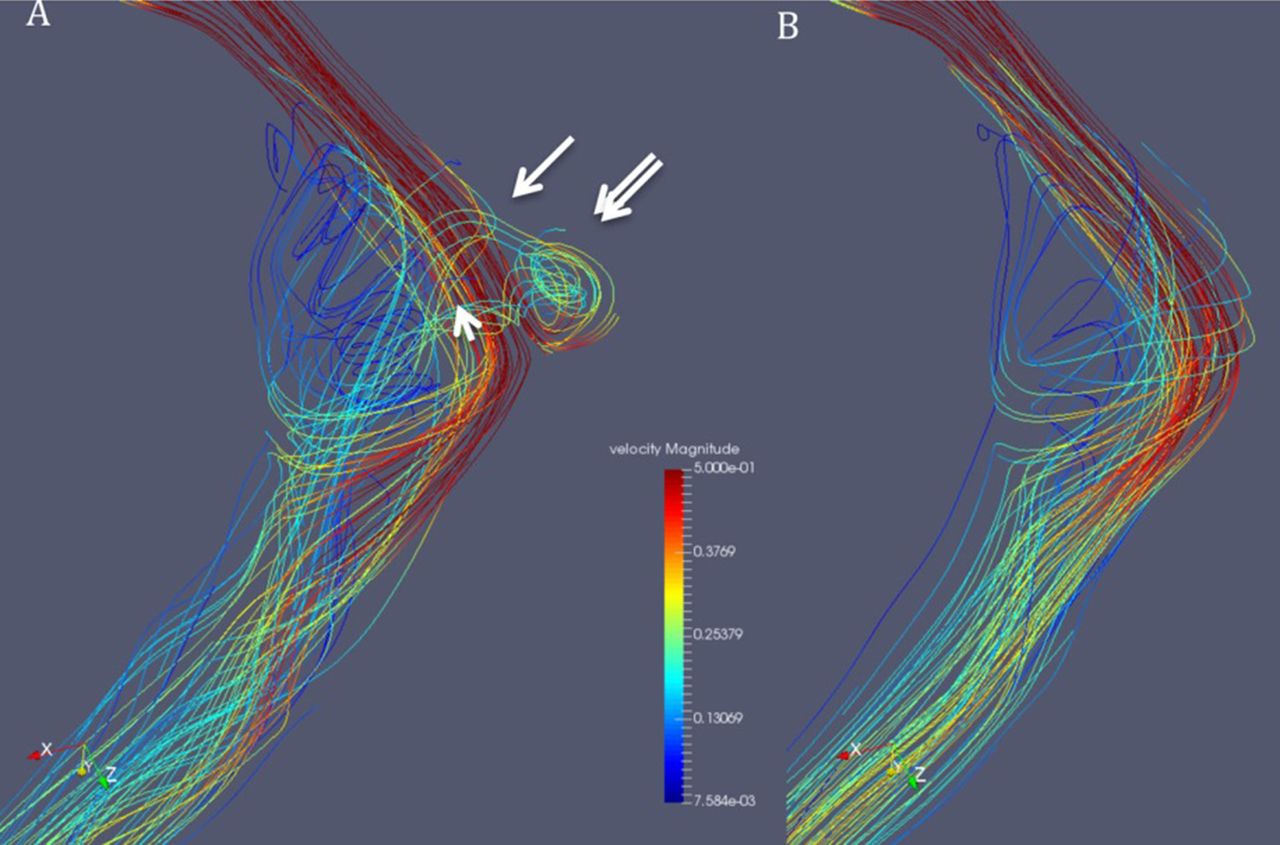
Townes projection CFD analysis of a patient with a left SSD and an upstream stenosis in the transverse sinus. The pretreatment analysis (A) shows a jet of flow from the stenosis into the SSD (arrow), vortex of flow in the SSD (double arrow), and a vortex component of flow in the sigmoid sinus down-stream from the SSD (arrow head). The post-treatment model (B) shows absence of the flow in the SSD as well as decreased vortex component of flow in down-stream sigmoid sinus
Disclosures M. Amans: None. E. Kao: None. S. Kefayati: None. K. Meisel: None. F. Faraji: None. C. Glastonbury: None. M. Ballweber: None. V. Halbach: None. D. Saloner: None.
This is an Open Access article distributed in accordance with the Creative Commons Attribution Non Commercial (CC BY-NC 4.0) license, which permits others to distribute, remix, adapt, build upon this work noncommercially, and license their derivative works on different terms, provided the original work is properly cited and the use is non-commercial. See: http://creativecommons.org/licenses/by-nc/4.0/