Article Text

Abstract
Objective To report our initial experience with the Medina Embolic Device (MED) in unruptured intracranial aneurysms either as sole treatment or in conjunction with additional devices.
Methods 15 consecutive patients (6 women, 9 men) with unruptured aneurysms were treated between September 2015 and April 2016. The aneurysm fundus measured at least 5 mm. We evaluated the angiographic appearances of treated aneurysms at the end of the procedure and at follow-up, the clinical status, complications, and requirement for adjunctive devices.
Results The MED was successfully deployed in all but one case and adjunctive devices were required in 10 cases. Aneurysm locations were middle cerebral artery bifurcation (n=3), internal carotid artery (ICA) bifurcation (n=1), supraclinoid ICA (n=5), posterior communicating artery (n=1), anterior communicating artery (n=2), cavernous ICA (n=2), distal basilar sidewall (n=1), basilar tip (n=1). Three patients had complications although none could be attributed to the MED. Immediate angiographic results were modified Raymond-Roy classification (mRRC) I=1, mRRC II=5, mRRC IIIa=3, mRRC IIIb=5, and one patient showed contrast stasis within the fundus of the aneurysm. Follow-up angiography was available in 11 patients, with four showing complete aneurysm exclusion, six with stable remnants and one patient with an enlarging neck remnant.
Conclusions The MED represents a major step forward in the treatment of intracranial aneurysms. It can result in rapid exclusion of an aneurysm from the circulation and has a good safety profile. We believe that the true value of the MED will be in combining its use with adjunctive devices such as endoluminal flow diverters that will result in rapid aneurysmal exclusion.
- Aneurysm
- Flow Diverter
- Coil
This is an Open Access article distributed in accordance with the Creative Commons Attribution Non Commercial (CC BY-NC 4.0) license, which permits others to distribute, remix, adapt, build upon this work non-commercially, and license their derivative works on different terms, provided the original work is properly cited and the use is non-commercial. See: http://creativecommons.org/licenses/by-nc/4.0/
Statistics from Altmetric.com
Introduction
The introduction of Guglielmi detachable coils (GDCs) in 1992 marked a seminal point in the history of interventional neuroradiology. The validity of this endovascular treatment option was subsequently proven 10 years later when the results of the International Subarachnoid Aneurysm Trial (ISAT) were published.1 The long-term follow-up data of patients in the ISAT cohort have since been published and further prove the durability of coil embolization for ruptured intracranial aneurysms.2 Despite the fact that more than a decade has passed, the essential therapy of putting coils into an aneurysm to induce thrombosis remains largely unchanged. Catheters may have changed, coils have become more complex in shape, softer and come with a variety of additive features that may improve intrasaccular thrombosis but, in essence, the principle of placing a coil into an aneurysm to encourage thrombosis has remained largely unadulterated. Recently, technological advances have allowed more complex intrasaccular devices to be produced, including the Woven EndoBridge (WEB, Sequent Medical) and, more recently, the Medina Embolic Device (MED, Medtronic).3 Both of these devices can be thought of as intrasaccular flow diverters; however, it is at this point that the similarities between the two devices effectively end.
In this paper we review our experience of the Medina system both as a solitary treatment strategy and in conjunction with additional treatment methods, our reasoning behind the various strategies, as well as the potential advantages and disadvantages of this new device.
Materials and methods
Device description
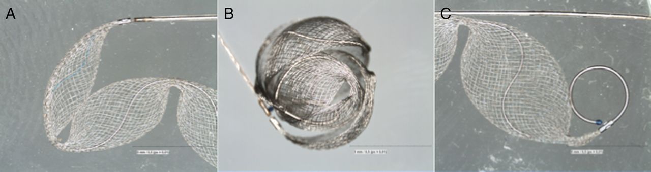
The MED is designed to treat saccular aneurysms. It has been granted a CE mark by the European Union. The device is a metallic three-dimensional layered structure made from a radiopaque shape set core wire and shape memory alloy filaments, which form a self-expanding mesh. This resembles multiple leaflets that can provide flow diversion. These leaflets lie along the long axis of the core wire and, when deployed, the device assumes a spherical shape. The MED is inserted via a 0.021 inch internal diameter (ID) microcatheter and can be resheathed and redeployed as with standard coils. It is mechanically detached with the same mechanism as Axium coils (Medtronic). The MED comes in two types—framing and filler. The filler variant is softer and designed to fill the internal space after a framing MED has been deployed to provide a suitable basket (figure 1).3 ,4

The Medina Embolic Device (MED) seen straightened out (A and C) and in its spherical form (B).
Patient selection
Between September 2015 and April 2016 a total of 216 aneurysms underwent endovascular treatment (85 coil occlusion, 131 flow diversion) in our department. From those we offered the use of MED to patients with unruptured aneurysms with a fundus diameter of 5 mm or more. The decision was based on individual aspects such as anticipated feasibility of this kind of treatment, propensity to coil compaction or aneurysm reperfusion. In several patients the use of the MED was from the very beginning part of a more complex treatment strategy including the usage of other implants (eg, flow diverters or bifurcation stents).
Procedure
Patients were selected based on aneurysm size, at least 5 mm, as this is currently the smallest size available. An 8 Fr system was used as standard in case adjunctive devices such as a pCONus, stents or flow diverters were required. With regard to the pCONus, this was deployed first and subsequently the aneurysm was re-catheterized to allow placement of the MED. In cases of adjunctive flow diverter use, the MED was placed in the aneurysm first and the flow diverter positioned in the parent vessel afterwards.
All patients gave informed consent for the procedure and received dual antiplatelet therapy for at least 3 days prior to treatment if adjunctive devices were to be used. Response to the antiplatelet medication was checked using the Multiplate analyser.
An intraoperative bolus dose of heparin (5000 IU) was given at the start of the procedure with subsequent bolus doses to maintain the activated clotting time (ACT) between 2 and 2.5 times normal.
For deployment of the MED a 0.021 inch ID Prowler Select Plus microcatheter (Codman) was used. The framing MED was chosen based on a similar sizing method to that used for standard coils. In spherical saccular aneurysms with a fundus diameter of 9 mm or less, the size of the first framing MED was purposely undersized by about 1 mm. After forming a spherical shape and adequate positioning of the framing MED, it was mechanically detached and the requirement for further occlusion either with a filling MED or standard coils was evaluated. In some instances we placed only a single framing MED into the aneurysm as we believed this would be adequate. In other cases we added a significant number of filling MEDs or conventional coils based on the operator’s discretion.
Follow-up
Angiographic follow-up was scheduled on an individual basis including a very early digital subtraction angiogram (DSA) a few days after the treatment if considered necessary by the circumstances and with an early DSA either 6 or 12 weeks after the initial procedure. Mid-term DSAs are typically scheduled for 6 or 12 months after treatment.
Evaluation
The histories of all patients treated with a MED were evaluated in retrospect. We recorded the procedural device function and the occurrence of any adverse events, both technical and clinical. The degree and stability of aneurysm occlusion was evaluated. On an individual basis we tried to determine the usefulness of the MED in the context of the specific treatment.
Results
Fifteen patients (six women and nine men) with 16 aneurysms were treated with the MED either alone or in conjunction with other devices. The average age of the patients was 62.2 years (range 46–79). Aneurysm locations were middle cerebral artery bifurcation (n=3), internal carotid artery (ICA) bifurcation (n=1), supraclinoid ICA (n=5), posterior communicating artery (n=1), anterior communicating artery (n=2), cavernous ICA (n=2), distal basilar sidewall (n=1), and basilar tip (n=1). All aneurysms were treated electively. The results are summarized in table 1.
Overview of the aneurysm characteristics, clinical and radiological follow-up and use of adjunctive devices
In one case (patient 2) we were unable to successfully deliver the MED into the aneurysm and therefore switched to coiling the aneurysm. This patient suffered a stroke related to coil migration and therefore the complication was secondary to the coil and not the MED. In all other cases the MED was successfully deployed. Patient 9 suffered from a left hemiparesis and MRI demonstrated multiple small diffusion-weighted imaging lesions within the right cerebral hemisphere consistent with a watershed type of infarction pattern. We are unaware of any significant falls in blood pressure during the procedure or during the induction of anesthesia.
In 10 patients adjunctive devices were used—for example, coils, flow diverters, or a neck-bridging device. There were no complications related to the use of these devices. The angiographic appearance at the end of the procedure was graded according to the modified Raymond-Roy classification scale.5
Illustrative cases
No adjunctive devices used
Complete occlusion with MED alone
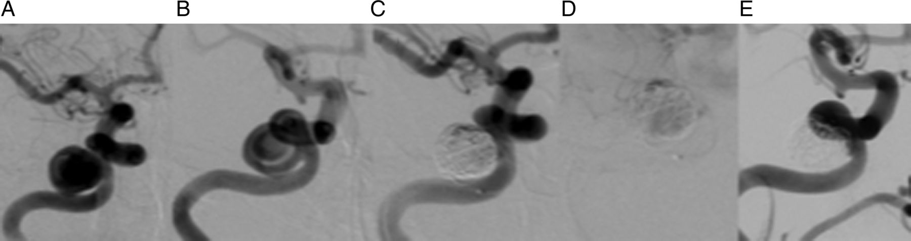
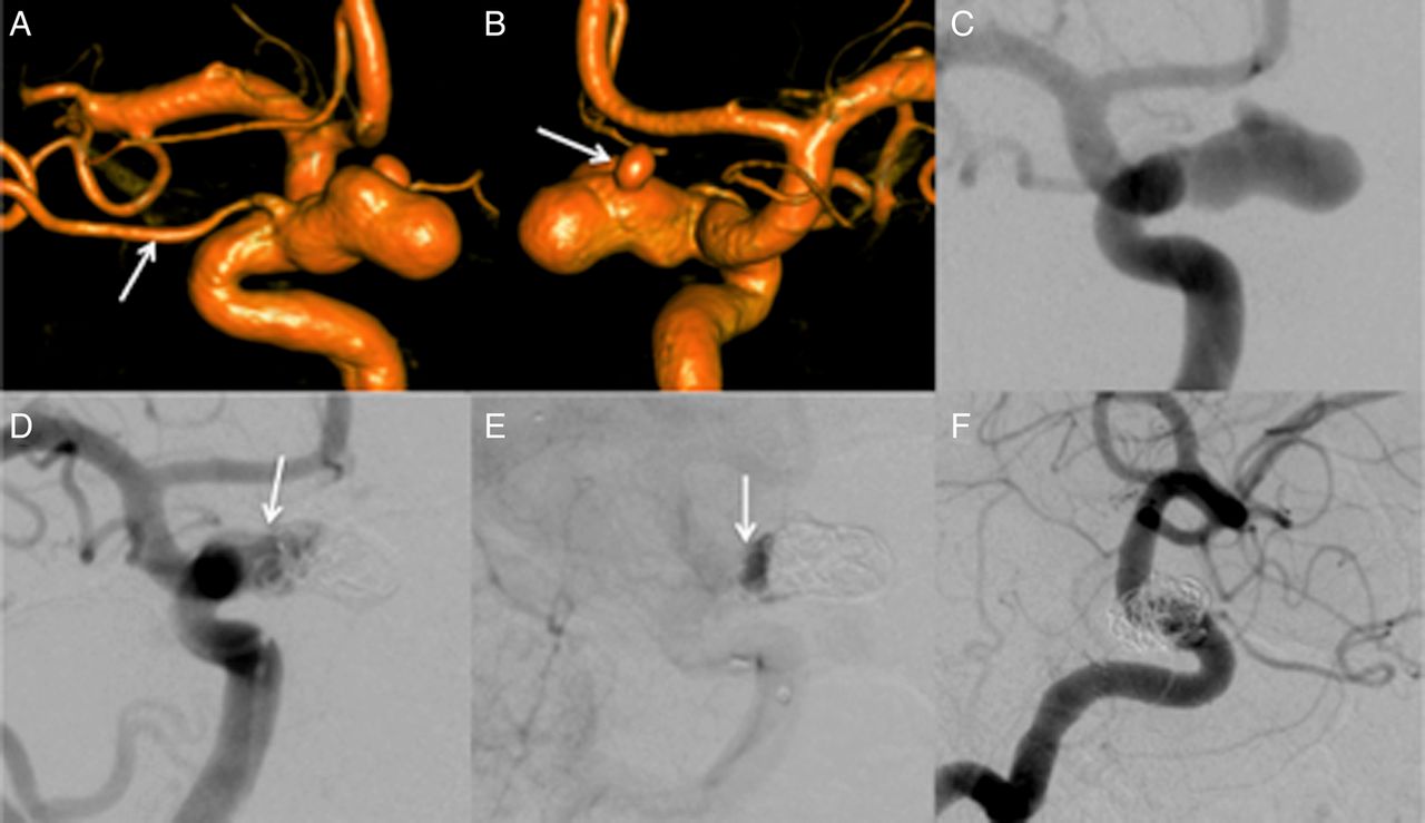
In certain anatomical situations the MED can effect extremely rapid aneurysm exclusion without the need for adjunctive devices. One such case is illustrated in figure 2 (patient 1). The patient was admitted to our institution for management of a large spherical extradural aneurysm found incidentally on MRI performed for a persistent headache. Multiple MEDs were placed into the aneurysm. The patient returned to the angiography suite 2 days later for a follow-up angiogram which showed complete obliteration of the aneurysm.

A right internal carotid artery angiogram shows a large cavernous aneurysm (A) with contrast swirling inside the aneurysmal dome (B). Angiography at the end of the procedure shows minimal opacification along the superior edge of the aneurysm (C) and pronounced stagnation (D). A follow-up angiogram 2 days after the intervention shows complete exclusion of the aneurysm from the circulation.
Neck remnant with MED alone
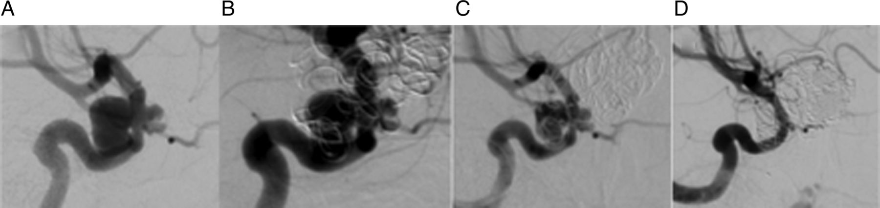
The classical spherical aneurysm is not often encountered in everday clinical practice. Instead, oblong or ‘beehive’-shaped aneurysms are frequently seen. These aneurysms can also be treated with the MED (±standard coils), but a neck remnant may remain if treated only with the MED. This situation was encountered in several of our cases and is represented in figures 3 and 4 (patients 11 and 6 in table 1). The further management of these patients will depend on the clinical status of the patient and also on the long-term appearance of the neck remnant. No further treatments are currently planned in either of these patients.

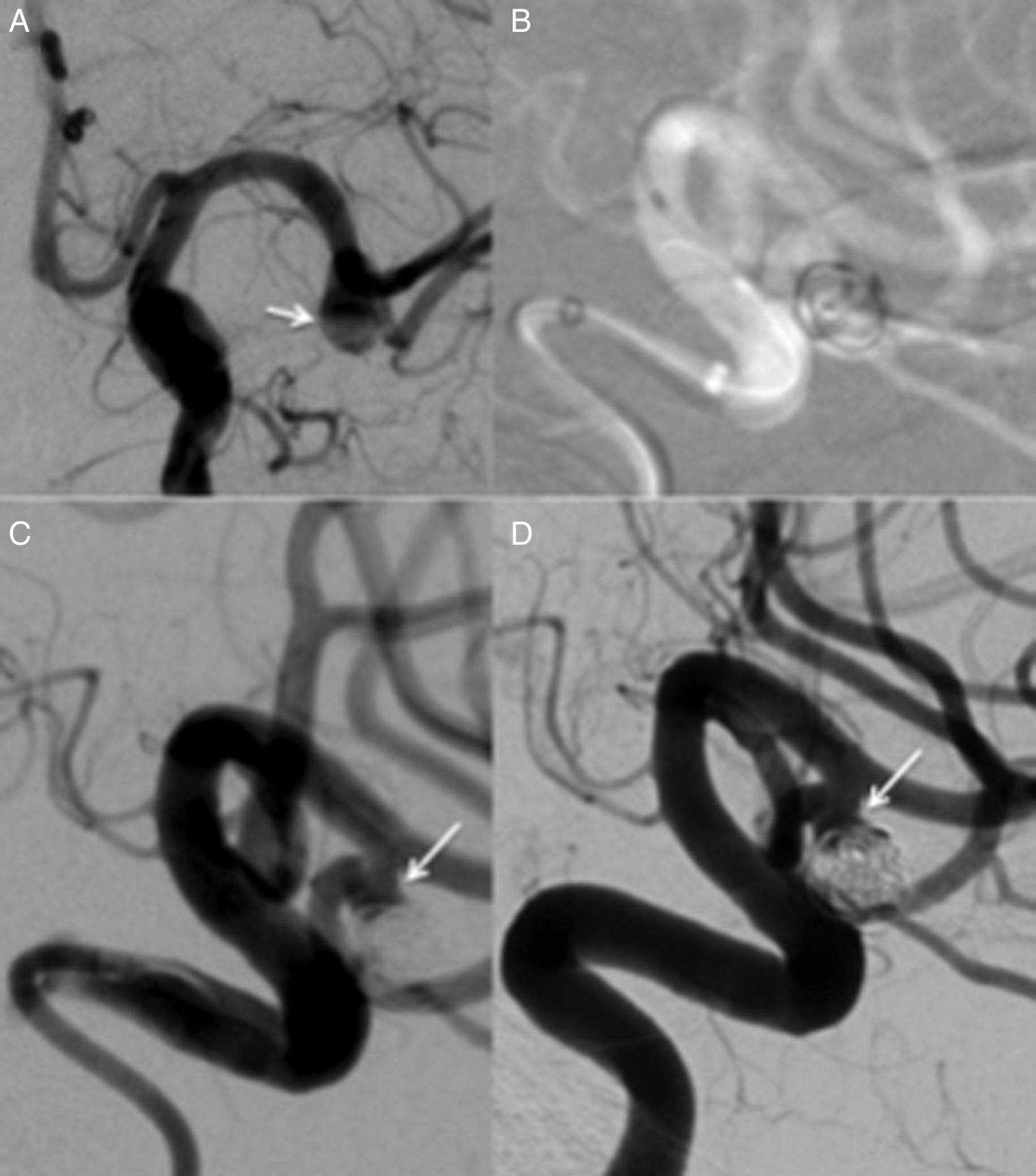
A patient in their 60s with a wide-necked ‘beehive’-shaped aneurysm arising from the inferior trunk of the left middle cerebral artery bifurcation (A). A single framing Medina Embolic Device was placed inside the aneurysm (B) followed by a single helical coil, and this resulted in the occlusion of the aneurysm fundus. A small neck remnant remained (C, white arrow). A follow-up angiogram performed 1 month later showed a stable result with a neck remnant (D).

A patient in their 70s with a wide-necked ‘beehive’ aneurysm at the right middle cerebral artery bifurcation (A) was treated with both the Medina Embolic Device and coils. The angiogram at the end of the procedure shows complete exclusion of the aneurysm dome from the circulation (B) and a small neck remnant. An early follow-up angiogram performed at 1 month shows a stable appearance to the neck remnant.
Combination of MED and adjunctive devices
Neck protection device and/or standard coils
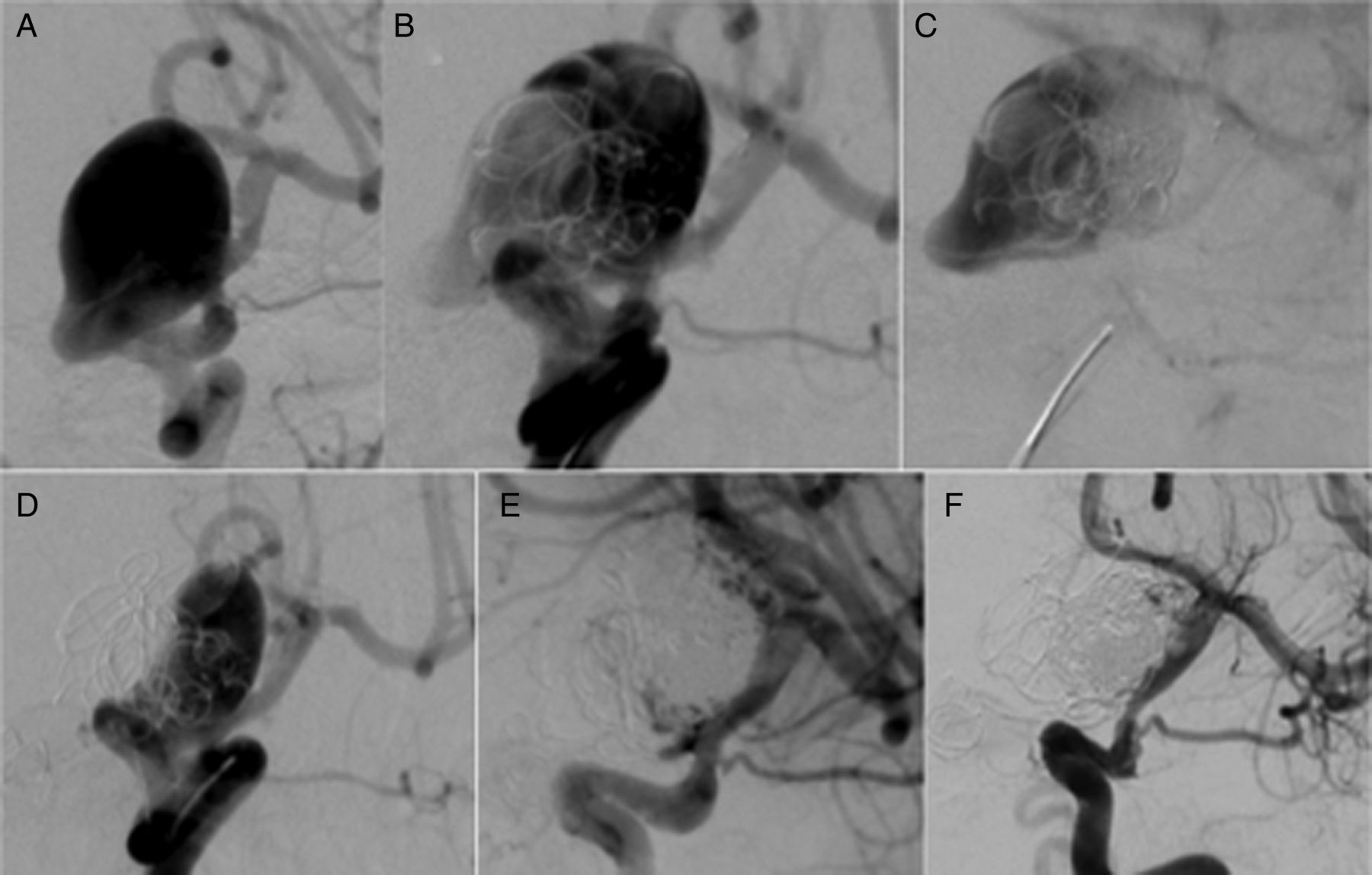
Treatment of wide-necked aneurysms and aneurysms where the vessels originate from the neck represent a significant challenge. Newer devices such as the pCONus and PulseRider allow the protection of these arteries. In our institution we have experience with the pCONus; however, the conjunctive use of the MED with the pCONus has proved successful in treating several aneurysms (figures 5⇓–7). As is standard practice, the pCONus is deployed first and not detached until satisfactory exclusion of the aneurysm is achieved. Additionally, the patients require standard dual antiplatelet treatment.

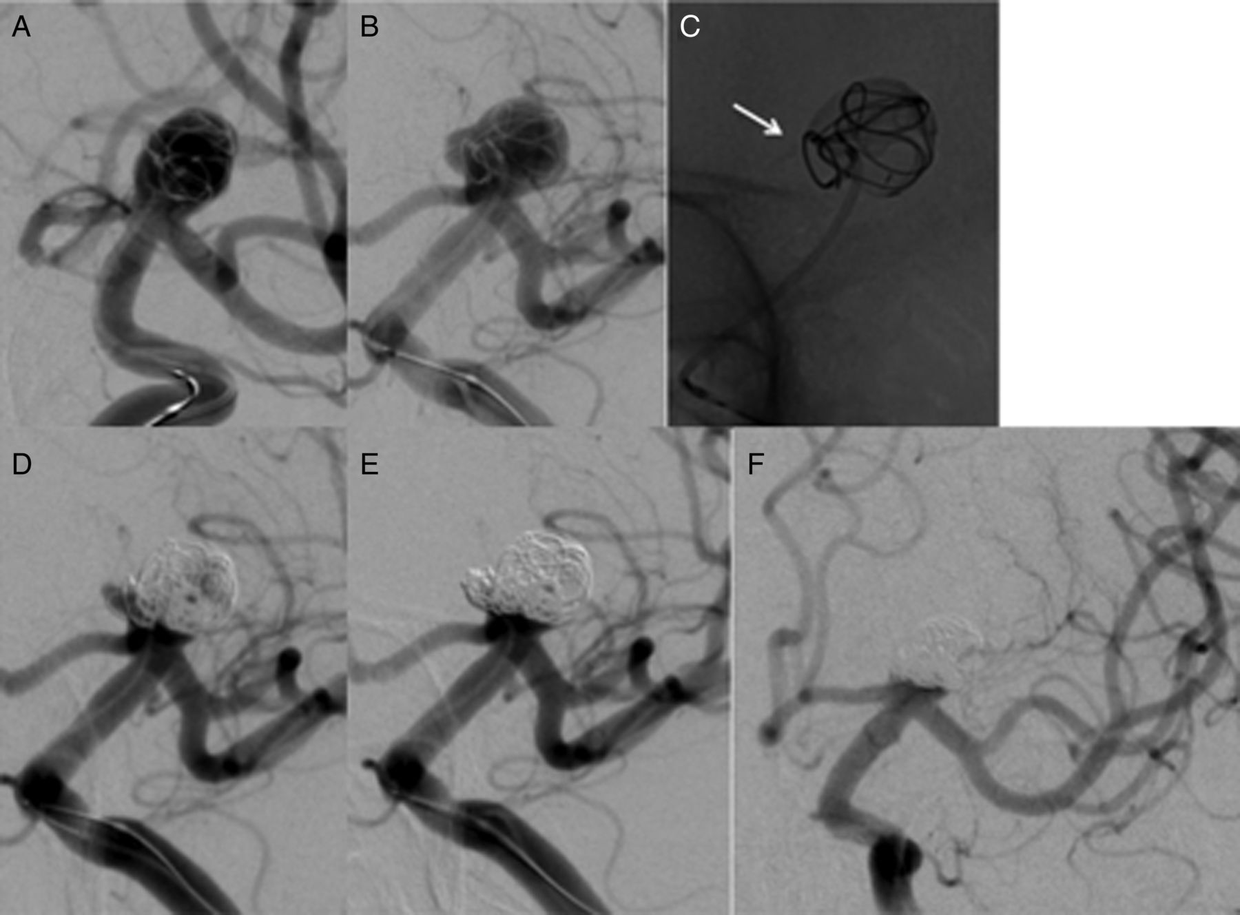
A wide-necked basilar tip aneurysm with planned treatment involving a pCONus and Medina Embolic Device (MED). On deployment of the MED framing coil, several gaps were noted between the leaflets (C, lateral unsubtracted image; D, frontal unsubtracted image) and therefore the device was resheathed and repositioned into what was believed to be a better position. Three filler MED devices were subsequently deployed into the aneurysm. Angiography revealed an unprotected area posteriorly (E) and this was therefore catheterized and six coils were deployed (G). Angiography at the end of the procedure showed modified Raymond-Roy classification IIIa occlusion status.
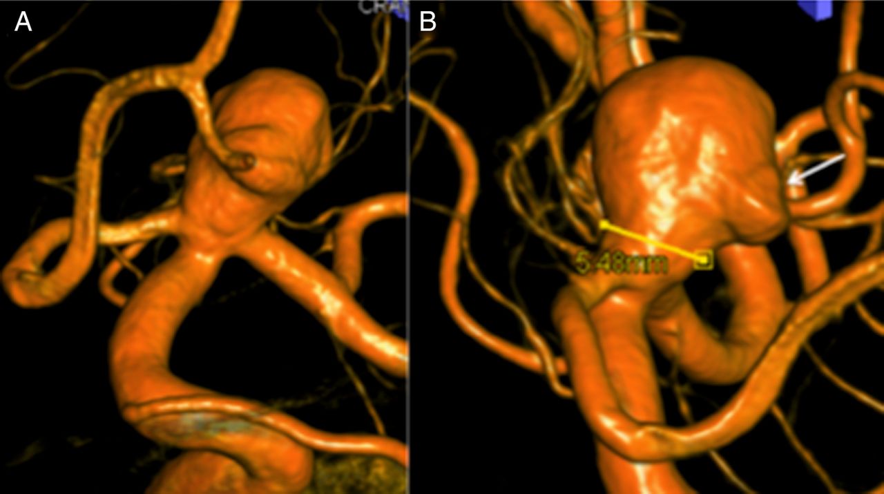
Rotational angiography demonstrating a large wide-necked lobulated aneurysm arising from the carotid bifurcation.

After successful delivery of a pCONus, a Medina framing device was placed inside the aneurysm (A and B). The position of the coil was believed to have been adequate at the time. However, careful inspection following the procedure shows that there was inadequate coverage of the bleb (C). Angiography after the framing and filling Medina Embolic Devices were placed in the dome of the aneurysm showed that flow had been redirected into the bleb (D) that was on the initial angiogram. This bleb was therefore catheterized and several coils were deployed (E), resulting in the complete exclusion of the aneurysmal dome from the circulation and only a small residual neck (F).
Both of these cases, patients 3 and 13, also highlight another extremely important point. The MED adopts a spherical shape; however, many aneurysms are not perfectly spherical. Extreme care must be taken to scrutinize the position of the MED and the petals to ensure they cover any particularly vulnerable segments of the aneurysm. Inadequate coverage of the aneurysmal blebs or inadequate apposition of the MED to the inner surface of the aneurysm can result in continued filling of the aneurysm or, in the worst case scenario, redirection of the flow by the MED into certain areas of the aneurysm (see figure 5E, D). In these situations it may be necessary to specifically catheterize these areas and deploy coils. Although none of our cases have involved ruptured aneurysms, we feel that this point is particularly pertinent to this scenario.
Endoluminal flow diversion
As mentioned above, there are often situations where the aneurysm neck may not be amenable to treatment solely with the MED. In these situations we used the MED to achieve rapid exclusion of the aneurysm dome and then endoluminal flow diversion to secure the residual neck and allow parent vessel reconstruction. In certain instances this has been done in a single setting, but in others the procedures have been performed sequentially.
Patient 10 (figure 8) had a large wide-necked paraophthalmic aneurysm and it was felt that endoluminal flow diversion alone would not be suitable. A single MED was therefore placed in the aneurysm dome followed by a p64 endoluminal flow diverter at the same sitting. Angiography at 2 months showed no filling of the aneurysm dome but residual filling of the aneurysm neck, which is expected to thrombose secondary to the endoluminal flow diverter.

A right-sided wide-necked paraophthalmic aneurysm (A). A single framing Medina Embolic Device (MED) was placed in the aneurysm (B, white arrow) followed by a p64 flow diverter (B, black arrows). Angiography at the end of the procedure demonstrated marked stagnation inside the aneurysm (C). Follow-up angiography 2 months later shows occlusion of the aneurysmal dome but persistent opacification of the aneurysm neck, which is expected to thrombose due to the impact of the endoluminal flow diverter (D, white arrow).
Similarly, patient 15 (figure 9) had a multilobulated aneurysm paraophthalmic aneurysm which we felt should not be treated solely with endoluminal flow diversion because of the size of the aneurysm and the hostile appearance of the daughter sac. MEDs were placed in the aneurysm dome which served to rapidly exclude this portion of the aneurysm from the circulation and showed no flow into the dome at the end of the initial treatment with the MED (figure 9D). Angiography at 2 weeks showed expectant filling of the neck and therefore a p64 flow diverter was implanted to allow parent vessel reconstruction.

A large oblong-shaped paraophthalmic aneurysm with the ophthalmic artery arising from the neck (A) and a prominent bleb (B and C). The aneurysm was initially treated solely with the Medina Embolic Device as the aim was to cover the bleb. At the end of the first treatment residual filling of the neck remained (D). The patient was scheduled for a repeat treatment with a p64 flow diverter to the parent artery and angiography after deployment of the p64 showed significant stasis in the neck of the aneurysm (E). The ophthalmic artery continued to fill at the end of the second procedure (F).
Complications and device failures
In our cohort only one MED failed to detach (patient 2). This was removed and the aneurysm was coiled.
Patient 14 (figure 10) had a large paraophthalmic aneurysm with a smaller, more subtle blister-type aneurysm on the supraclinoid ICA. The treatment strategy was to treat the larger aneurysm with a MED and then place a p64 flow diverter into the ICA to cover both the aneurysms. After the procedure the patient had a subarachnoid hemorrhage, which we felt was due to the fact that she was a hyper-responder to ticagrelor, so the dose was reduced by half. Not taking ticagrelor in the evening resulted in a complete loss of effect and subsequently the patient developed an acute in-stent thrombosis and signs of right hemisphere stroke. The thrombus was successfully aspirated with a subsequent return to normal baseline neurological status. Angiography performed at 2 months revealed complete occlusion of both aneurysms.

A spherical paraophthalmic aneurysm with the ophthalmic artery arising from the aneurysmal neck. A further very small blister-type aneurysm was also noted (A, long white arrow). The paraophthalmic aneurysm was treated with a single Medina Embolic Device (MED) and stasis in the aneurysm was noted following the deployment of the MED (not shown). In order to treat the small blister-like aneurysm, a p64 flow diverter was deployed that also covered the neck of the paraophthalmic aneurysm (B). Five days later the patient showed signs of a right-sided stroke with in-stent thrombosis seen on the angiogram (C). Complete restoration of flow was achieved with no clinical sequelae. A follow-up angiogram performed at 2 months shows complete occlusion of both aneurysms and a patent ophthalmic artery (D).
Patient 9 (figures 11 and 12) was one of the initial patients treated with the MED and had bilateral aneurysms. On the right, multiple aneurysms could be seen; however, only one was suitable for treatment with the MED. Initial treatment consisted of a solitary MED framing device. Angiography at the end of the procedure did not show any significant stasis within the aneurysm.⇓

Multiple aneurysms can be seen arising from the supraclinoid internal carotid artery (ICA). The largest aneurysm was initially treated with a single Medina Embolic Device (B). However, no significant stasis was seen at the end of the procedure (not shown). Angiography 2 weeks later showed an intra-aneurysmal thrombosis (C) and therefore a p64 flow diverter was placed in the ICA to reconstruct the parent vessel and manage all the aneurysms simultaneously. Delayed angiography showed no further filling of any of the aneurysms (D).

Angiography on the left showed a large wide-necked conical aneurysm. The available Medina Embolic Device (MED)s were too small to create a single framing cage so it was decided to compartmentalize the aneurysm. At the end of the procedure there was significant stasis in the aneurysm dome (C). Angiography 2 weeks later showed occlusion of the aneurysm dome but compaction of the MEDs closer to the neck of the aneurysm so the aneurysm was recatheterized and coiled. A p64 flow diverter was also placed across the neck of the aneurysm and delayed angiography shows complete aneurysmal occlusion (F).
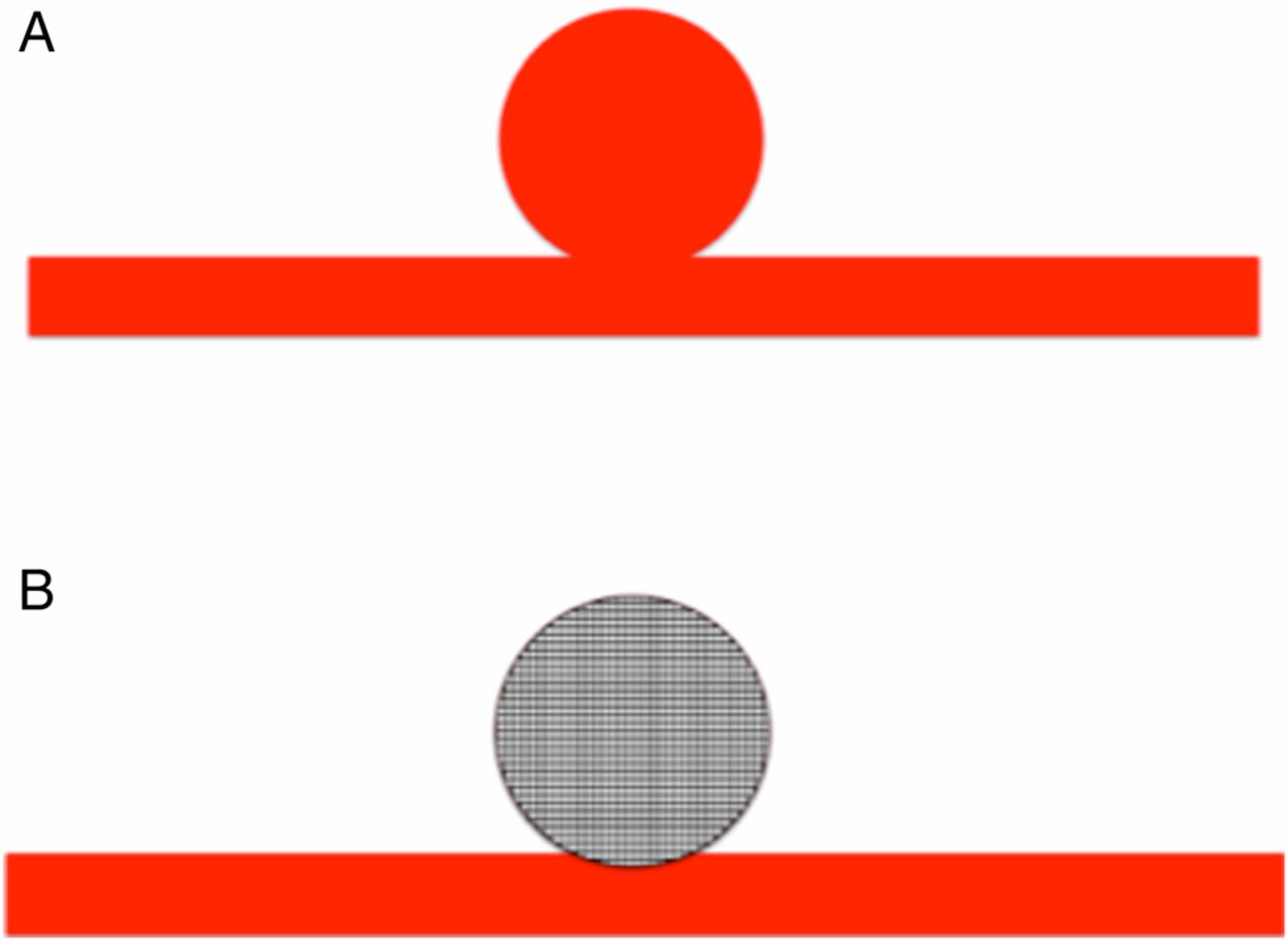
Pictorial representation of a spherical sidewall aneurysm (A) and the Medina Embolic Device (MED) within the aneurysm (B). This is the perfect scenario where the MED completely fills the aneurysm, there is no significant neck and no significant protrusion of the MED into the parent vessel. This scenario is similar to our patient 1.
On the left there was a large wide-necked conical aneurysm. The aneurysm was too large for a single MED so our strategy was to compartmentalize the aneurysm with multiple MEDs in addition to using a Solitaire device to protect the aneurysmal neck. Angiography at the end of the procedure revealed significant stasis within the aneurysm. Follow-up angiography 2 weeks later showed no significant change in the appearance of the aneurysm on the right. On the left there was thrombosis within a large part of the aneurysmal dome, where stasis had previously been seen. In addition, compaction of the MEDs was noted and therefore the aneurysm remnant was coiled. A flow diverter was placed across the neck of the aneurysm on both sides to encourage aneurysmal thrombosis. Follow-up angiography at 7 months revealed complete occlusion of all the aneurysms.
From this case we deduced that, when deploying MEDs, it is important to obtain contrast stasis within the aneurysm. If stasis is not seen it is possible that thrombosis will not occur. Furthermore, compaction of the MEDs can occur and, although compartmentalizing an aneurysm with MEDs can be done, we would advise against this strategy now.
Discussion
Since the introduction of detachable coils in 1991 and the publication of the ISAT study, endovascular coiling of intracranial aneurysms has become the preferred treatment option in many neurosurgical centers.1 ,6 ,7 However, major aneurysm recurrences can be found in 15–19% of patients by 3–6 months, which can rise to 21% at 16 months.8 ,9 The long-term follow-up of the ISAT study also showed that rebleeds from endovascularly coiled aneurysms were higher than in patients with clipped aneurysms,2 with 13 patients (n=809) in the endovascular treated cohort having a rebleed and 4 patients (n=835) in the surgically clipped group. The Cerebral Aneurysm Re-rupture After Treatment (CARAT) study also showed that the risk of early re-bleeding after treatment from ruptured aneurysms was strongly related to the occlusion status of the treated aneurysm.10 Therefore, a major goal for endovascular techniques has been to maintain the improvement in clinical outcomes but to decrease the risk of rebleeding so that it falls in line with, or is superior to, microsurgical clipping. To achieve this aim, coils with a variety of properties have been developed—for example, hydrogel coatings that expand and cause an increase in volumetric filling of the aneurysm. More recently, newer devices such as endosaccular flow diverters such as the WEB have entered the market. These devices take advantage of modifications in blood flow at the aneurysmal neck to encourage intra-aneurysmal thrombosis and with subsequent neo-endothelialization over time. The publication of the recent WEBCAST study showed that 23 of 41 patients (56.1%) had complete aneurysm occlusion at 6 months and 12 (29.3%) had a neck remnant. However, the long-term efficacy of this device is still unknown.
Cognard and Januel11 published the results of 15 aneurysms treated with the WEB (13 unruptured and 2 ruptured). In this series, radiological deterioration in the appearance of the aneurysm was noted in 10 of 14 aneurysms at initial follow-up (3–6 months) and, of those with longer-term follow-up, four had deteriorated. This group also showed that compression of the WEB is possible and is likely involved in aneurysm recurrences. However, in a cohort of eight patients reported by Sivan-Hoffman et al,12 only two had evidence of radiological deterioration at 12 months. These disparities highlight the need for further long-term evaluation of this device.
The MED is an alternative device to the WEB, which also employs intrasaccular flow diversion technology. The MED is deployed in a very similar manner to a coil with a similar technique used for accurately sizing the device to the aneurysm, unlike the WEB which requires a different sizing technique and is ideally done with on-table rotational angiography. The MED, in its current iteration, can be delivered via a 0.021 inch ID microcatheter which is an advantage over the WEB, some of which require a 0.027 inch microcatheter. The ability to place multiple MEDs in a Russian doll fashion or standard coils into the aneurysm is also helpful as this, in our opinion, will likely cause an increase in the degree of intra-aneurysmal stasis that will hopefully translate into earlier aneurysmal occlusion. We feel that this is a major advantage over the WEB and, in terms of efficacy, we have seen extremely rapid occlusion of large aneurysms (patient 1).
The delivery of the MED, while similar to that of standard coils, is significantly stiffer and the retraction and redeployment of the device is, in our experience, less accurate than with standard coils. This is probably a result of the increased surface area of the MED inside the microcatheter and in the aneurysm and, hopefully, with later derivations of the device this feature will improve.
An additional advantage (or disadvantage, depending on your standpoint) is that the leaflets of the MED are relatively radiolucent. This can make it easier to see through the device to determine if there is ongoing filling of the aneurysm dome, which can prove difficult with standard coils, especially at higher packing densities. Additionally, if bilateral aneurysms require treatment, gaining a suitable projection can sometimes be problematic. However, this is likely to be less of an issue with the MED because of its relative radiolucency. The disadvantage of the radiolucency of the leaflets is that one must take extreme care to determine where the leaflets are and that they are covering the aneurysm sufficiently (see patient 5).
On deployment, the framer MED adopts a spherical shape (figure 13). This in our experience can pose problems. In the study by You et al,13 of the various characteristics of both ruptured and unruptured aneurysms (n=290), only 21% of ruptured aneurysms and 35% of unruptured aneurysms were spherical. The device may therefore be optimally suited to a minority of aneurysms, and certainly not to ruptured aneurysms if aneurysm shape is the main deciding factor.
However, as the majority of aneurysms do not conform to the spherical shape, use of the MED becomes more complicated. In its simplest form, those aneurysms with a neck are likely to retain a small neck at the end of the procedure (figure 14). This may not be ideal but can be tolerated, especially in the acute phase where the deposition of stents may not be desirable. However, experience shows that aneurysm remnants can remain stable for long periods of time and then undergo sudden rapid spurts of growth. Statistical modeling has also shown that aneurysm growth rates are not linear but rather discontinuous and irregular, resulting in periods of high and low growth rate as well as associated higher and lower risks of rupture. Late aneurysm re-rupture is a well-known phenomenon, and Tsurumi et al14 reported that re-rupture occurs after coiling when there is an aneurysm remnant but is also possible if a neck remnant remains. This has also been reported by other groups.15 The effect of the addition of a flow-modulating device placed within the aneurysm on this process is unknown at present, although it is not inconceivable that significant changes in flow and haemodynamic stress could occur in a neck remnant.
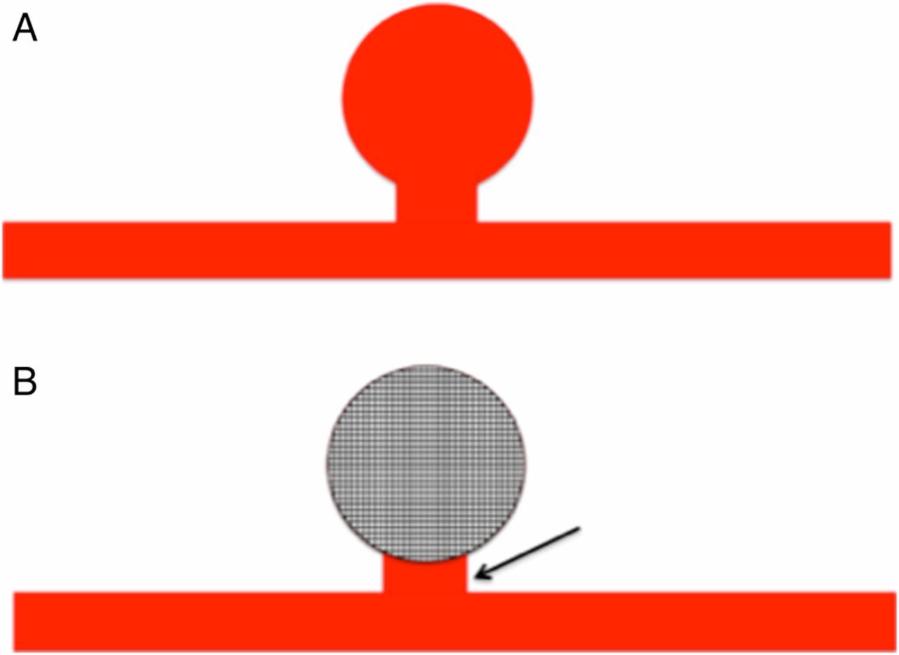
A much more likely scenario is an aneurysm with a neck, as in patients 2 and 4. The dome of the aneurysm is relatively spherical and a Medina Embolic Device may be a suitable device for the dome. An alternative strategy is required to treat the neck remnant (B, black arrow).
A further problem, not entirely unrelated to that described above, relates to lobulated aneurysms (figure 15). Irregularity of the aneurysm wall and the presence of daughter sacs have previously been associated with a higher risk of rupture.16–19 Further reports have documented the formation of blebs on conservatively managed aneurysms,20 ,21 and the formation of these lobulations has been implicated in the rupture process of aneurysms. Irregular shape was strongly associated with rupture in the observational series of Elsharkawy et al22 and was also an independent predictor of rupture in the Japanese natural history study.23 Most recently, Lindgren et al24 again showed that irregular shape is strongly associated with rupture. The exact reason why irregularity is associated with rupture is unknown, but it is postulated that a focal weakness in the aneurysm wall results in the formation of the bleb. Recent studies using dynamic DSA and dynamic CT angiography showed that deformation of the bleb of an aneurysm was twice as great as that of the rest of the sac.25 ,26 It would therefore seem reasonable either to protect any blebs during treatment or completely exclude the aneurysm as quickly as possible. One problem we have faced with the MED is in lobulated aneurysms (patient 5). As the MED framer is spherical, it may not definitely cover any blebs or appropriately adapt to a complex shape. Worse still, once in position it may actually redirect flow into the bleb, as happened in patient 5. In this scenario we were able to successfully catheterize the bleb and pack it with coils; however, this may not be possible in all situations. Therefore, one should consider placing coils into any blebs prior to deploying the MED or consider using a jailing method. Accessing any blebs after deploying the MED can be extremely challenging, as tracking a catheter past the leaflets of the MED is difficult. A similar method of coiling and WEB placement has previously been described.27
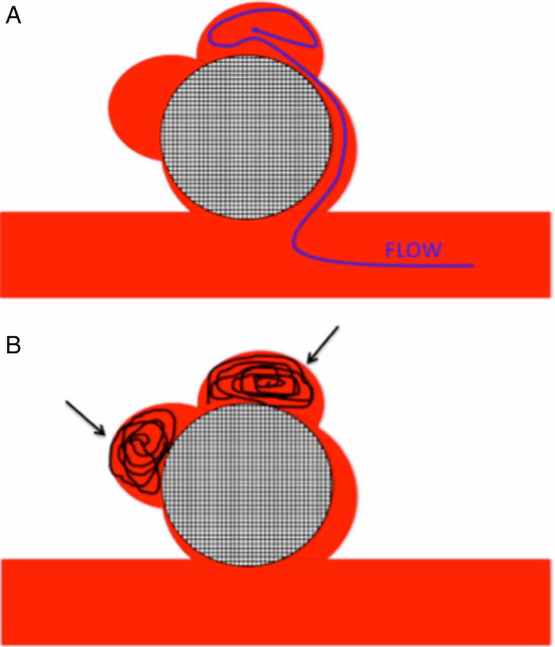
In lobulated aneurysms a channel for blood flow may persist (A) and, if this flow is directed towards an area of presumed weakness such as a bleb, the consequences could be disastrous. This is also possible if the leaflets do not conform to their pre-specified spherical shape and gaps between the leaflets would then allow flow into the aneurysm. One may consider coiling any blebs prior to deployment of the Medina Embolic Device (B, black arrows) or using a jailing technique; however, we have no experience of the latter.
In order to deal with these potential pitfalls with the MED we have, in certain anatomical situations, adopted a strategy of using both endosaccular flow diversion with the MED and endoluminal flow diversion (figure 16, examples include cases 4 and 8). Our reasoning for this strategy is that we believe it will cause a significant retardation in the flow into the aneurysm since at least two layers of flow diverter are in situ. The endoluminal flow diverter will have a flow-redirecting effect, so the flow into the actual aneurysm will have decreased significantly before it is in contact with the endosaccular flow diverter. Indeed, in case 8 there is a marked stagnation of flow in the neck of the aneurysm between the endoluminal and endosaccular flow diverters. In addition, this tactic allows us to minimize the number of endoluminal flow diverters and thereby decrease the coverage across branches, as well as avoiding the technique of compacting the endoluminal flow diverter which can cause complications.28 Although some may argue that we could use endoluminal flow diversion alone, we believe the combination of these two techniques will result in a more rapid exclusion of aneurysms from the circulation and also prevent potential destabilization of any mural thrombus that could result in aneurysmal rupture.29 In the retrospective analysis of delayed aneurysm ruptures after flow diversion (RADAR) study, delayed rupture was found in 1% of patients; however, in the subgroup of aneurysms >10 mm, the rate of delayed rupture increased to 2.1%.30 Although delayed rupture has occurred in aneurysms treated with both coiling and flow diversion, this has been reported in only a handful of cases and in all reported cases the aneurysms were >15 mm in diameter.31–35

By placing an endoluminal flow diverter across the neck of the aneurysm (A, dashed arrow) as well as a Medina Embolic Device inside the aneurysm, we can negate the potential problems outlined earlier. The endoluminal flow diverter will help to redirect flow away from the aneurysm as well as act as a scaffold for neo-endothelialization that is in continuity with the vessel itself. The endosaccular flow diverter will act as a secondary flow-modulating device that will inhibit flow into the aneurysm dome. The relatively slow and likely turbulent flow between the two layers of flow diverter material will act to promote thrombosis (B). In addition, the minimization of flow into the dome will hopefully prevent aneurysm rupture.
It is also important to discuss the mechanism of action by which aneurysm occlusion with endoluminal flow diversion occurs. Recent work by Kadirvel et al36 suggests that endothelialization of the endoluminal flow diverter is the more important step in aneurysm exclusion rather than thrombus formation within the aneurysm itself, when only endoluminal flow diversion is used. Endothelialization of the aneurysm neck proceeds from the parent artery, so it is likely that wide-necked aneurysms will require longer to be completely excluded from the circulation. In these circumstances the use of an intrasaccular flow diverter is likely to be beneficial since it would allow the rapid exclusion of the aneurysm dome from the circulation, as seen in several of our cases. The slower reconstruction of the parent vessel can then continue with a lower risk to the patient.
We prefer not to place a large number of MEDs into the aneurysm when we intend to use conjunctive flow diversion as this may lead to an increase in thrombogenicity and potential thrombus encroachment into the vessel lumen.34 Similar strategies of endoluminal and endosaccular flow diversion treatment have been performed previously.27 Clajus et al37 recently reported their initial and mid-term results from 108 consecutive patients treated with the WEB. In their cohort, 11.8% of patients required adjunctive endovascular techniques and, among the unruptured aneurysms, 17.2% (10 of 58) required retreatment. Consistent with this finding, a recent systematic review conducted by Armoiry et al38 noted the need for additional treatment after the WEB was deployed in 8–15%. We also feel that the addition of an endoluminal flow diverter will help prevent compression of the MED, which has previously been reported to occur with the WEB in 25% of cases reported by Sivan-Hoffman et al12 and in 57.2% of cases at early follow-up in the series by Cognard and Januel.11
Conclusion
The MED represents a novel device for the treatment of intracranial aneurysms by combining flow-diverting technology and the familiarity of standard coils. It can result in rapid exclusion of aneurysms from the circulation and has a good safety profile, although long-term outcome data are required. We believe that the true value of the MED will be in combining its use with adjunctive devices such as endoluminal flow diverters that will result in rapid aneurysm exclusion.
Acknowledgments
The authors are grateful for the intellectual contribution of L Bloom.
References
Footnotes
Contributors MAP, PB and RMM: manuscript preparation and data collection; OG and HB: manuscript review, editing; HH: manuscript review.
Competing interests MAP and RMM serve as proctors and consultants for phenox GmbH with moderate financial compensation. HH is a co-founder and shareholder of phenox GmbH.
Provenance and peer review Not commissioned; externally peer reviewed.