Article Text

Abstract
Background The Derivo Embolization Device (DED) is a novel flow diverter stent that provides increased x-ray visibility, an improved delivery system, and potentially reduced thrombogenicity. The objective of this study was to evaluate the early safety and efficacy of the second-generation DED.
Methods We retrospectively analyzed all patients with unruptured intracranial aneurysms (UIAs) treated with the DED between November 2015 and December 2017 in three German tertiary care centers. Procedural details, complications, and morbidity within 30 days after treatment, as well as the aneurysm occlusion rates after 6 months (O’Kelly–Marotta scale, OKM), were evaluated.
Results Implantation of the DED was attempted in 42 patients with 42 aneurysms. All procedures were technically successful. Multiple DEDs were used in three aneurysms (7.2%) and adjunctive coiling in 11 (26.2%). Procedure-related complications occurred in four cases (9.5%) including three thromboembolic events and one aneurysm perforation. The morbidity rate was 2.4% and there was no mortality. One patient suffered an ischemic stroke with persistent aphasia at 30-day follow-up due to a thromboembolic infarct (modified Rankin Scale score 1). Among 33 patients (78.6%) available for angiographic follow-up, complete (OKM D) and favorable (OKM C+D) aneurysm occlusion was obtained in 72.7% (24/33) and 87.9% (29/33), respectively.
Conclusions Endovascular treatment of UIAs with the DED is associated with high procedural safety and adequate occlusion rates. Examinations at 1- and 2-year follow-up will provide data on the long-term safety and angiographic outcomes of this device.
- aneurysm
- angiography
- device
- flow diverter
- intervention
Statistics from Altmetric.com
Introduction
During the past decade, flow diversion has emerged as a first-line treatment option for a broad range of intracranial aneurysms.1 Since the approval of the Pipeline Embolization Device (PED, Covidien, Mansfield, Massachusetts, USA) in 2008, flow diverter devices (FDDs) have proved advantageous for the endovascular treatment of wide-necked, large, and fusiform aneurysms that are otherwise difficult to treat.2–4 Flow diverter treatment is associated with comparably high occlusion rates and acceptable morbidity, as documented by large series and meta-analyses.5–7 Owing to these promising results, the indication for FDDs is continuously expanding.8
However, there are reports of thromboembolic complications, particularly for the treatment of fusiform and posterior circulation aneurysms.9 Further restrictions of first-generation devices are poor fluoroscopic visibility and problems with deployment of the device in difficult anatomic situations.
To address these limitations, FDDs are continuously being revised technically and novel devices such as the Flow Re-Direction Endoluminal Device (FRED, Microvention, Tustin, California, USA),10 the Pipeline Flex (Covidien),11 and the p64 (Phenox, Bochum, Germany)12 have been introduced to the market. However, while experience with the PED has been extensively reported in the literature, evidence for newer FDDs is still limited.
The Derivo Embolization Device (DED; Acandis, Pforzheim, Germany) is a novel second-generation FDD. It consists of 48 nitinol composite wires with an inner platinum core and three additional radiopaque markers at each end which provide better visibility under fluoroscopy. Moreover, a thin deep-blue colored surface layer of oxides and oxynitrates (BlueXide®) is supposed to reduce the thrombogenicity of the device. Other main features include a flexible structure and an improved delivery system that facilitates recapturing and repositioning of the DED in case of misplacement.
To date, only one clinical study has been published, which focused on the early safety and short-term angiographic results of the first-generation DED.13 Clinical data on the new version of this device have not been reported to date. The objective of this study was to retrospectively evaluate the early safety and efficacy of the second-generation DED.
Methods
This is a retrospective, observational, single-arm study conducted at three neurovascular tertiary care centers in Germany. All patients with unruptured intracranial aneurysms (UIAs) treated electively with the DED between November 2015 and December 2017 were included. According to institutional guidelines, no ethics committee approval was required for this retrospective observational study. All imaging and patient data were blinded and independently reviewed by three experienced consultant neuroradiologists (BK, FD, CK). Discrepancies were resolved by consensus.
Data collection
Patient demographics and aneurysm characteristics were obtained at baseline. Conventional four-vessel digital subtraction angiography (DSA) with three-dimensional rotational angiography of the target vessel was performed in all patients in order to confirm the location, size, and morphology of the aneurysm. The largest diameter of the aneurysm dome (aneurysm size), neck width, and the dome-to-neck ratio were recorded. The aneurysm size was categorized as small (<10 mm), large (10–20 mm), and very large (≥20 mm). Aneurysm morphology was classified as saccular, fusiform, and dissecting.
Procedure
All interventional procedures were performed under general anesthesia via a transfemoral approach. Selection of the DED for flow diversion was at the discretion of the individual neurointerventionalist operator. An intravenous bolus of heparin (5000 IU) was given after groin puncture, followed by aliquots of 1000 IU/hour until the end of the procedure. An 8F guiding catheter was introduced through a short femoral sheath into the internal carotid artery or a 6F guiding catheter into the vertebral artery. In the anterior circulation, a triaxial approach was attempted using an intermediate catheter (Navien 058, Medtronic, Irvine, California, USA or Sofia Plus, Tustin, California, USA). The DED was delivered through a 0.027 inch microcatheter (Headway 27, MicroVention, Tustin, California, USA or Neuroslider 27, Acandis, Pforzheim, Germany) in all cases.
The appropriate size of DED was chosen according to the proximal parent artery diameter. The decision to use multiple DEDs or adjunctive coiling was left to the discretion of the operator. Correct vessel wall apposition of the DED was assessed by DSA and non-subtracted images. Device deployment was considered successful when the aneurysm neck was completely covered by the DED.
Device visibility on DSA images was rated by an ordinal rating scale ranging from ‘invisible’ (0) to ‘excellent’ (4), as summarized in table 1.
Device visibility during the procedure. The rating scale includes the visibility of the contour and the radiopaque markers at both ends of the Derivo Embolization Device
Antiplatelet regimen
All patients were treated with 100 mg acetylsalicylic acid (ASA) and clopidogrel 75 mg for 5–7 days prior to the procedure. Platelet inhibition testing was performed using ASA, P2Y12 (Verify Now, Accumetrics, San Diego, California, USA), and vasodilator-stimulated phosphoprotein-phosphorylation (VASP) assays.14 Levels of 350–550 ASA Response Units (ARU) for ASA and 30–60% for clopidogrel were defined as sufficient platelet inhibition. An insufficient response to either drug was treated by dose escalation (eg, clopidogrel 150 mg/day) or substitution with prasugrel (60 mg bolus, 10 mg/day). Dual antiplatelet medication consisting either of ASA and clopidogrel or ASA and prasugrel was administered for at least 4 months after treatment, followed by permanent single antiplatelet treatment with ASA 100 mg/day.
Complications and clinical outcome
All patients received neurological examinations at baseline, immediately after the procedure, and at discharge. Clinical follow-up was performed 30 days after the procedure by an office visit or telephone call. Procedure-related complications such as thromboembolic and hemorrhagic events were recorded. Complications were classified as symptomatic when they were associated with transient or permanent neurological deficits. Stroke severity was assessed by the National Institutes of Health Stroke Scale (NIHSS). A persistent NIHSS score of ≥4 points was defined as major stroke.
Functional outcome was evaluated by the modified Rankin Scale (mRS). Morbidity was defined as any increase in the mRS score within 30 days after the procedure.
Evaluation of aneurysm occlusion
Angiographic follow-up was scheduled 6 months after the procedure using DSA, magnetic resonance angiography (MRA), or CT angiography (CTA). The O’Kelly–Marotta (OKM) grading scale for flow diversion was used to assess aneurysm occlusion after the procedure and during follow-up as follows: A, total filling (>95%); B, subtotal filling (5–95%); C, entry remnant (<5%); and D, complete occlusion.15 Favorable aneurysm occlusion was defined as OKM C+D. Furthermore, the extent of intra-aneurysmal contrast stasis was categorized as follows: 1, no stasis; 2, moderate stasis; and 3, significant stasis.
Statistical analysis
Categorical variables were presented as numbers and percentages. Continuous variables were presented as means±SD. All statistical tests were performed with SPSS Version 25.0 (SPSS, Chicago, Illinois, USA).
Results
Patient and aneurysm characteristics
We identified a total of 90 UIAs treated with FDDs during the study period, of which 42 patients with 42 UIAs were treated with the DED. The mean patient age was 54.8±11.9 years and 37 patients (88.1%) were female. Details of patient and aneurysm characteristics are shown in table 2.
Baseline patient and aneurysm characteristics
The location of the aneurysms was the internal carotid artery (ICA) in 38 (90.5%) patients (paraophthalmic 31 (73.8%), posterior communicating artery (Pcom) 4 (9.5%), terminus 3 (7.1%)), basilar artery in 2 (4.8%), vertebral artery in 1 (2.4%), and posterior cerebral artery in 1 (2.4%). The mean aneurysm size was 8.9±6.2 mm (range 2–28 mm); 31 (73.8%) aneurysms were small (<10 mm), 8 (19.0%) were large (10–20 mm) and 3 (7.1%) were very large (≥20 mm). The mean neck width was 5.3±3.1 mm and the average dome-to-neck ratio was 1.5±0.8. Aneurysm morphology was saccular in 34 (81.0%) cases, fusiform in 6 (14.3%), and dissecting in 2 cases (4.8%).
Treatment
Procedural details are summarized in table 3. Delivery and deployment of the DED was successful in all patients. Thirty-nine patients (92.9%) were treated with a single DED, whereas multiple devices were used for three aneurysms (7.2%; two DEDs in 2 cases, three DEDs in 1 case). The mean number of devices per aneurysm was 1.1±0.37. Adjunctive coiling was used in 11 aneurysms (26.2%) due to large aneurysm size. Secondary balloon angioplasty was necessary in four cases (9.5%) in order to ensure appropriate wall apposition of the DED. Other adjunctive endovascular devices were not used. Arterial side branches were covered in 38 cases (90.5%) and all of them were patent at the end of the procedure.
Procedural details, adverse events, and clinical outcome within the 30-day follow-up period
All DEDs were clearly visible during the procedure: visibility was rated ‘excellent’ in 33 cases (78.6%), ’good' in 8 cases (19.0%), and ‘fair’ in 1 case (2.4%). The treatment with a ‘fair’ visibility was for recurrence of a giant (22 mm) Pcom aneurysm that had been treated with stents, coils, and another flow diverter (FRED). In this complex case, the implanted stents and the coil package led to restricted visibility of the contour and the radiopaque markers of the DED. Additional balloon angioplasty was necessary after deployment in order to achieve full deployment and appropriate wall apposition.
Illustrative cases of the DED procedure and its enhanced visibility are given in figures 1 and 2.

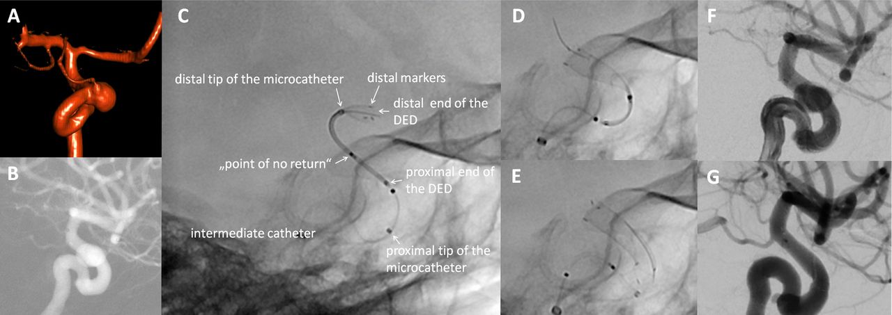
Treatment of a paraopthalmic aneurysm of the internal carotid artery (ICA) with the Derivo Embolization Device. 3D rotational angiogram (A), roadmap of the working position (B), positioning and partial opening of the device in the middle cerebral artery (C), during (D) and after complete deployment in the ICA (E), and final control (F). Angiographic follow-up after 6 months demonstrates complete occlusion of the aneurysm sac (G).
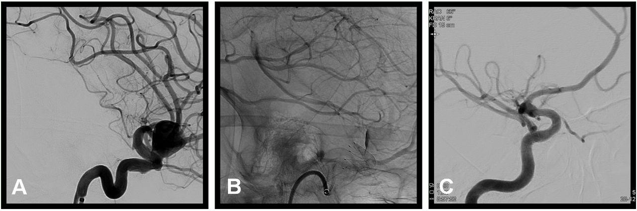
A large paraophthalmic aneurysm of the right internal carotid artery (16×12 mm). Digital subtraction angiograms before placement of the Derivo Embolization Device (DED) (A), immediately after placement of the DED (B), and at 6-month follow-up (C). The unsubtracted image immediately after DED placement (B) shows the superior radiopacity and visibility of the device. After 6 months the aneurysm is completely occluded (OKM D) while the ophtalmic artery remains patent.
Immediate angiographic outcomes are presented in table 3. Complete immediate occlusion (OKM D) was achieved in two aneurysms (4.8%), whereas all other aneurysms had at least a neck remnant (OKM A-C). Among the aneurysms with incomplete occlusion, intra-aneurysmal contrast stasis (OKM 2+3) was observed in 32 aneurysms (76.2%) at the end of the procedure. There was no stasis in the aneurysm sac at all in six aneurysms (14.3%) (OKM 1).
Complications and clinical outcome
Complications and clinical outcome are presented in table 3. Within the first 30 days after treatment, procedure-related adverse events occurred in four patients (9.5%), all of them during the hospital stay.
Periprocedural thromboembolism occurred during two procedures. Both patients presented with a motor aphasia after the intervention although complete thrombus resolution was achieved by the immediate administration of tirofiban (Aggrastat, Correvio, Bielefeld, Germany) intravenously. Aphasia resolved completely in one patient and persisted at 30-day follow-up in the other one (NIHSS 1).
There was one incident of periprocedural in-stent thrombus formation. In this case, thrombus formation occurred after incomplete proximal opening of the DED in the recurrent Pcom aneurysm with ’fair' DED visibility that was pretreated with coils, stent, and a FRED flow diverter. After balloon angioplasty, complete opening of the device and good wall apposition was achieved and no residual thrombus was seen after administration of tirofiban intravenously. The patient did not have any neurological symptoms.
We further recorded one case of aneurysm perforation by a microwire that was not associated with detectable intraparenchymal or subarachnoid hemorrhage and had no clinical consequences. There were no further ischemic or hemorrhagic events within the first 30 days after treatment. The overall morbidity rate during the 30-day clinical follow-up was 2.4% (1/42), with no incidence of major ischemic strokes or deaths.
Angiographic follow-up
Six-month aneurysm occlusion is presented in table 4. A total of 33 (78.6%) patients were available for angiographic follow-up at a mean of 177.2±97.4 days. Of these, 26 patients underwent DSA (78.8%), 4 MRA (12.1%), and 3 CTA (9.1%). Complete occlusion (OKM D) was achieved in 24 of the 33 aneurysms (72.7%), neck remnants (OKM C) in 5 (15.2%), and subtotal filling (OKM B) in 4 (12.1%) aneurysms. Thus, favorable aneurysm occlusion (OKM C+D) was obtained in 29/33 aneurysms (87.9%). No aneurysm had total filling (OKM A) at follow-up. Among the 29 cases with DED coverage of artery side branches, only one patient with a paraophthalmic ICA aneurysm had an asymptomatic occlusion of the ophthalmic artery (3.4%) at follow-up. Moreover, 32/33 (97.0%) DEDs remained patent at the last follow-up. In one patient with thrombotic stent occlusion, we did not observe any new neurological deficits and further interventional procedures were not required.
Aneurysm occlusion at 6-month follow-up.
Discussion
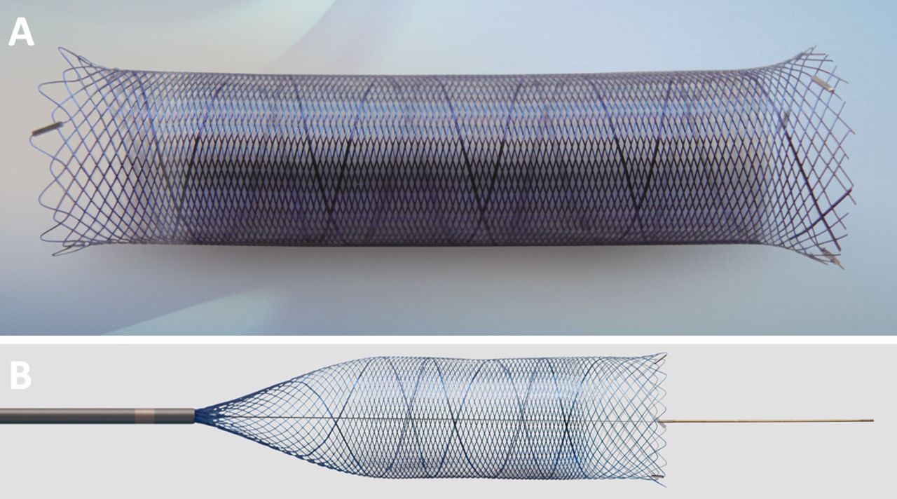
The Acandis DED is a new generation flow diverter stent that provides enhanced device visibility on x-ray imaging, a flexible and self-expandable structure, and reduced thrombogenicity (figure 3). These features contribute to a smooth device deployment and an improved safety profile. The DED is available in diameters of 3.5–6 mm and in lengths of 15–50 mm, with the intention to treat vessel diameters ranging from 2.5 to 6.0 mm. It has a porosity of approximately 62–65% and is compatible with any standard 0.027 inch microcatheter.
Ex vivo photographs of the Derivo Embolization Device (DED) (A) and its delivery system (B). The bluish surface layer of oxides and oxynitrates (BlueXide®) is supposed to reduce the thrombogenicity of the device. The flared ends of the DED improve wall apposition and the radiopaque markers at both ends increase device visibility under fluoroscopy. The distal tip of the delivery system provides support during deployment of the DED. Photographs provided by Acandis.
Similar to other studies on FDDs, we used the DED primarily for saccular wide-necked sidewall aneurysms located at the ICA. However, in our series the DED was also effectively used for fusiform and posterior circulation aneurysms, which are in general more difficult to treat by FDDs and are associated with a higher morbidity than ICA aneurysms.16 In our series we demonstrated a high rate of technical success, with successful delivery and deployment of the device in all cases.
The DED has flared ends which are closed during delivery and unfold after detachment. This mechanism ensures a smooth positioning of the device and a secure wall apposition after detachment. Due to the flexible and self-expandable structure of the device, subtotal resheathing and repositioning of the device can be performed smoothly and did not result in adverse events in any case.
The major advantage of the DED compared with other FDDs is the advanced visibility under fluoroscopy, which allows for accurate positioning and an appropriate assessment of the opening and the final wall apposition. The contour of the DED was clearly visible in all of our procedures and periprocedural device visibility was rated as ‘excellent’ or ‘good’ in all but one case.
In general, flow diverter coverage of side branches should be avoided in order to prevent occlusion and ischemic complications. In our series, all but one covered side branch were patent immediately after the implantation and at follow-up (97.6%). Thus, the occlusion rate of covered side branches was low when compared with other flow diverter studies. A recent study by Bhogal et al reported that 20% of the covered side branches were occluded in their series on 147 aneurysms treated mainly with the PED.17 Other series on the PED reported occlusion rates similar to those of Bhogal et al.18 19
Complete and favorable occlusion rates at short-term angiographic follow-up were 72.7% and 87.9%, respectively. These findings correspond to the angiographic results reported by previous studies on second-generation FDDs.12 13 20
In general, flow diverter treatment is associated with comparably high mortality and morbidity. To date, the best data on adverse events and associated morbidity after flow diverter implantation is available for the PED. The IntrePED study, which is the largest series on PED, reported a 30-day morbidity and mortality rate of 5.4% (39/717) for UIAs.5 In the Aneurysm Study of Pipeline in an Observational Registry (ASPIRe), neurological morbidity and mortality rates for the treatment of UIAs were 6.8% (13/191) and 1.6% (3/191), respectively.21 Moreover, Brinjikji et al performed a meta-analysis of 1451 patients with 1654 aneurysms treated by different types of FDDs and reported morbidity and mortality rates of 5% (95% CI 4% to 7%) and 4% (95% CI 3% to 6%), respectively.6
Data on the success and safety of the DED are sparse. The only clinical study to date was published by Akgul et al.13 The authors analyzed the immediate and short-term clinical and angiographic results mainly of the first-generation DED in 24 patients with 36 aneurysms and reported a morbidity rate of 4.2% within the first 30 days after treatment.
In our series on the second-generation DED, the overall morbidity within 30 days after treatment (2.4%) was comparably favorable to the aforementioned studies with the PED and we had no associated mortality. The only symptomatic patient at the 30-day follow-up had persistent aphasia caused by an embolic ischemic stroke and was mildly disabled (NIHSS 1, mRS 1). Thus, our data confirm the high safety profile of the second-generation DED.
Morbidity after FDD treatment is mainly related to thromboembolic and ischemic events.5 22 The IntrePED study cited major ischemic stroke rates of 4.7%, with half of the events (2.4%) occurring within the first 72 hours after PED implantation.5 In the current study we treated the patients under standard double antiplatelet medication and observed thromboembolic events in 7.1%. However, we did not observe any incidence of major ischemic stroke and only one case was associated with persistent aphasia (mRS 1). These results are in line with the findings by Akgul et al who reported thromboembolic events in 8.3% and the absence of major ischemic stroke in patients treated with the first-generation DED.13 They only mentioned one case (4.2%) with stent stenosis that was associated with mild hemiparesis (mRS 1).
A major reason for thrombus formation after flow diverter deployment is insufficient wall apposition. Balloon angioplasty is helpful in improving the wall adaptation after FDD deployment. However, device deployment and appropriate wall apposition are difficult to assess due to the limited visibility of most FDDs. The increased visibility of the DED under fluoroscopy can presumably help to identify cases with incomplete wall adaptation and thereby increase the safety of the procedure. Moreover, the DED is provided with a thin surface layer (∼50 nm) of oxides and oxynitrates (BlueXide®), which reduces the friction during delivery and expansion of the device.13 This surface finishing technology is intended to further reduce the thrombogenicity of the device and may add to the relatively low rate of ischemic complications in our series.
It has been shown that the complication rate correlates with the number of implanted FDDs.9 In this study the majority of cases could be treated with only one device, while fewer than 10% of the aneurysms required more than one flow diverter. This percentage is low when compared with the IntrePED study, in which multiple devices were used in 34.2%.5 We suggest that this is at least partially due to the easy and reliable resheathability of the device, which allows for an accurate positioning. Moreover, the DED is available in long lengths up to 50 mm, which may further reduce the need for multiple devices.
Limitations of the study
The short follow-up periods and the low rate of patients with angiographic follow-up examinations are limitations of this preliminary study. However, the long-term clinical and angiographic outcome is currently being evaluated.
A further limitation is that angiographic outcomes were not assessed by a core laboratory. This might bias the interpretation of the imaging results.23 To reduce this potential bias, at least in part, all imaging data were reviewed blinded and independently by three experienced consultant neuroradiologists. Discrepancies were resolved by consensus.
Although the present study showed promising results regarding short-term safety and aneurysm occlusion rates, further studies with larger patient samples and longer follow-up periods will provide a definite conclusion about the safety and efficacy of the DED.
References
Footnotes
FD and CK contributed equally.
BK and LG contributed equally.
Contributors BK, BT, JB, MS, FD and CK acquired the data. LG, BK and CK analyzed the data and drafted the manuscript. CK and FD developed the project. All authors revised the paper critically for important intellectual content and provided final approval of the version published. All authors agree to be accountable for all aspects of the work in ensuring that questions related to the accuracy or integrity of any part of the work are appropriately investigated and resolved. BK and LG contributed equally as first authors. FD and CK contributed equally as last authors.
Funding The authors have not declared a specific grant for this research from any funding agency in the public, commercial or not-for-profit sectors.
Competing interests FD and CK serve as consultants for Acandis.
Patient consent Not required.
Provenance and peer review Not commissioned; externally peer reviewed.
Data sharing statement All data will be made available upon request in an anonymized manner.