Article Text

Abstract
A patient with a giant partially thrombosed basilar apex aneurysm was treated with balloon-assisted coil embolization. At the conclusion of the embolization, an Enterprise stent was placed from the upper basilar artery to the left P1 segment. Follow-up angiography 4 months later showed that the distal stent had pulled out of the posterior cerebral artery and was in the coil mass within the aneurysm. Angiography clearly showed that the stent had migrated down the basilar artery and that the artery was now kinked proximal to the stent. Nonetheless, the left posterior cerebral artery and the basilar artery were still widely patent. This is the first documented case in which a stent ‘compacted’ into a completed coiled aneurysm in a delayed fashion.
- Aneurysm
- brain
- cerebral aneurysm
- device
- Enterprise stent
- stent
Statistics from Altmetric.com
Self-expanding nitinol stents are a useful, recent development for the treatment of wide-necked and large aneurysms. These stents allow successful coil embolization of many aneurysms that were previously deemed uncoilable. We report a case of delayed migration of an intracranial Enterprise stent (Cordis Neurovascular, Miami Lakes, Florida, USA). Similarities with three other published case reports are discussed.
Case report
A patient with a large basilar apex aneurysm was treated electively with coil embolization (figure 1). The day before the procedure the patient was loaded with aspirin (650 mg) and Plavix (clopidogrel, 600 mg). The aneurysm had a very large neck that involved the origin of the left posterior cerebral artery (PCA). Therefore, a 4×7 mm Ascent compliant balloon (Micrus Corporation, San Jose, California, USA) was inflated from the proximal left PCA and into the distal basilar artery to aid in packing the aneurysms. At the conclusion of coiling, there was concern about coil impingement on the origin of the left PCA. To mitigate this possibility, a 4.5×14 mm Enterprise stent was placed from the upper basilar artery to the left P1 segment (figure 2). The diameter of the basilar artery was 3 mm and the diameter of the left P1 was 1.9 mm.
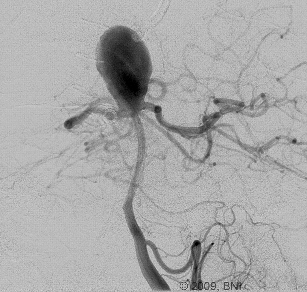
Left vertebral artery angiogram, oblique view, shows a giant basilar tip aneurysm with a wide neck. Used with permission from Barrow Neurological Institute.
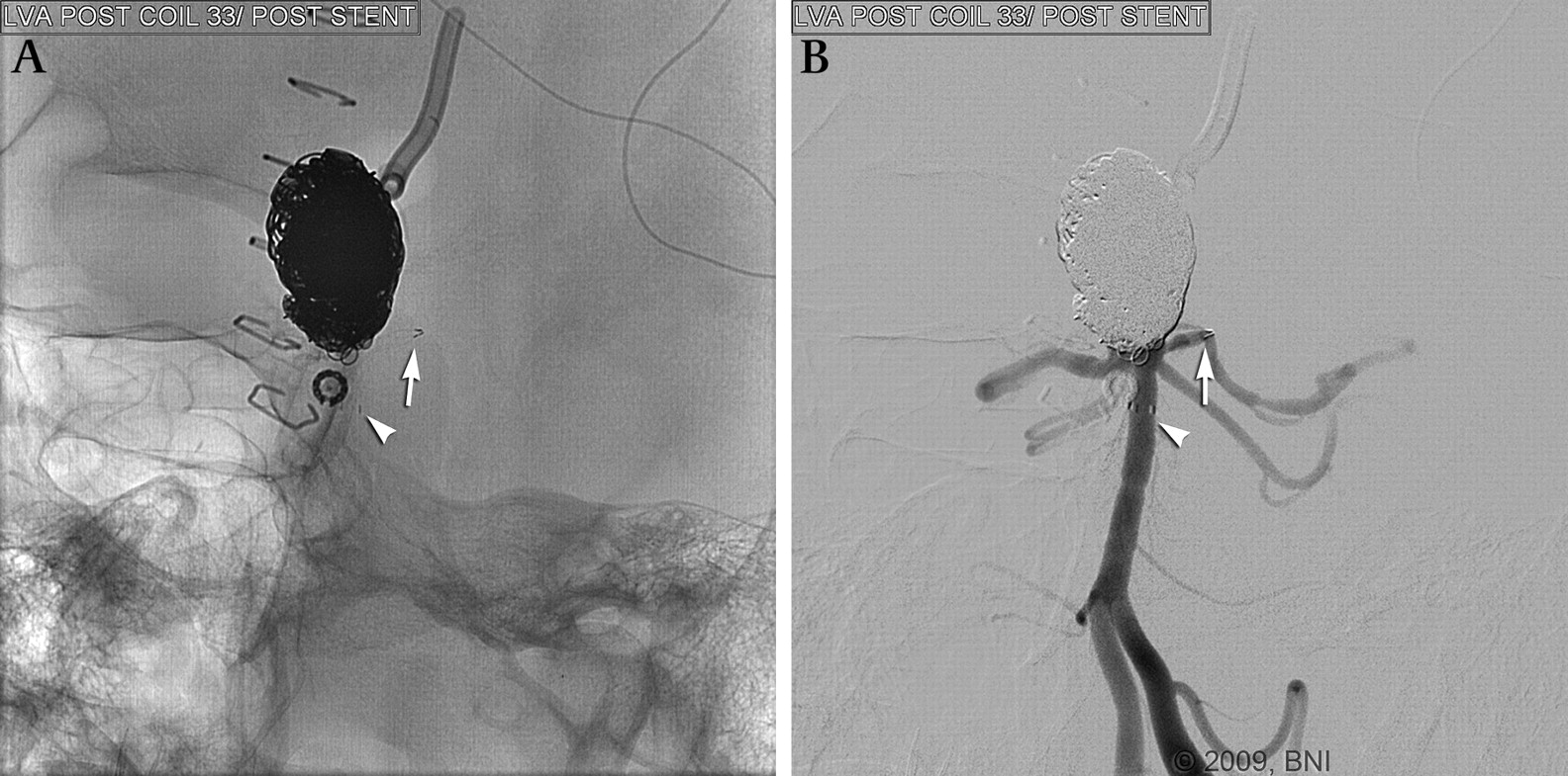
Left vertebral artery angiogram, native (A) and subtracted (B) views, show the Enterprise stent from the upper basilar artery to the mid-portion of the P1 segment of the left posterior cerebral artery. The proximal and distal markers of the stent are denoted by arrows. Note that the distal tines of the stent in the posterior cerebral artery are not fully open. Used with permission from Barrow Neurological Institute.
Blood tests at this time confirmed that the patient was an aspirin-responder, with 88% inhibition on Plavix. Postoperatively, the patient remained on dual antiplatelet therapy. The patient's immediate postoperative course was complicated by a retroperitoneal hematoma, which necessitated several blood transfusions. Four days after the procedure, the patient was discharged home in good condition. Two weeks after the procedure, the patient had an episode of dizziness that resulted in a trip to the local emergency department. This episode lasted several hours and then resolved. The patient had no further episodes.
Four months later, the patient returned for follow-up examination. Repeat angiography at that time showed that the distal stent had pulled out of the PCA and was now in the coil mass within the aneurysm (figure 3). Despite wide patency of the left PCA and basilar artery, there was an obvious kink on the basilar artery just proximal to the stent. Furthermore, the stent had migrated proximally down the basilar artery. Because of obvious coil compaction, the patient was treated in that setting with additional coiling (figure 4). The Plavix was discontinued, and the patient was sent home the next morning on aspirin only.
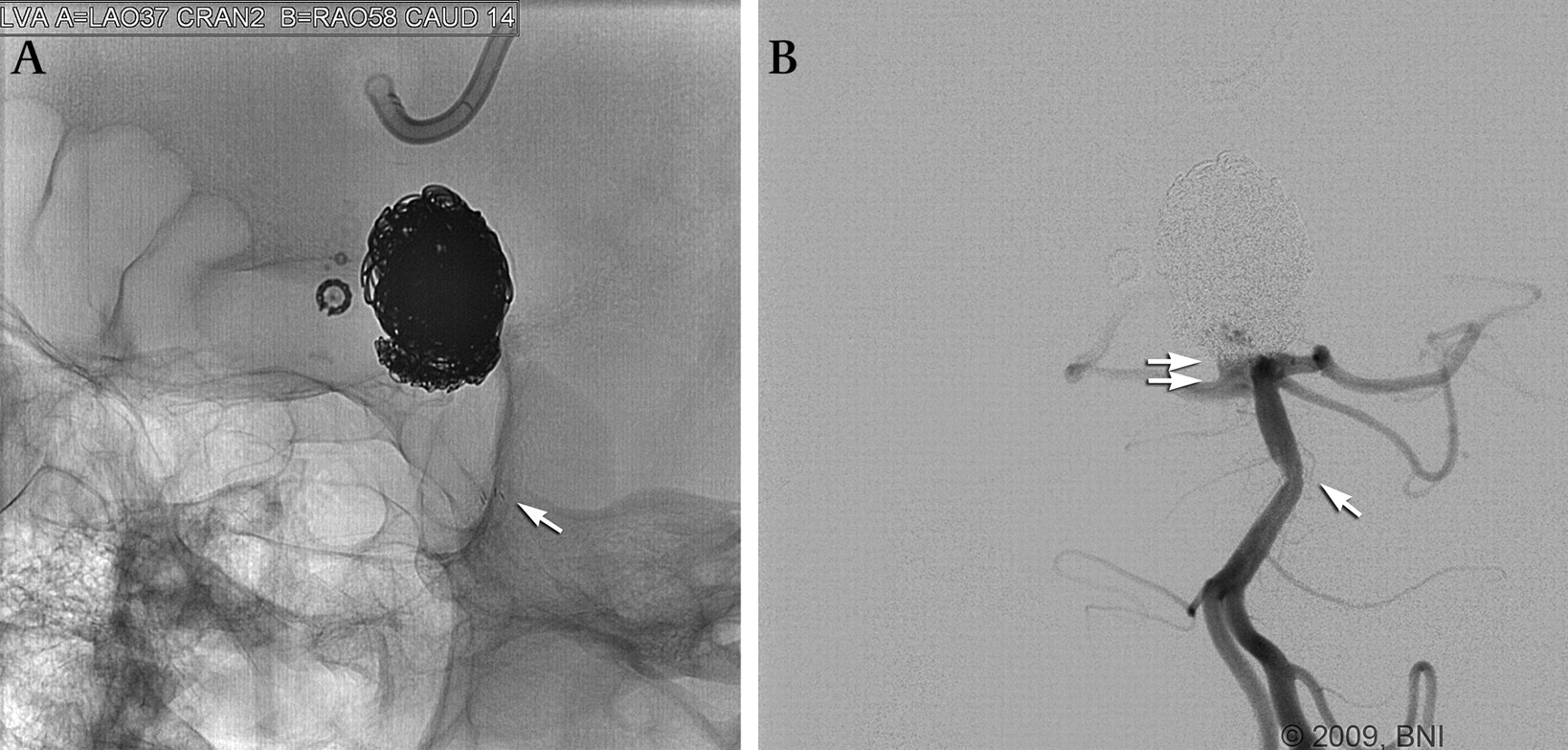
Follow-up left vertebral artery angiogram, native (A) and subtracted (B) views, showing retraction of the distal stent marker into the coil mass. The proximal stent marker appears to have moved down the basilar artery. The basilar artery and the posterior cerebral artery are widely patent, but the basilar artery is clearly kinked proximal to the stent. A small recurrence is at the right anterior aspect of the base of the aneurysm. Used with permission from Barrow Neurological Institute.

Left vertebral artery angiogram, oblique view, shows successful coil embolization of the aneurysm recurrence. Used with permission from Barrow Neurological Institute.
Discussion
The Enterprise stent is a relatively new device approved by the US Food and Drug Administration under an Investigative Device Exemption (IDE) for use in the treatment of cerebral aneurysms. It is used primarily as an adjunct for the treatment of wide-necked aneurysms. In comparison with open-cell counterparts, the closed-cell design of the Enterprise stent provides superior scaffolding across the neck of an aneurysm in preventing coil loop herniation into the parent vessel. In our aneurysm practice, the Enterprise stent has largely replaced the Neuroform stent (Boston Scientific Corporation, Fremont, California, USA), which has an open-cell design. It is interesting that there has never been a reported case of delayed migration of a Neuroform stent. Such an open-cell stent may have less propensity for migration because the unconnected tines along the stent may help grasp the arterial wall.
The Enterprise stent is extremely flexible, allowing easy navigation in the intracranial arteries. It provides very little outward radial force and can easily be dislodged during the procedural manipulations. Consequently, we usually use balloon remodeling for large-necked aneurysms and place an Enterprise stent at the end of the procedure if we believe that the parent vessels are at risk of coil impingement or to lessen the likelihood of an aneurysmal recurrence. Alternatively, we occasionally place the stent in one setting, allow it to ‘scar’ in place with endothelialization, and then coil the aneurysm in a delayed fashion. There is also the option of jailing the microcatheter with the Enterprise stent, thus avoiding manipulation of the stent after placement.
There have been three previous reports of Enterprise stent migration.1–3 These reports share many similarities with our patient (table 1). All cases involved the same anatomic scenario: stenting from the mid-basilar artery to the P1 segment during treatment of a basilar tip aneurysm. Our case is unique in that it represents the first report of delayed ‘compaction’ of a stent into a coiled aneurysm.
Characteristics of previously reported cases of delayed Enterprise stent migration, and of the current case
As with our patient, two of the three reported patients suffered delayed transient symptoms of posterior circulation ischemia. One patient developed a transient visual disturbance 2 weeks after stent placement.1 A second patient suffered multiple episodes of vertigo 4 months after the procedure, soon after discontinuation of clopidogrel. These episodes ceased when the patient resumed clopidogrel treatment.1 We hypothesize that these episodes were caused by thromboembolic phenomena secondary to the stent movement and possibly from kinking of the basilar artery. It makes more sense that stent migration would occur within a few weeks of placement, before endothelialization and scarring have occurred.
It is important to note that no instances of stent migration have been reported in cases where the proximal and distal stents were deployed in vessels of the same size (ie, stenting of the supraclinoid internal carotid artery (ICA) for coil embolization of wide-necked ICA aneurysms). Our case, along with the three previously reported cases of stent migration involved the same anatomic scenario: PCA-to-basilar artery stenting, where there is a significant mismatch in the size of the vessels within which the proximal and distal stents were deployed. The Enterprise stent is available in only one diameter: 4.5 mm. In our case, there was a 1.1 mm mismatch in the size of the vessels (1.9 mm PCA diameter compared with 3.0 mm basilar artery diameter).
Multiple factors predisposed this case to such a ‘watermelon-seeding phenomenon’. First, the stent is polymer coated and very lubricious, allowing easy movement with small forces. Second, the outward radial force of the stent in contact with the P1 segment is significantly less than the rated radial force for the Enterprise stent. That is, a 14-mm stent consists of one 5-mm flare at each end, which means the body of the stent is only 4 mm long. The flared ends have ∼30% less radial force than the body of the stent (personal communication, Juan Lorenzo, Director Product Development, Codman Neurovascular, December 2009). In our case, that would translate to the flared end (with its lower radial force) being the only portion in contact with the P1 segment, with its associated lower anchoring force. This point is accentuated by the fact that all three reported cases of Enterprise stent migration involved the shorter stents (two 14-mm stents and one 22-mm stent). Third, there is a natural tendency for the stent to achieve a lower energy state by expanding to its full diameter in the larger vessel.
Furthermore, the Enterprise stent has elastic properties and acts as a spring along its longitudinal axis. In this clinical scenario, when the stent is delivered into vessels that are much smaller than its diameter, some lengthening occurs (ie, it is 15.1 mm long when constrained in the delivery. This is analogous to attaching a weight at the end of a spring, which increases the length (L) of the spring by a distance x. Doing so results in a recoil force (F) directed along the longitudinal axis that is equal to the product of the spring constant (k) and the change in length (x). This force seeks to foreshorten the stent, contributing to the watermelon seeding (F =−kx).
In conclusion, when possible, a longer stent should be used when there is a large mismatch in the diameters of vessels, as is often the case in basilar artery-to-PCA stent placement. This will increase the length of the body of the stent along with its relatively higher radial force serving to anchor the stent to the vessel wall.
Footnotes
Competing interests None declared.
Ethics approval This study was conducted with the approval of our institutional review board.
Provenance and peer review Not commissioned; externally peer reviewed.