Article Text

Abstract
Introduction Dissecting and wide-necked aneurysms that incorporate a large portion of the parent artery can be challenging to treat with currently available devices. This study reports three cases treated with a new hybrid stent design that incorporates a smaller cell size and more pliable design than current generation stents and results in some flow diversion characteristics.
Methods In all three cases, use of the low-profile visible intraluminal support (LVIS) device in conjunction with coil embolization was determined to provide the best opportunity to achieve aneurysm occlusion while mitigating adverse events. The institutional review board reviewed all cases and approval was obtained. All cases were performed under emergent use exemption from the US Food and Drug Administration.
Results All three patients were successfully stent coiled with the LVIS device. One patient was completely occluded initially and remained so at follow-up, one patient progressed to complete occlusion at follow-up, and the last patient had stable incomplete occlusion of their fusiform aneurysm. There were no complications related to the procedures and the patients were maintained on dual-antiplatelet therapy.
Conclusion The LVIS device offers promise as a stent-assisted coil device with certain characteristics that may be advantageous over currently available microstents.
- Aneurysm
- artery
- balloon
- CT
- embolization
- intracranial
- LVIS
- MRI
- pseudoaneurysm
- spine
- stent
- stroke
- subarachnoid
- thrombectomy
- thrombolysis
- vein
Statistics from Altmetric.com
- Aneurysm
- artery
- balloon
- CT
- embolization
- intracranial
- LVIS
- MRI
- pseudoaneurysm
- spine
- stent
- stroke
- subarachnoid
- thrombectomy
- thrombolysis
- vein
Dissecting and wide-necked aneurysms that incorporate a large portion of the parent artery can be challenging to treat with currently available devices. Microstents can assist in reconstructing the vessel wall, but may result in suboptimal coiling of the underlying aneurysm.1–3 Multiple microstents may be required in some cases.4 We report three cases treated with a new hybrid stent design that incorporates a smaller cell size and more pliable design than current generation stents and results in some flow diversion characteristics.
Materials and methods
The use of a non-US Food and Drug Administration (FDA)-approved device is a rigorous process requiring significant involvement of outside experts and diligent follow-up with regular reports to the FDA and the regulatory officer of the manufacturer. Great lengths were taken to comply with all regulations set forth by the FDA, the device manufacturer, and the institutional review board (IRB) to ensure the welfare of our patients was the utmost priority. All cases were initially discussed at a multidisciplinary conference. In addition, non-treating, independent physicians with expertise and knowledge in the treatment of these types of aneurysms reviewed all cases and provided written opinions. The independent physicians, after thorough review, determined that the use of the low-profile visible intraluminal support (LVIS) device (Microvention, Tustin, California, USA) in conjunction with coil embolization provided the best opportunity to achieve aneurysm occlusion while mitigating adverse events, and that the alternative treatment options had an unacceptably high risk of complications and/or failure. The IRB reviewed all cases, which included documentation from the expert outside physicians, and approval was obtained. All cases were performed under emergent use exemption from the FDA according to the regulations set forth by the FDA and the manufacturer's regulatory officer. In all cases, the patients granted informed consent to use the non-FDA-approved investigational device.
Case no 1
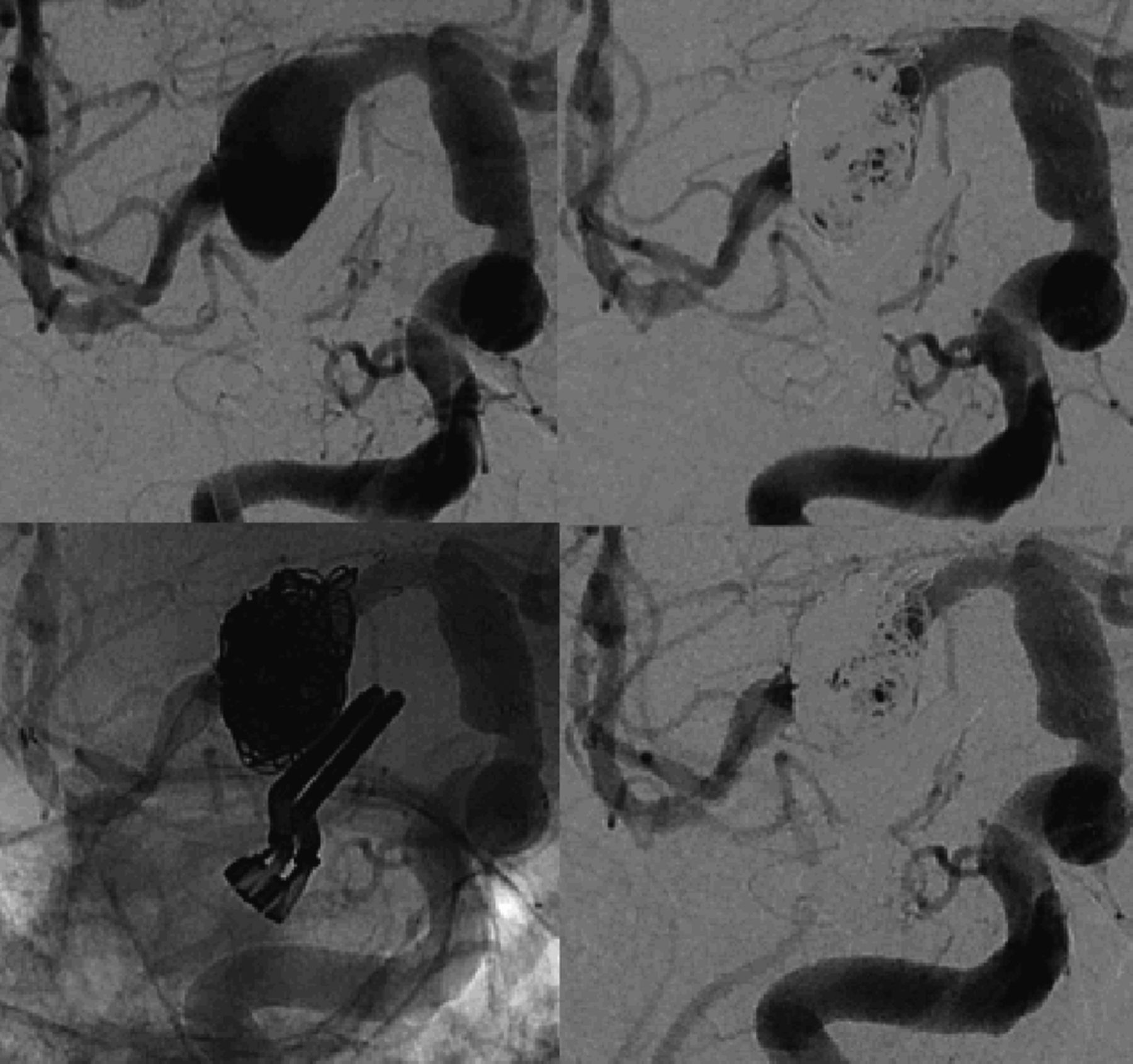
The first case was a 57-year-old woman with a history suggesting subarachnoid hemorrhage (SAH) 7 weeks ago based on lumbar puncture, and a vertebral aneurysm was identified. The aneurysm was found to measure 9 mm long × 5.5 mm deep with a 7.7 mm neck that circumferentially involved the vertebral artery and adjacent distal segment. The patient was pretreated with Plavix and aspirin. The vertebral artery was reconstructed with the LVIS device (microvention) while jailing a microcatheter in the aneurysm. Dyna-CT with 20% dilute contrast demonstrated good opposition of the stent to the vessel wall. Coil embolization of the aneurysm resulted in a small remnant at the neck. The microcatheter was kicked out of the aneurysm during the coiling. The aneurysm remnant was catheterized via the true lumen of the device and through the stent tines and further coil embolization resulted in complete occlusion. The patient went home the following day at baseline clinical condition. Three-month follow-up angiography demonstrated stable complete occlusion of the aneurysm (figure 1).

Demonstrates in two views, the pre-embolization, immediate post-embolization, and 3-month follow-up (subtracted and native) of the right vertebral artery dissecting aneurysm. Of note is the progressive remodelling of the dissected vessel around the aneurysm.
Case no 2
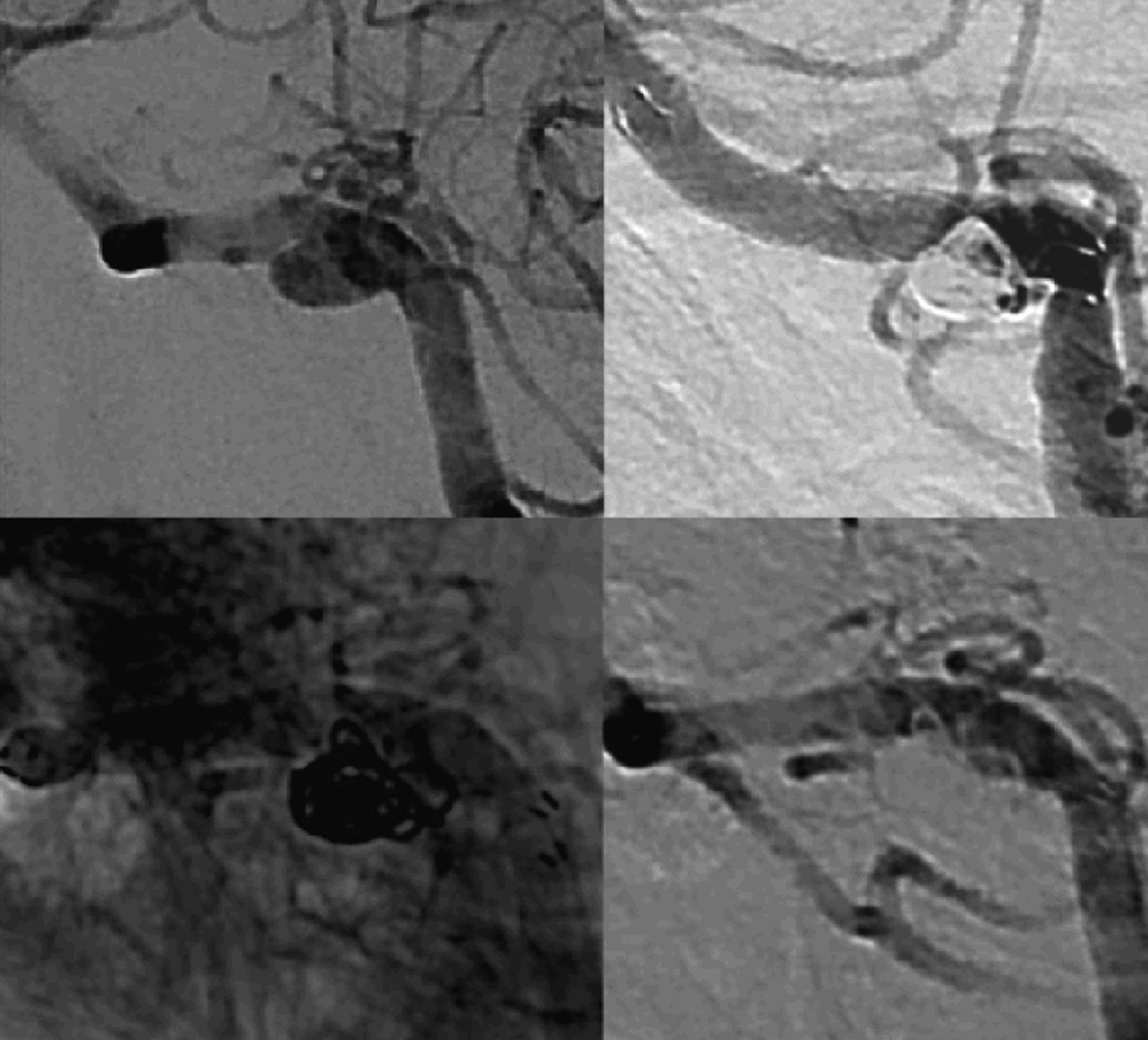
A 67-year-old man with a history of chronic headaches underwent elective clipping of a large, wide-necked middle cerebral artery (MCA) aneurysm in 2005, with incomplete aneurysm occlusion on postoperative angiography. He was lost to follow-up until he presented with a SAH (Hunt–Hess 2, Fisher 3) from the partially clipped right MCA aneurysm. Over the 6 years, the aneurysm enlarged over time and was now fusiform in nature. The anterior temporal artery originated from the distal portion of the aneurysm. The distal right internal carotid artery (ICA) was dysplastic in nature. Due to the complex nature of the ruptured aneurysm and previous surgery, definitive treatment was postponed and he was allowed to recover. The patient remained neurologically intact and after several weeks, the patient was readmitted and pretreated with Plavix and aspirin. The MCA aneurysm was found to measure 15 mm × 14 mm and to involve the M1 trunk circumferentially. The MCA was reconstructed with the LVIS device while jailing a microcatheter in the aneurysm. Dyna-CT with 20% dilute contrast demonstrated the LVIS device was well opposed to the MCA distally and proximally; however, there was no vessel lumen adjacent to the device in the region of the fusiform aneurysm. Coil embolization was performed through the jailed microcatheter until position in the aneurysm was lost. At the conclusion of the coiling, persistent aneurysm filling was noted in the proximal aspect of the aneurysm and this was catheterized through the LVIS device and further coil embolization performed. Subsequently, two additional telescoping LVIS devices were deployed to reconstruct the fusiform supraclinoid ICA. The patient was discharged at baseline neurological condition. Short-term follow-up demonstrated the patient no longer had daily chronic headaches. At 3 months, no further headaches have been reported and the angiography demonstrated stable incomplete occlusion (figure 2).
Demonstrates a large fusiform right middle cerebral artery aneurysm that was previously clipped (top left). In the anteroposterior view, notice the immediate post-stent coiling result (top right) and the 3-month follow-up native and subtracted views (bottom left and right), which appear stable over time.
Case no 3
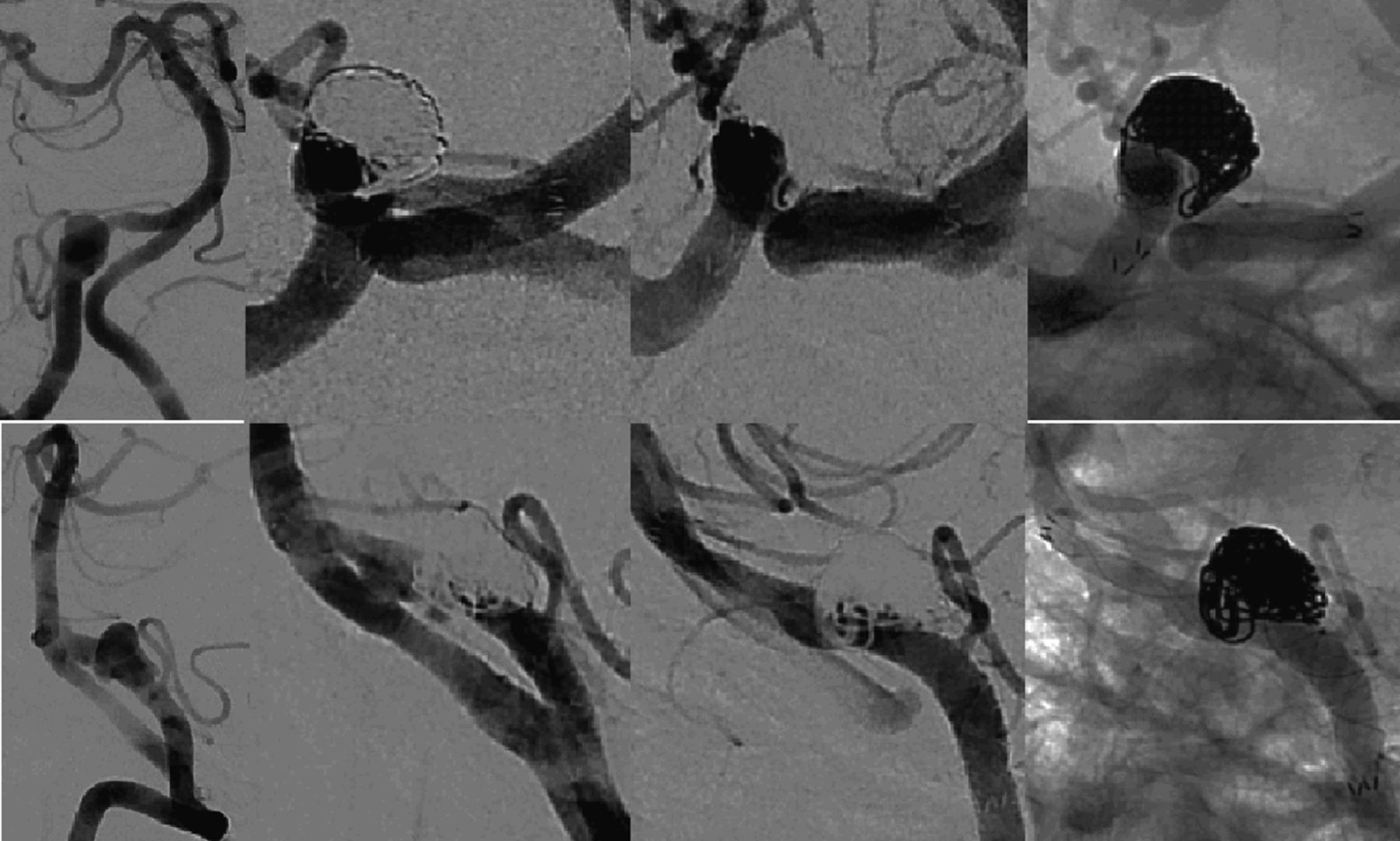
A 62-year-old woman presented with neck pain and tinnitus. Work-up revealed a right vertebral artery aneurysm that was dissecting in nature. The aneurysm measured 6 mm × 4 mm in size with a 5 mm neck. The patient was pretreated with aspirin and Plavix. The LVIS device was deployed from the distal right vertebral artery to just proximal to PICA, across the neck of the aneurysm. Dyna-CT demonstrated good stent opposition. The aneurysm was catheterized through the lumen and stent tines with a SL-10 microcatheter and coil embolization resulted in incomplete occlusion. The patient was discharged in a stable condition. At 1 month, angiography demonstrated progression to complete occlusion (figure 3).
Demonstrates a right vertebral artery dissecting aneurysm (top left). In consecutive oblique views, notice the immediate post-stent coiling result (top right) and the 1-month follow-up native and subtracted views (bottom left and right), which demonstrate progressive occlusion.
Discussion
Dissecting and wide-necked aneurysms often require stent reconstruction of the parent artery.1–3 Symptomatic dissecting aneurysm in particular pose a risk of SAH and infarction.5 Currently available devices are poorly visualized with large interstices that may allow coil herniation into the stent lumen.6 These devices are currently available from the FDA under humanitarian device exemption. Multiple stents may be needed to reconstruct the parent artery.1 ,3 ,4 Newer flow-diverting devices offer promise in treating these types of aneurysms.1 There is currently limited access to flow diverters in the USA and they are only indicated for wide-necked unruptured ICA aneurysms. At the time of treatment, flow diverters for these cases were not available and would have been considered off label.
The LVIS device is a self-expanding braided stent device. It is well visualized throughout its course due to two radio-opaque helical strands. The four distal device markers are evenly spaced around the circumference of the device while the four proximal markers are paired together. The smaller cell structure (∼0.9 mm) provides greater protection across the aneurysm neck and improved flow diversion compared with currently available coil-assist stents. The braided wire structure enables the strands of wires to slide on each other, allowing catheterization through the interstices. The device delivers through a 0.021-inch microcatheter (figure 4).

Photos of the low-profile visible intraluminal support device, demonstrating its helical wind design. The distal stent markers are evenly dispersed on the distal end of the stent, while the proximal markers are paired. On fluoroscopy, there are two radio-opaque helical strands to assist in the detection of full expansion of the device within the vessel.
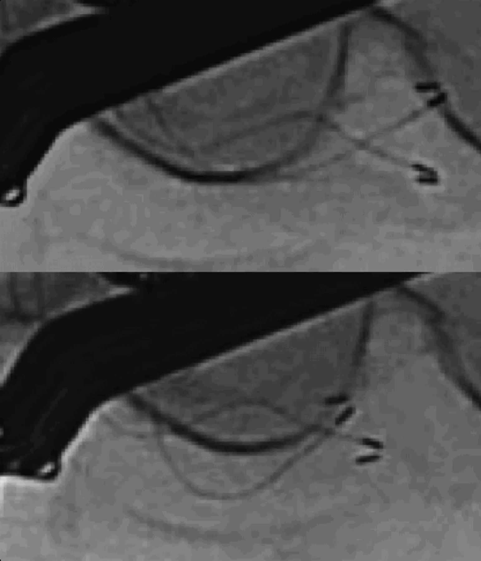
The LVIS device delivered through the 0.021-inch microcatheter without difficulty. There is a short lead coil attached to the delivery wire system. The device unsheaths initially and then is pushed out. The device can be reconstrained up to 80% of its deployment. Once deployed, the delivery wire is removed and the microcatheter can access the true lumen of the device using a traditional microwire. The proximal and distal markers splay apart, demonstrating that the proximal and distal ends of the device are open. The two helical strands also spread into a double helix configuration, with alternating wall opposition visually on angiography. A 20% dilute contrasted Dyna-CT was performed in all cases to demonstrate wall opposition of the device. During case 2, the proximal body of the third stent (from supraclinoid ICA to cavernous ICA) was not well opposed on angiography despite the proximal tines fully expanding. This was probably the result of unsheathing the device rather than pushing the device out. The poor wall opposition was demonstrated on conventional angiography by incomplete opening of the radio-opaque helical stands. Dyna-CT imaging demonstrated that the proximal body of the stent was not fully opposed despite the proximal end fully deploying. A microcatheter and microwire were used to manipulate the stent proximally resulting in the device foreshortening and opening to maximal diameter. A repeat Dyna-CT confirmed excellent wall opposition throughout the entire course of the device (figure 5).

A native fluoroscopy of the low-profile visible intraluminal support device overlying the sella turica. In the top image, the helical strands of the device are narrowed beneath the aneurysm clips and the device extends to the clivus. Following maneuvers to expand the device, a bottom image demonstrates that the helical strands are now fully expanded and the device has foreshortened to the posterior sella.
During the treatment of the first two cases, the microcatheter was initially jailed in the aneurysm, while in the third case, the microcatheter crossed the stent tines in a more traditional fashion. At some point during all cases the aneurysm was accessed through the stent. The ability to access aneurysms through the stent construct is of benefit because the flow-diverting properties of this device are minimal compared with other flow diverters. However, the smaller cell size of this coil-assist device does provide robust protection of the parent artery, more so than currently approved coil-assist stents. During coiling of the vertebral artery aneurysms, the parent artery was profiled well and there were no instances of coil loop herniation into the parent artery. While treating the MCA fusiform aneurysm, it was impossible to profile the parent artery; however, despite this, coil embolization was performed without the complication of coil loop herniation (proved on Dyna-CT). It remains possible to have a proximal coil tag herniate into the stent lumen if the catheter position is lost, as in the third case. A 20% dilute contrast-enhanced Dyna-CT was performed in all cases, confirming that the true lumen of the device was not compromised by coil loops; however, the tag end of a coil was present in case 3.
Conclusions
All patients were able to undergo aneurysm stent-assisted coiling of very challenging aneurysms with the LVIS device with good angiographic and clinical outcomes. There were no adverse events. This device has attributes that are unmet by currently available approved devices and offers promise in the treatment of challenging aneurysms.
Footnotes
-
Competing interests None.
-
Patient consent Obtained.
-
Ethics approval IRB approval was obtained to use a non-FDA-approved device under emergency use exemption from the FDA. IRB approval was required only for the device usage.
-
Provenance and peer review Not commissioned; externally peer reviewed.
-
Data sharing statement All data related to this research/case report are available in the published manuscript. There are no other unpublished data.