Article Text

Abstract
Background A study was undertaken to determine the typical length, diameter and taper of vessels in the anterior cerebral circulation.
Methods The sample size was calculated at 100 patients based on similar measurements in the literature and divided into cohorts based on gender and side. These patients were consecutively collected from a population that had undergone CT angiography and did not have any vascular abnormality. The arterial diameter was measured at the proximal cavernous internal carotid artery (ICA), the ICA terminus, the middle cerebral artery (MCA) origin and an M2 origin. The length between these endpoints was calculated along the center line. The vessel taper was calculated for the ICA as the change in caliber per unit length.
Results The mean length of the ICA from the proximal cavernous segment to the ICA terminus was 33.1±6.1 mm. The mean diameter at the cavernous ICA and the ICA terminus was 5±0.6 mm and 3.6±0.4 mm, respectively. The mean ICA taper was 0.04±0.02 mm/1 mm. For the MCA, the diameter at the MCA and M2 origins measured 3.1±0.4 mm and 2.4±0.4 mm, respectively. The mean MCA length was 22.5±8.1 mm. There was no significant difference based on gender or between right and left sides. Patients aged >60 years had longer ICAs (p=0.02), larger cavernous ICA (p=0.003), ICA terminus (p<0.0001) and MCA origin (p=0.01) diameters than those aged 40–60 years. The ICA vessel taper did not change with age.
Conclusion ICA and MCA vessel size did not change based on gender or side. Older patients had more redundant vessels based on diameter and length. The ICA has a gentle taper from its proximal cavernous segment to the ICA terminus. This information can be important in planning interventions or designing endovascular devices.
- Brain
- MRI
- drug
- thrombectomy
- catheter
- CT angiography
- intervention
- stroke
Statistics from Altmetric.com
Introduction
The anterior cerebral circulation, particularly the carotid ophthalmic segment and the carotid siphon, is a frequent site for endovascular interventions. For cerebral aneurysm, these interventions are evolving from a purely endosaccular approach to treatment aided by endoluminal assist devices such as stents and possibly to purely endoluminal treatment such as with flow-diverting devices. An intimate knowledge of the vascular geometry is helpful in planning treatment, assessing device behavior and potentially impacting on device designs. Some vascular dimensions for the cerebral circulation are available in the literature1–4; these were reported before the era of neuroendovascular medicine. We approached our analysis with an understanding of the current endovascular therapies as well as upcoming novel devices and innovations. The goal of the study was to generate measurements for the anterior circulation only. We specifically focused on the intracranial internal carotid artery (ICA) and the middle cerebral artery (MCA) because of the higher propensity of endovascular interventions performed in these arterial segments. Because of a greater variation in the anatomy and perhaps an influence based on the completeness of the circle of Willis, the posterior circulation measurements and the anterior cerebral artery are not included in this study and can be a focus of a separate analysis.
Materials and methods
The study was conducted under an institutional review board approved protocol.
Sample size
A sample was calculated to represent men and women and right and left hemispheres. The age cut-off was set at 40 years based on our experience that most patients undergoing neurovascular interventions are above this age. We reviewed the literature regarding previous measurements of intracranial blood vessels.1–4 The SD of the measurements at the ICA and the MCA in these studies is between 0.4 and 0.5 mm.1–4 In order to have a sample that is sufficient to account for inherent variations in the population based on intracranial vascular geometry, we conservatively chose the upper limit of SDs previously recorded for these measurements.1–4 Thus, setting the SD at 0.5 mm and using a 95% CI with a margin of error of 0.2 yielded a sample size of 25 patients for each representative subgroup; male-right, male-left, female-right and female-left. These 100 patients formed the cohort analyzed.
Patient selection
The population from which this sample of 100 patients was collected included patients who had undergone an intracranial CT angiogram (CTA) study in either an outpatient clinic center or the emergency room. Patients with any vascular abnormality on the CTA were excluded. These abnormalities included atherosclerotic disease, arterial stenosis, aneurysm or vascular malformations, arterial dissections or other traumatic vascular injuries. Based on these criteria, which were evaluated by two experienced neuroradiologists, we included 50 consecutive men and 50 consecutive women aged ≥40 years. For each of the male and female subgroups the side of the measurements was alternated resulting in 25 male-left, 25 male-right, 25 female-left and 25 female-right subgroups. We also divided the sample into two groups based on age (40–60 years and >60 years). The sample was spread over a 4-month period.
Technique
Imaging protocol
A consistent protocol was used for all CTA studies, which were performed on an Aquilion-64 CT scanner (Toshiba America Medical Systems, Tustin, California, USA). The protocol specifies injection of 50 ml Optiray 350 (Covidien, Hazelwood, Missouri, USA) through a large bore antecubital venous access at a rate of 3–4 ml/s. An initial localizer is obtained at the C2–C3 cervical level and the scan is manually initiated when the carotid arteries are opacified. A small field of view of 240 mm is used with a 1.0×1.0 image thickness and reconstruction interval. Multiplanar reconstruction is performed on a Vitrea workstation (Vital Images, Minnetonka, Minnesota, USA). All source data are stored on a server from which it can be accessed via a picture archive and imaging system as well as the workstation.
Target measurements
Two experienced neuroradiologists performed the image processing and analysis. The intracranial ICA and the MCA constituted the target vessels with a goal of obtaining the following parameters:
-
The length of the ICA from the start of the proximal horizontal segment of the cavernous ICA to the ICA terminus.
-
The diameter of the ICA at the proximal cavernous segment, just distal to the petrous-cavernous junction.
-
The diameter of the ICA just proximal to the ICA terminus.
-
The length of the MCA from its origin to an M2 origin.
-
The diameter of the MCA at its origin.
-
The diameter of the M2 at its origin.
-
The tortuosity index (TI) of the ICA along its measured length.
-
The vessel taper for the ICA segment.
Image processing and vessel analysis
In order to obtain these measurements, the source CTA data for the 100 studies were imported into a workstation running the Vitrea Core software V.6.2.1 (Vital Images). Using the software's vascular package, a three-dimensional model of the cerebral vasculature with automated removal of the bone and soft tissues was generated. The vessel of interest was segmented out from the rest of the model. A center line was obtained along the long axis of the blood vessel, typically starting from the distal petrous segment of the ICA to an M2 branch of the MCA. In addition to the three-dimensional model, a curved planar reformation (CPR) of the target blood vessel along its center line was also obtained.
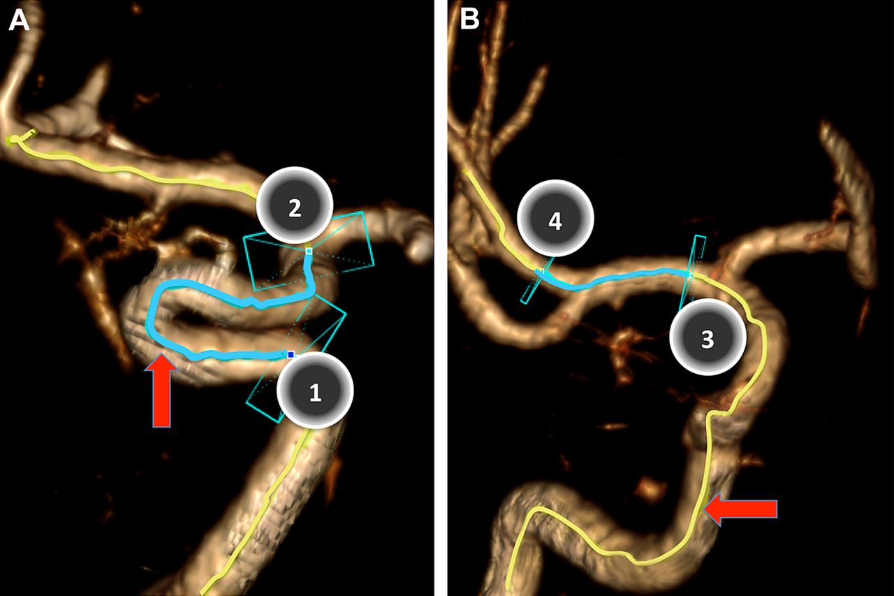
The proximal and distal ends of the vessel under evaluation were manually selected (figure 1). For the ICA, the proximal end was identified within 3 mm of the start of the cavernous ICA segment and the distal end was identified within 3 mm of the ICA terminus. The cross-sectional area of the blood vessel perpendicular to its long axis was obtained at both the proximal and distal ends and the diameter was calculated based on πR2. The length of the blood vessel between the proximal and distal ends (in mm) as well as a TI was then obtained. These measurements (except the TI and maximum tortuosity) were repeated for the MCA with the proximal marker at the MCA origin and the distal marker at the M2 origin.

Three-dimensional model of the right internal carotid artery (ICA) in (A) oblique and (B) frontal projections. The center line is drawn in yellow (horizontal arrow). Also shown are the locations at which the cross-sectional area measurements were obtained for the proximal cavernous ICA (1), the ICA terminus (2), the middle cerebral artery (MCA) origin (3) and an M2 origin (4). The blue line (vertical arrow) represents the vessel length between (1) and (2) for ICA and between (3) and (4) for MCA.
Prior to obtaining the actual measurements, the image analysis technique was applied on ‘practice’ cases to assess its reproducibility among the two neuroradiologists. A high degree of consistency was obtained in the measurements for the ICA but, for the MCA, there was significant variation in defining the distal extent of the MCA, especially due to a difference in selecting the M2 branch at which the measurement was applied. We therefore obtained independent measurements for the ICA, but the MCA analysis and measures were obtained by consensus between the two neuroradiologists. Our purpose was to obtain as accurate a measurement as possible and we felt a consensus allowed us to do that.
Tortuosity index (TI) and vessel taper
The TI measured on the CPR image is a ratio of the curved length of the blood vessel to the straight line distance between the two markers. Thus, a higher TI will indicate a more tortuous vessel. For example, a curved length of 4 cm and a straight line distance of 2 cm between two points gives a TI of 2, or the curved length is twice as long as it would be if the vessel was straight. A maximum tortuosity measurement along the ICA segment was also obtained in degrees/cm, which typically occurred at the anterior bend of the carotid siphon, and was an estimation of its curvature.
The vessel taper was defined as the decrease in caliber per unit length. This was obtained by dividing the difference between the proximal and distal diameter over the length of the measured segment giving a taper/mm. Additionally, the taper was calculated for 1 cm, 15 mm, 20 mm, 25 mm and 30 mm segments for vessel length. The vessel taper was only calculated for the ICA as the proximal and distal diameter was obtained along a continuous segment of blood vessel. We did not measure the taper for the MCA since the distal diameter measurement was obtained at the M2 origin rather than the distal M1 segment. We felt that, because of the terminal bifurcation of the MCA, the change in vessel caliber from M1 to M2 could represent more of a step-off than a true taper and the smaller length of the MCA may not yield a significant taper.
Statistical analysis
The data were analyzed using JMP statistical software Version 9 (SAS Institute, Cary, North Carolina, USA). Mean values and SDs for all the measurements were generated along with the respective 95% CIs. We performed a bivariate analysis of age with the vessel parameters to determine any association of vessel size with age and used the Student t test to compare the means based on gender and side.
Results
Vessel dimensions: length and diameter
There was a strong positive correlation between the measurements obtained by the two neuroradiologists for ICA length (R2=0.9), proximal cavernous ICA diameter (R2=0.9) and ICA terminus diameter (R2=0.9). For the ICA, the mean of the two neuroradiologists' measurements and, for the MCA, the consensus measurements constituted the units analyzed. The baseline measurements for the entire sample are shown in figure 2. The greatest variation was seen in the length of the MCA. We did not find any significant difference between the values based on either gender or right and left sides (table 1). We arbitrarily chose an age cut-off of 60 years to assess any difference in vascular dimensions. Patients aged >60 years had significantly longer ICA segments between the proximal cavernous carotid artery and the ICA terminus. Additionally, the diameter at both the cavernous carotid artery and the ICA terminus was significantly larger in older patients. For the MCA, only the diameter at the M1 origin was significantly larger in older patients (table 1).

Drawing of the right internal carotid artery (ICA) in an oblique projection showing the mean values for diameter and length for all patients (n=100). D-C, proximal cavernous ICA diameter; D-T, ICA terminus diameter; L-C, length of the ICA segment between D-C and D-T; D-M1, middle cerebral artery (MCA) origin diameter; D-M2, M2 origin diameter; L-M, length of the MCA segment between D-M1 and D-M2. The vessel taper for the ICA was along L-C.
Vessel dimensions based on gender, side and age
Vessel dimensions: taper and tortuosity
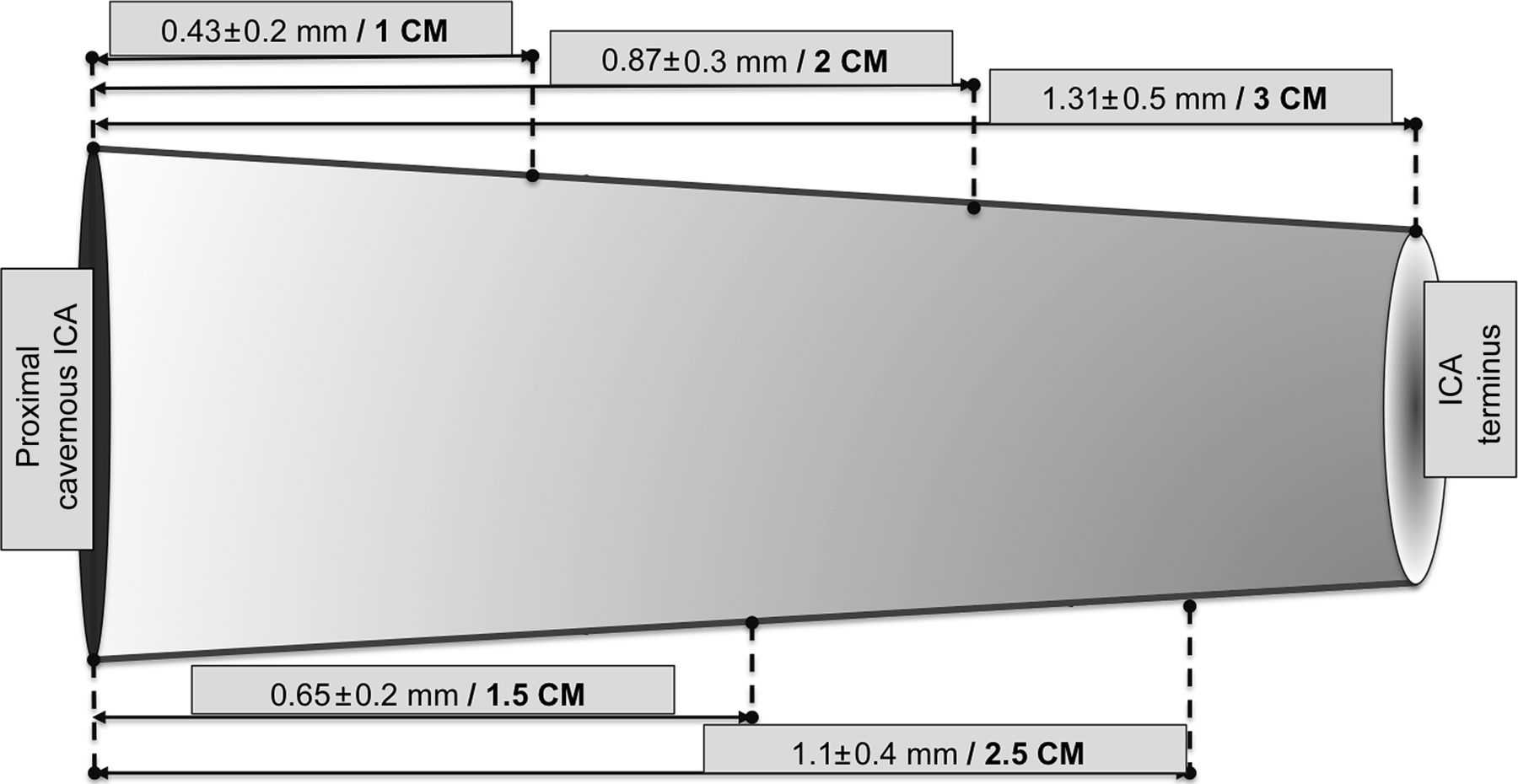
The mean vessel taper for the ICA from the proximal cavernous segment to the ICA terminus was 0.04±0.02 mm of diameter decrease per 1 mm of vessel length. The caliber change for 1 cm, 1.5 cm, 2 cm, 2.5 cm and 3 cm is shown in figure 3. The mean ratio of the diameter of the ICA terminus to the diameter of the cavernous ICA was 0.72±0.08 and of the M2 diameter to the MCA diameter was 0.77±0.1. There was no correlation between vessel taper and gender, side or age of the patient. The mean TI was 2.9±0.8 and the mean curvature along the anterior genu of the cavernous ICA was 85.6±12.5 degrees/cm.

Vessel taper along the long axis of the internal carotid artery (ICA) graphically represented as the decrease in caliber over different segments of vessel length from 1 cm to 3 cm.
Discussion
Knowledge of the cerebrovascular dimensions is integral to the performance of neurovascular procedures. These procedures may require placement of an endoluminal device, and understanding the vascular geometry in terms of caliber, taper and tortuosity can aid in device selection and modification, potentially making them more conformable to the arterial anatomy. The dimensions of the intracranial circulation have been published previously.1–4 These measurements were based on both post mortem and angiographic analysis.1 ,4 We used CTA because of its non-invasive and ubiquitous nature. The role and accuracy of CTA in the cerebral vasculature is well established,5 and it is increasingly being used for the screening and investigation of neurovascular pathologies.6–9
The reported diameter of the ICA terminus ranges from 3.7 mm to 4.57 mm1–4 depending on the distance from the terminus at which these measurements are obtained. In our study, the mean diameter of 3.6±0.4 mm measured within 3 mm of the ICA terminus is at the lower end of this range, but is close to the 3.7 mm diameter in the study where the measurement was similarly obtained 3 mm from the terminus.1 In studies reporting a larger ICA diameter3 ,4 the measurements were obtained more proximally, typically ≥5 mm from the terminus. The MCA diameter in the same studies ranges from 2.5 mm to 3.82 mm, again depending on the point of measurement and its proximity to the MCA origin.1–4 In our study, the mean MCA origin diameter of 3.1±0.4 mm measured within 3 mm of the origin is within this range of measurements. We found neither a measurement for the M2 branches nor for the cavernous or proximal intracranial ICA.
The association of arterial caliber with gender, right or left side or age is not consistent in the literature. There is a reported increase in the right MCA caliber in men aged 51–60 years compared with those aged 41–50 years.4 A transcranial color Doppler ultrasonography study on 120 patients showed a trend of increasing vessel caliber in older patients, but this was not statistically significant.10 In the same study, arteries on the left side were larger than the right.10 In our data there was no significant association between the arterial caliber and gender or the side of measurement. There was a trend for men to have longer internal carotid arteries and a marginally increased caliber of the proximal cavernous ICA than women (table 1). However, we did find that patients aged >60 years had significantly longer ICA segments and larger diameters at the proximal cavernous segment, the ICA terminus and the MCA origin. Previous studies have shown an increase in caliber of large arteries with ageing.11 ,12 A comprehensive review by Van Bortel and Spek reported the influence of ageing on blood vessels.13 They found an increase in pulse pressure with ageing which they suggest is secondary to a decrease in arterial compliance, specifically those of the larger arteries. Compliance represents the buffering property of the blood vessel while elasticity reflects its distensibility. Using echo-tracking techniques, it has been shown that the properties of arterial compliance and elasticity in the common carotid artery decrease with advancing age. At the same time the arterial diameter increases with ageing, which could be a mechanism to compensate for the decrease in compliance and the increase in pulse pressure.13
The vessel taper along the long axis of the intracranial ICA has not previously been reported. We chose the segment between the start of the cavernous ICA and the ICA terminus to measure the vascular taper and tortuosity. This segment represents a location where most neurovascular interventions for sidewall aneurysms are performed. An increasing number of such interventions could in the future be performed with stent assistance or with flow diversion devices. This segment also represents a contiguous length of blood vessel where the caliber decreases gradually rather than a bifurcation or branch point where the change in caliber may be more abrupt. Our mean TI of almost 3 as measured on the CPR images indicates that, on average, this vascular segment is three times longer than it could be if the vessel was absolutely straight. This is therefore a measure of its redundancy or curvature. The parameter of maximum tortuosity in our study represents the maximum degree of curvature that was present along the anterior bend of the cavernous ICA and measured 8.56±1.25 degrees/1 cm or almost 9 degrees/mm. Of note, while our data show lengthening of the ICA and an increase in caliber at both the cavernous ICA and the ICA terminus, the vessel taper did not change with age indicating a uniform increase in caliber across the length.
Our calculations for vessel taper are shown in figure 3. The mean taper of 0.04 mm/mm of vessel length is indicative of a relatively gentle change in caliber. Figure 3 also shows that the caliber reduces by about 0.5 mm/1 cm of vessel length, 0.65 mm/1.5 cm vessel length and that it takes almost 2.5 cm of vessel length to effect a change of 1 mm. The effect of vessel taper and other geometric variations on the mechanical stability of large arteries has been studied.14 Tapered vessels have lower critical pressures than non-tapered arteries,14 ,15 which makes them more vulnerable to instability or buckling than normal cylindrical vessels.14 Furthermore, in these tapered vessels the shift of the buckling is towards the distal aspect resulting in increased tortuosity.14 Tapering is one phenomenon that can affect vessel buckling along with other factors such as elliptical shape and areas of stenosis.14 Apart from these inherent properties related to vessel geometry, external factors such as hypertension and diabetes can affect vessel dynamics by changing the material properties of the vessel wall.
All other parameters of stent design being equal, a cylindrical stent with a uniform caliber and radial force along its long axis can potentially cause more deformation at the distal tapered end of the blood vessel than a tapered stent conforming to the caliber change.16 This is especially true for stents with higher stiffness than those with lower stiffness or radial force.16 The higher the stiffness, the more the force exerted on the tapered end of the blood vessel and the more the vessel deformation. One way to overcome the impact of non-tapered stents on the distal tapered segment of a blood vessel is to have stents with very low stiffness or radial strength.16 Indeed, the currently available stent devices—that is, the Neuroform EZ (Stryker Neurovascular, Fremont, California, USA) and the Enterprise (Codman Neurovascular, Raynham, Massachusetts, USA)—claim a low stiffness profile. For the Enterprise reconstruction device, a diameter of 4.5 mm is thus designed to accommodate vessels ranging from 2.5 mm to 4 mm. The Neuroform EZ comes in a variety of diameters with a range of expansion for each diameter to cover vessels of different calibers. A retrospective study of tapered versus non-tapered stents for carotid artery stenosis found that, while not showing any difference in the 30-day stroke or death rate, non-tapered stents had a higher incidence of re-stenosis than tapered stents.17 There is no similar literature with respect to the intracranial circulation.
In the carotid siphon and the ophthalmic ICA segment, incomplete apposition of stents is documented and is not without consequence.18 ,19 Vessel size and geometry may influence adequate stent apposition.20 Additionally, cell design and other biomechanical properties of the device also influence its expansion and wall apposition. Whether incorporating vessel taper in intracranial stents will significantly affect stent apposition is speculative. However, theoretically, the addition of a gentle taper to the stent, certainly for stents longer than 15 mm (figure 3), may allow it to have a slightly higher radial force potentially making the stent more conformable, especially along a vessel bend such as the carotid siphon.
Conclusion
The mean length of the ICA from the proximal cavernous segment to the ICA terminus was 33.1±6.1 mm while the mean diameters at the cavernous ICA and the ICA terminus were 5±0.6 mm and 3.6±0.4 mm, respectively. The mean MCA length was 22.5±8.1 mm and the mean diameters at the MCA and M2 origins were 3.1±0.4 mm and 2.4±0.4 mm, respectively. There was no difference based on gender or side, but patients aged >60 years had longer ICAs and larger diameters of both the ICA and the MCA than those aged 40–60 years. There is a definite taper along the ICA from the cavernous segment to the ICA terminus measuring 0.04±0.02 mm/1 mm. This taper becomes more pronounced for vessel segments above 15 mm. However, the clinical or technical consequence of this taper is speculative at this time.
References
Footnotes
-
Competing interests None.
-
Ethics approval Ethics approval was provided by the Institutional Review Board.
-
Provenance and peer review Not commissioned; externally peer reviewed.