Article Text

Abstract
Background and significance We describe a method by which to efficiently and atraumatically achieve distal positioning of a flexible guiding catheter beyond extreme cervical tortuosity using a hypercompliant temporary occlusion balloon.
Methods A retrospective review of a prospective neuroendovascular database was used to identify cases in which a hypercompliant balloon catheter (Hyperform or Hyperglide, ev3/Covidien, Irvine, California, USA; Scepter or Scepter XC, Alisa Viejo, California, USA) was used to achieve distal positioning of a flexible guiding catheter (Navion, ev3/Covidien, Irvine, California, USA; Neuron, Penumbra Inc, Alameda, California, USA). After achieving a stable guiding sheath position within the proximal cervical carotid artery, a hypercompliant balloon catheter was manipulated beyond the tortuous cervical internal carotid segment into the distal carotid artery. The balloon was then inflated to anchor it distally within an intracranial (cavernous or petrous) segment of the internal carotid artery. The guiding catheter was then advanced beyond the tortuous cervical segment, over the balloon catheter, as gentle counter traction was applied to the balloon.
Results Balloon-assisted guiding catheter placement was used to perform endovascular treatments of 12 anterior circulation aneurysms. One patient underwent coiling alone. Five patients underwent balloon-assisted coiling. One patient underwent balloon and stent assisted coil embolization. Four patients with five carotid aneurysms (one with bilateral carotid aneurysms) underwent vascular reconstruction with the pipeline embolization device. All patients had severe tortuosity of the extracranial carotid system. Three patients had findings consistent with cervical carotid fibromuscular dysplasia. The technique was successful each time it was attempted. No parent artery dissections or catheter induced vasospam were noted in any case.
Discussion Hypercompliant balloon catheters can be reliably used to facilitate safe and rapid distal positioning of flexible guiding catheters beyond severe cervical tortuosity.
- Aneurysm
- Balloon
- Catheter
- Navigation
- Technique
This is an Open Access article distributed in accordance with the Creative Commons Attribution Non Commercial (CC BY-NC 3.0) license, which permits others to distribute, remix, adapt, build upon this work non-commercially, and license their derivative works on different terms, provided the original work is properly cited and the use is non-commercial. See: http://creativecommons.org/licenses/by-nc/3.0/
Statistics from Altmetric.com
Introduction
Flexible intracranial guiding catheters have greatly facilitated neuroendovascular interventions.1 ,2 However, in cases of severe tortuosity of the cervical vasculature, intracranial positioning of these catheters can be impossible or hazardous.3–7 We describe our experience in a series of cases using a balloon-assisted technique designed to expedite the safe distal positioning of flexible guiding catheters beyond extreme extracranial tortuosity.
Methods
A retrospective review of our prospectively maintained cerebrovascular database identified 12 cases (in 11 patients) performed between June 2011 and December 2012 in which a balloon catheter was used in patients with challenging cervical carotid anatomy to achieve a stable distal position with a flexible distal access guiding catheter for the treatment of intracranial aneurysms. All patients had severe tortuosity of the extracranial cervical carotid artery.
Technique
Initial angiography was typically performed through a 5 F diagnostic catheter positioned within the proximal cervical internal or common carotid artery. This diagnostic catheter was then exchanged, along with the femoral sheath for a 6 F long (80 or 90 cm) sheath (NeuronMax; Penumbra Inc, Alameda, California, USA) or KSAW Shuttle (Cook Inc, Indianapolis, Indiana, USA), which was positioned within the common or proximal internal carotid artery. The distal access catheter (070 Neuron (Penumbra Inc, Alameda California, USA); 058 or 072 Navien (ev3/Covidian Irvine, California, USA)) was then introduced into the NeuronMax sheath and advanced to the distal tip. In some cases, small doses of intra-arterial verapamil or nicardipine were infused prophylactically to prevent catheter induced vasospasm. Next, a hypercompliant balloon catheter (Hyperform, Hyperglide (ev3/Covidian, Irvine, California, USA) or Scepter, Scepter XC (Microvention/Terumo, Tustin, California, USA)) was prepared and introduced under fluoroscopic roadmap control and navigated over a microwire to a location just distal to the desired position of the distal access guiding catheter. The balloon was then inflated to oppose the vessel wall at that point, functionally anchoring the balloon into position distally. Next, while placing gentle counter traction on the balloon catheter, the distal access catheter was advanced over the balloon catheter, beyond the area of carotid tortuosity and to the targeted landing zone—typically the petrous segment of the internal carotid artery (figures 1⇓⇓–4). Once the guiding catheter was in position and the balloon deflated, a contrast injection was performed to verify preserved antegrade flow and to assess the parent artery. After aneurysm embolization was completed, angiography or fluoroscopic contrast injections were performed with filming over the cervical segment of the catheterized parent artery to assess for arterial injury or catheter induced vasospasm.

Female patient with a large, multilobulated left internal carotid artery (ICA) aneurysm (A). Left common carotid angiogram demonstrates challenging arch anatomy, with a backwards facing common origin of the left common carotid artery from the innominate (ie, ‘bovine’ arch anatomy) (B). Left internal carotid angiogram demonstrates marked tortuosity of the extracranial ICA with irregularity of the proximal vessel wall (arrows), compatible with fibromuscular dysplasia (C). A 7×7 mm Hyperform balloon was navigated past the tortuosity and inflated in the distal cavernous segment of the ICA over a 0.010 inch Xpedion microwire (D, E). Then, while applying counter tension to the balloon catheter, a Navien 058 guiding catheter was manipulated to the proximal cavernous segment of the ICA (F–H). Note that as the catheter passes through the regions of abrupt tortuosity, the balloon counter traction centers the catheter, moving it away from the outer curvature (arrows, F–G). Throughout the positioning of the guiding catheter, there is little, if any, distortion of the native carotid anatomy (D–H). This catheter position supported reconstruction of the left ICA with the pipeline embolization device (I–J).

Female with a ruptured left carotid–ophthalmic segment aneurysm and marked fibromuscular dysplasia of the entire mid cervical carotid artery with numerous serial webs and diffuse luminal irregularity (A). A Scepter 4×15 mm balloon catheter was navigated over an Avigo 0.014 inch microwire (ev3/Covidien, Irvine, California, USA) into the cavernous segment of the internal carotid artery (ICA) (B). Using the distal anchoring technique, a 072 Navien guiding catheter was then manipulated into the vertical petrous segment of the ICA (C, D). From this guiding catheter position, the Scepter 4×15 mm balloon was used to perform balloon-assisted coil embolization of the aneurysm (F). Following treatment, angiography performed from the proximal cervical ICA demonstrated no evidence of carotid dissection or injury (F).

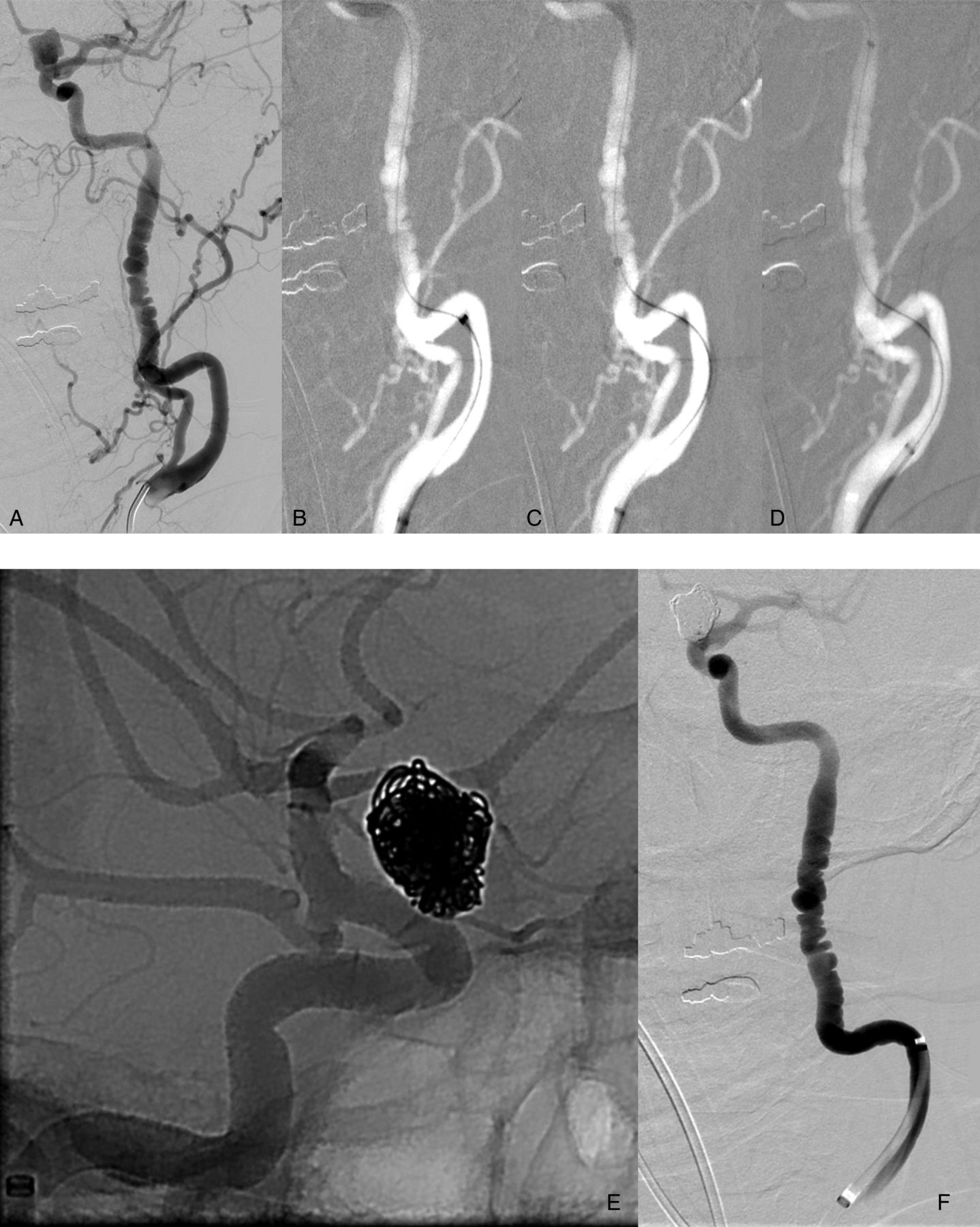
Young male patient with sickle cell disease, bilateral paraclinoid segment aneurysms and a remote history of subarachnoid hemorrhage. The proximal cervical segment of the right carotid artery takes serial abrupt 180° turns (A). A 7×20 mm Hyperglide balloon was manipulated over a 0.010 inch Xpedion microwire into the proximal cavernous segment of the internal carotid artery (ICA) (B). A Navien 058 guiding catheter was then atraumatically manipulated over the Hyperglide balloon into the proximal cavernous segment of the ICA (C–E). Counter tension applied to the balloon catheter drew the guiding catheter away from the outer curvature of the vessel at the apex of both abrupt turns (C, D, arrows). Throughout the positioning of the guiding catheter, there is little, if any, distortion of the native carotid anatomy (B–D). Injection of contrast in the working angle for coil embolization demonstrates no flow limitation, vascular injury, or vasospasm (F). The aneurysm treatment was successfully completed with deployment of a pipeline embolization device across the lesion (G).

Elderly female with a ruptured anterior communicating artery aneurysm. Initial angiography demonstrated marked tortuosity of the left common carotid artery as it arose from the aortic arch (not shown). Left internal carotid artery (ICA) angiography demonstrates marked tortuosity of the proximal cervical ICA (A). An Avigo 0.014 inch microwire (A) and then a Scepter XC 4×11 mm balloon catheter (B) were easily navigated beyond the tortuous segment. The Scepter balloon was inflated (C) and while applying counter tension to the balloon, the Navion 058 guiding catheter was advanced over the balloon catheter (D–G). The counter tension applied to the balloon catheter functioned to center the guiding catheter in the parent artery, pulling the catheter tip away from the outer curvature of the vessel wall (D–E, arrows). In the final guide catheter position, there is minimal distortion of the native carotid anatomy (G).
Results
The balloon-assisted guiding catheter placement technique was used to perform endovascular treatments of 12 anterior circulation aneurysms in 11 patients (aged 44–85 years, mean 59.6 years; 10 females) (table 1). One patient underwent coiling alone (figure 5). Five patients underwent balloon-assisted coiling (figure 2). One patient underwent balloon- and stent-assisted coil embolization. Four patients with five carotid aneurysms (one with bilateral carotid aneurysms) underwent vascular reconstruction with the pipeline embolization device (figures 1 and 3). All patients had tortuosity of the cervical carotid artery. Three patients had findings consistent with cervical carotid fibromuscular dysplasia (figure 2). Arch types were type I (n=5), type II (n=5), and type III (n=2). In seven of the 12 cases, prophylactic calcium channel blockers were administered prior to attempted distal guide catheter positioning to help prevent catheter induced vasospasm (table 1).
Patient and procedural details
The technique was successful in all cases on the initial attempt. No dissections or guide catheter induced vasospasm were encountered during any of the cases. In no case did the guiding catheter have to be retracted as a result of poor distal flow resulting from catheter induced arterial spasm. All aneurysm embolizations were completed successfully.
Discussion
The present case series demonstrates a novel technique designed to allow the efficient and atraumatic positioning of a distal access guiding catheter in patients with a tortuous and/or fragile cervical carotid anatomy.
Distal guiding catheter access can greatly facilitate endovascular therapy by providing a stable platform from which to manipulate, deliver, and deploy devices through the tortuous cerebrovascular anatomy.1 ,2 ,8 As the distance from the distal tip of the guide catheter to the distal tip of the microcatheter increases, so do the number of potential points for friction between the microcatheter and the parent vessel walls. Increasing friction limits the ability of the operator to control the behavior of the devices in that manipulations performed at the proximal aspect of the devices are not reliably transmitted to the distal aspect in a one to one manner.9 This circumstance adds considerable technical complexity to the cases, increasing operator time and likely also the risk for complications. The newest generation of flexible, compliant, distal access guiding catheters have substantially advanced the ability to achieve distal guiding catheter position.
Typically the delivery of these flexible distal access catheters is established by advancing the guiding catheters (with or without tapered internal introducer catheters) over 0.035 inch guidewires. In the setting of tortuous or fragile (eg, fibromuscular dysplasia) cervical vasculature, this technique is frequently challenging and requires multiple attempts, has a tendency to induce severe, often flow limiting catheter induced vasospasm, as well as parent artery dissections. In some cases a distal position cannot be achieved and the operator is forced to attempt intracranial therapy from a more tenuous proximal cervical guiding catheter position. In other cases, after distal position is achieved, the vasospasm induced within the parent artery is so severe that the guiding catheter ultimately has to be withdrawn more proximally. Finally, if excessive forward pressure is exerted to advance the guiding catheter system, the entire system can herniate out of the parent artery and into the aortic arch. These technical challenges inevitably add time, complexity, and risk to the procedure.
These technical challenges can be attributed to several mechanisms. First, navigating a 0.035 inch wire across very tortuous segments of the carotid artery not infrequently induces dissections, particularly when the wire catches the wall of the vessel at an abrupt 180° or 360° turn. Advancing larger guiding catheter systems through tortuous vessels over a 0.035 inch wire essentially forces the catheters along the outer curvature of the vessel at each turn, creating a great deal of stress on the vessel wall, providing a stimulus for vasospasm and a potential mechanism for dissection. Finally, forward vectors applied to advance a guiding catheter system are also transmitted through the junction of the great vessels with the aortic arch. In tortuous or unfavorable (ie, common carotid catheterization with a ‘bovine’ arch configuration) arch anatomy, these forces create a tendency for the entire guiding catheter system to herniate out of the target vessel and into the ascending aorta.10
The balloon-assisted technique overcomes each of the above technical limitations. First, the balloon is navigated beyond the tortuous cervical anatomy over a 0.010 or 0.014 inch microwire with a distal J configuration. This microwire–microcatheter navigation through the tortuous segment creates significantly less stress on the vessel wall than a 0.035 inch wire. These smaller wires are far less likely to create any kind of traumatic injury or vasospasm, even in the setting of severe tortuosity or fibromuscular dysplasia. Next, when the balloon is inflated, it creates friction against the vessel wall distally, providing a distal anchor for the entire guiding catheter system. Advancing the guiding catheter system while applying gentle counter tension to the balloon catheter functions to add a distal ‘pulling’ force to the proximal ‘pushing’ force. The counter tension also functions to center the guiding catheter system within the parent artery, pulling it away from the outer curvature of the vessel and eliminating or diminishing the forces applied by the guiding catheter system to the vessel wall. During this maneuver, no inner transitional catheter is used. As such, the tortuous cervical segment is negotiated by the flexible distal access catheter alone, resulting in much less distortion of the native anatomy than is typically observed when traversing severe cervical tortuosity with a coaxial guiding catheter introducer system. Preservation of the native carotid anatomy throughout the positioning of the guiding catheter is likely the single greatest factor in eliminating catheter induced vasospasm with this technique (figures 1, 3 and 4). Finally, the ‘pulling’ force at the distally anchored balloon reduces the amount of forward ‘pushing’ force that must be applied to advance the guide system, thereby reducing the vectors which might favor herniation of the system into the aortic arch. In addition, the balloon anchors the entire guiding system (long sheath and guiding catheter) within the parent artery, further reducing the risk for arch herniation.
A similar concept has been described in acute stroke treatment, taking advantage of the counter tension applied during the retrieval of a distally deployed MERCI device to advance a proximal aspiration catheter.11 The hypercompliant balloon represents a much safer means by which to optimize guide catheter positioning in the elective or semi-elective setting. The current generations of hypercompliant balloons are very low profile and atraumatic, and the risks associated with inflation of these balloons within the extradural, intracranial carotid circulation are minimal. The degree of balloon inflation can also be dynamically modified to achieve a level of distal anchoring sufficient to successfully advance the guiding catheter system. In addition, anchoring the balloon catheter within the petrous segment of the internal carotid artery during this maneuver further increases the safety profile by minimizing the risk for, and consequences of, any balloon induced vascular injury. The maneuver typically only requires a few seconds and, as such, the duration of parent artery occlusion during balloon inflation is inconsequential.
The reduced level of vascular trauma associated with this technique is supported by our observation that following guiding catheter placement, no catheter induced vasospasm was observed. Thus even in cases with extreme tortuosity (figures 1 and 4), the distal guiding catheter never had to be retracted once a distal position was achieved.
In summary, the balloon-assisted guiding catheter placement technique represents a straightforward technique by which to facilitate and expedite the endovascular treatment of intracranial aneurysms in the setting of tortuous cervical carotid anatomy.
References
Footnotes
-
Contributors Both authors made substantial contributions to the conception and design, acquisition of the data, or analysis and interpretation of the data.
-
Competing interests None.
-
Ethics approval The study was approved by the institutional review board.
-
Provenance and peer review Not commissioned; externally peer reviewed.