Article Text

Abstract
We present a computational fluid dynamics (CFD) analysis of the hemodynamic environment of an anterior communicating artery that spontaneously ruptured immediately following three-dimensional rotational angiography. Subsequent digital subtraction angiography allowed for the localization of the point of rupture within the aneurysm dome. CFD analysis demonstrated a concentrated jet that impinged directly at the site of rupture. Peak systolic pressure and wall shear stress were both maximal near the rupture location.
- Aneurysm
- Angiography
- Blood Flow
- Hemorrhage
- Subarachnoid
Statistics from Altmetric.com
Background
While numerous studies on hemodynamic simulations of intracranial aneurysms have been published, few previous reports have included knowledge of the rupture site. In the current report we performed a detailed hemodynamic analysis of an anterior communicating artery that ruptured during cerebral angiography, providing knowledge of the hemodynamic environment at the site of the rupture.
Case presentation
The detailed clinical history of the patient has been published previously.1 Briefly, the patient had a subarachnoid hemorrhage leading to conventional angiography. Three-dimensional rotational angiography (3DRA) and standard digital subtraction angiography were performed under conscious sedation and showed a right anterior communicating artery aneurysm 4 mm high and 3 mm wide. Immediately following 3DRA the patient had a seizure and was placed under anesthesia. Immediate follow-up angiography showed new development of extravasation from the previously noted aneurysm (figure 1). The aneurysm was coiled and the patient made full recovery and was discharged home the following week.

Angiograms taken before (left) and immediately after (right) rupture. The rupture site is indicated with a red arrow.
Investigations
The volume reconstruction of the computational model was obtained by delineating the lumen boundary from 3DRA images using image segmentation software (Mimics, Materialise, Leuven, Belgium). To let the flow develop physiologically at the neck of the aneurysm, we included a long inflow branch about 14 times the neck diameter.
Using ICEM CFD (ANSYS, Canonsburg, Pennsylvania, USA), we generated a high quality hexahedral grid inside the model boundary with higher density near the wall to better model the boundary layer. To obtain an accurate grid independent solution, we successively refined four more times the first generated grid and conducted a grid sensitivity study as described in our previous study.2
At the inlet boundary we imposed the Womersley velocity profile with a peak flow rate of 8 ml/s and mean flow rate of 4 ml/s. At the outlets we adopted traction-free boundary conditions.2
Blood was modeled as a Newtonian fluid with a density of 1 g/cm3 and dynamic viscosity of 0.04 Poise, and the walls were approximated as rigid.
Solutions were generated with Fluent 13.0 (ANSYS), with a second-order implicit solver in both time and space. The simulation presented here was based on the finest grid of 7 million elements with a maximum element size of 0.06 mm inside the bleb and 0.15 mm inside the arteries. Four cardiac cycles with a time period of 1 second were numerically simulated and the results of the fourth cycle were used to compute the flow variables.
We used velocity, pressure and wall shear stress (WSS) values at peak systole to characterize the intra-aneurysm flow behavior. To investigate the changes in the WSS vector within the cardiac cycle we used the oscillatory shear index (OSI) which attains a maximum value of 1 for a reversing flow and a minimum value of 0 for a unidirectional flow.3–5 To describe the inflow jet inside the aneurysm, we used the impact area defined by Cebral et al.5
Outcome and follow-up
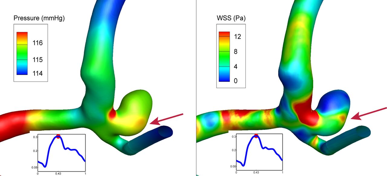
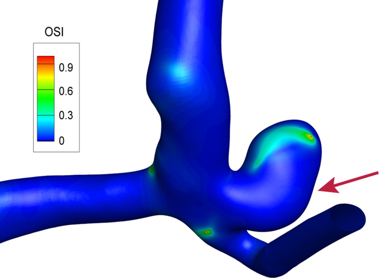
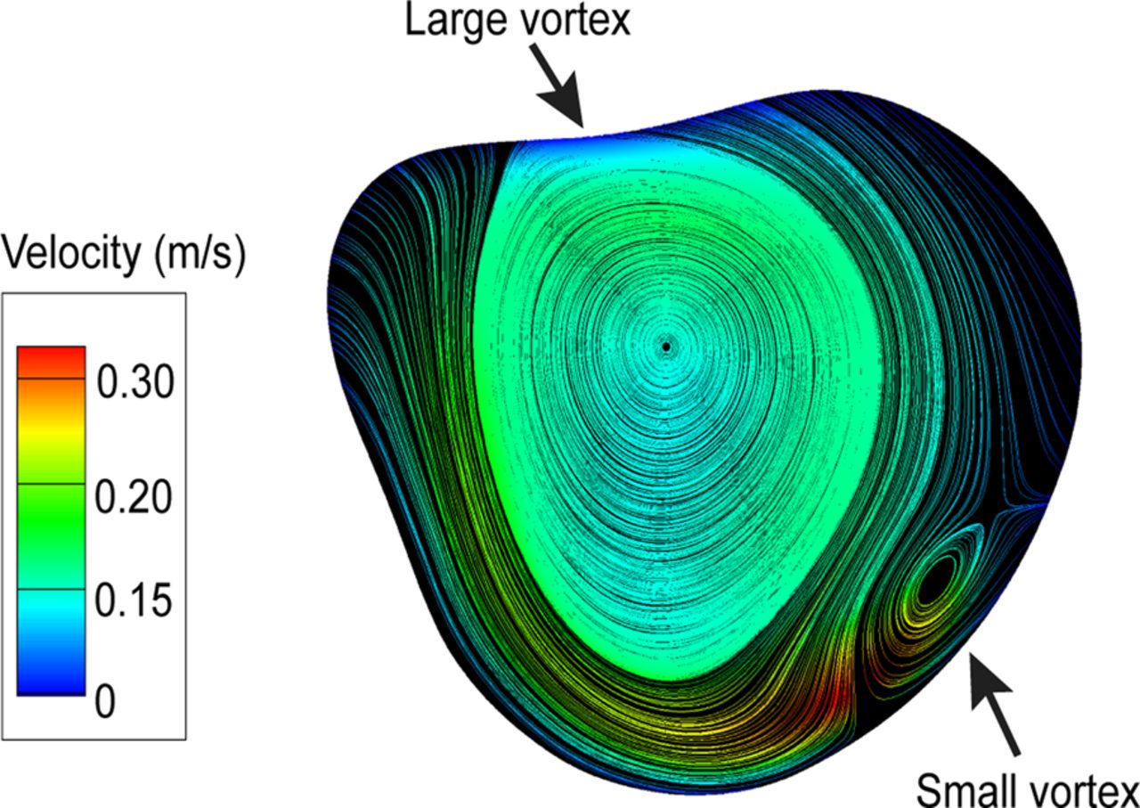
The angiograms immediately before and after rupture are shown in figure 1. The location of the rupture was identified as the area where blood leaked out of the aneurysm (figure 1). The velocity isosurface corresponding to 0.3 m/s and the velocity contour in a cutting plane, presented in figure 2, showed that a concentrated inflow jet was directed straight at the rupture location. The impaction area was relatively small, approximately 30% of the aneurysm surface. This agrees with other findings where small impingement areas were noticed in ruptured cases.5–7 Both the peak systole pressure and WSS contours (figure 3) showed maxima near the rupture location. The systemic pressure was mostly uniform across the aneurysm surface with a 1% elevation near the impingement location. Figure 4 illustrates the OSI which showed a maximum value of 1 on the aneurysm surface, similar to that usually found in ruptured cases.4 ,5 ,8 One major vortex whose axis of rotation was along the longest axis of the aneurysm and a much smaller vortex that formed upstream of the impingement area were observed (figure 5).

Velocity isosurface and velocity contour of the inflow jet on a cutting plane at peak systole. The rupture site is indicated by a red arrow.

Contours of pressure (left) and wall shear stress (right) at peak systole. The rupture site is indicated by a red arrow.

The oscillatory shear index distribution during one cardiac cycle with a maximum value on the dome surface. The rupture site is indicated by a red arrow.

Streamlines at peak systole projected on a plane. A large vortex emerged downstream of the impingement area while a small vortex formed upstream.
Discussion
This case report shows that the site of rupture of an anterior communicating artery corresponds to the site of the impingement zone of the simulated inflow jet; the perirupture environment was also characterized by elevated WSS and pressure. The small elevation in WSS and pressure can hardly explain the rupture mechanism, but it helps confirm the flow impingement location in the proximity of the rupture point. The inflow jet was stable throughout the cardiac cycle and concentrated to a small impact zone.
Previous authors have correlated the sites of aneurysm rupture with CFD results. Kono et al9 studied a pericallosal aneurysm which also ruptured during angiography. Similar to our findings, those authors noted that the rupture site corresponded to the jet impingement zone and elevated pressure area. However, unlike our case, the authors noted low WSS and unstable inflow jet during the cardiac cycle.
In two separate studies, Cebral et al7 ,10 showed a concentrated inflow stream into a large basilar aneurysm immediately prior to their spontaneous rupture, but the authors did not have detailed information about the rupture sites in either case. As such, correlation of the inflow jet location and the rupture location was not possible. Our case confirms their findings, as the concentrated inflow jet and elevated WSS correlated with the aneurysm rupture event. In addition, in our case we found the rupture location and the jet impingement area to be the same.
The current study has some limitations that need to be addressed in future studies. We simplified the model by truncating the other small arteries because of the observed small blood flow rates. In addition, we identified the rupture location from a two-dimensional image and imposed generic and mathematically-defined (rather than patient-specific) flow boundary conditions. We recognize that the injection of contrast agent may affect the flow field in the artery, but this flow change is difficult to evaluate.
Key message
-
Although the rupture of an aneurysm may result from a complex interplay of multiple hemodynamics and wall remodeling factors, we found that the calculated hemodynamic environment was characterized by a concentrated impingement jet with elevated pressure and wall shear stress (WSS) near the rupture location. One major and stable vortex developed inside the aneurysm. During the cardiac cycle the calculated WSS reversed its direction at certain points on the aneurysm surface as also noted in prior research studies on ruptured aneurysms.
References
Footnotes
-
Republished with permission from BMJ Case Reports Published 7 March 2013; doi:10.1136/bcr-2012-010596
-
Competing interests None.
-
Provenance and peer review Not commissioned; externally peer reviewed.