Article Text

Abstract
Background Infectious intracranial aneurysms (IIAs) are rare and potentially devastating. First-line management involves intravenous antibiotics, with surgical or endovascular management reserved for cases of failed medical treatment or aneurysmal rupture. Endovascular therapy has become the primary approach for treating these small, distally located aneurysms. Liquid embolic agents are well suited for use because of their ability to fill the aneurysm and parent vessel. We present our experience in treating these aneurysms via Onyx embolization and review the literature.
Methods We retrospectively reviewed the endovascular treatment of IIAs at our institution from 2010 to 2012. Eight patients with 16 IIAs ranging in size from 1 to 16 mm underwent treatment. Seven of the patients initially presented after aneurysmal rupture. Onyx was pushed until the aneurysm and parent artery were filled. Confirmation of aneurysmal occlusion was made by repeat cerebral angiography.
Results One symptomatic stroke occurred after embolization. Fourteen of the 16 aneurysms have been evaluated with follow-up angiography and remain occluded.
Conclusions Treatment of IIAs using an endovascular approach with Onyx is safe and effective.
- Aneurysm
- Artery
- Embolic
- Infection
- Liquid Embolic Material
Statistics from Altmetric.com
Introduction
Infectious intracranial aneurysms (IIAs) comprise 2.5–4.5% of all intracranial aneurysms. Rupture of these lesions carries a mortality risk of up to 80%. Endovascular treatment of intracranial aneurysms is an alternative to open microsurgery and clip ligation. Owing to their often irregular morphology, distal location, friable nature and disruption of the parent vessel microarchitecture, IIAs are particularly well suited for endovascular treatment using liquid embolic agents, with or without concomitant coil embolization.1–9 We present our experience with Onyx embolization for the treatment of IIAs and review the literature.
Methods
Embolization technique
Following informed consent, all procedures were carried out under general anesthesia with neurophysiologic monitoring of somatosensory evoked potentials and transcortical EEG. Percutaneous femoral arterial catheterization was used to achieve access in all patients. Typically, 6 Fr guide catheters were navigated into the distal internal carotid or vertebral artery. Selective catheterization of the diseased vessel was performed with Echelon or Marathon microcatheters (Covidien, Irvine, California, USA). Once the target parent vessel was catheterized and deemed to be appropriate for embolization, every attempt was made to advance the microcatheter into or just beyond the aneurysm or at least up to the aneurysm neck. The microcatheter was then prepared in standard fashion by filling the dead space with dimethyl sulfoxide (DMSO). Operating under the assumption that IIAs typically incorporate the parent vessel, Onyx 18 or Onyx 34 was subsequently injected until the angiogram demonstrated obliteration of the IIA and at least 5 mm of the proximal and distal parent artery. An early (7 days) and late (1–3 months) post-embolization angiogram was obtained in each case to assess the durability of treatment and screen for new IIAs.
Data collection
We retrospectively reviewed the hospital and endovascular treatment records of patients with IIAs treated via Onyx embolization at the University of Pittsburgh Medical Center from 2010 to 2012. Collected data included patient age, gender, presentation, aneurysm location, types of embolic agents used and complications related to the endovascular embolization procedure.
Results
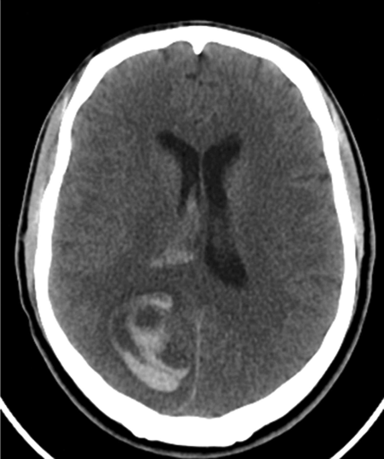
Eight patients with septic endocarditis with 16 IIAs were treated (table 1). One case was performed after failed medical management and documented aneurysm enlargement. The remaining patients initially presented with aneurysmal rupture, resulting in subarachnoid (n=4), intraparenchymal (n=1) or a combination of intraparenchymal and intraventricular hemorrhage (n=2) (figure 1). We were able to fill the aneurysm and parent vessel successfully with Onyx in all cases. Infected aneurysms ranged in size from 1 to 16 mm (mean 3.9 mm) and were located distally in the middle cerebral (n=10), anterior cerebral (n=4) and posterior cerebral (n=2) territories. One patient experienced a periprocedural cardiac arrest with recovery. This patient had severe cardiac disease which was compounded by his endocarditis. During the procedure he became bradycardic and subsequently arrested. After treatment he recovered. All parties involved believed the event was not a result of the embolization but rather a consequence of his pre-existing cardiac myopathy. One patient had an intraparenchymal hemorrhage on day 4 after the procedure which was later confirmed to be the result of a newly formed IIA. This same patient experienced a symptomatic stroke which involved an expected and fortunately transient distal right upper extremity monoparesis. On follow-up imaging, seven of the eight patients had radiographic evidence of new asymptomatic infarctions or edema. The presumed infarcted territory was small, with an average infarct size of 5.5 cm3.
Characteristics and outcomes of patients with infectious intracranial aneurysms treated with Onyx embolization

Initial head CT scan showing a right parietal intraparenchymal hemorrhage and intraventricular hemorrhage in a patient with a ruptured infectious intracranial aneurysm.
Of the 16 treated IIAs, 7 (44%) were discovered in three patients after the initial Onyx embolization procedure an average of 6.2 months (range 0.1–18.9) later. All three patients had transesophageal echocardiograms which showed infectious valvular pathology that had not resolved despite intravenous antibiotic therapy. In two of the patients the new IIAs were found on catheter angiography performed during routine follow-up. One patient experienced a symptomatic intracranial hemorrhage which required emergency craniotomy for clot evacuation 4 days after initial treatment of an IIA. A subsequent angiogram revealed four new IIAs which were then embolized. Less than a week after this second embolization another new IIA was found and treated on follow-up.
To date, 14 of the 16 aneurysms (88%) have been evaluated in follow-up with catheter-based angiography. All 14 remain occluded. No patient had a subsequent hemorrhage or brain abscess during mean radiographic follow-up of 5.8 months (range 1.5–18.9). Four of the seven patients (57%) who presented after aneurysmal hemorrhage have regained the ability to function independently (modified Rankin scale 0–2) but all still have at least a mild clinical deficit.
Discussion
The first report of an IIA was in 1869 by Church who presented a case of a 13-year-old patient with infective endocarditis affecting the mitral valve.10 In 1885, Sir William Osler called these lesions mycotic aneurysms because they appeared as ‘fresh fungus vegitations’,11 a misnomer considering this term came to be ascribed to all aneurysms of infectious origin, regardless of their underlying microbial etiology. Recently, the more appropriate term ‘infectious intracranial aneurysm’ was popularized in the literature.12 IIAs are pseudoaneurysms of infectious etiology which comprise 2.5–4.5% of all intracranial aneurysms.13–15 The true incidence of IIA rupture is unknown, but it is the key prognostic factor in determining patient outcome. Aneurysm rupture predicts a mortality rate of up to 80% compared with 30% in those who do not present after hemorrhage.16–18
The principal risk factor for the development of IIAs is endocarditis, particularly those infections involving prosthetic valves.19 Up to 80% of patients with IIAs have an underlying endocarditis, while IIAs occur in 1–4% of patients with infective endocarditis.13 ,18 ,20–23 Other less common etiologies include meningitis, cavernous sinus thrombophlebitis, cerebral abscess, subdural empyema, osteomyelitis of the skull and sinus infections.24 The pathophysiologic mechanism by which IIAs arise involves destruction of the vasa vasorum and subsequent inflammation and necrosis of the adventitia of blood vessels, which then proceeds inwards, causing the normal histologic architecture of the muscularis layer and internal elastic lamina to break down.3 ,25
The majority of cases are caused by the Gram-positive bacteria Streptococcus viridians or Staphylococcus aureus.26 In our series all eight patients had endocarditis, with seven patients having positive blood cultures; the most common isolate was Staphylococcus aureus. One patient was found to have a fungal infection with Candida tropicalis, a pathogen that has not previously been reported to be associated with infectious aneurysms of the intracranial or systemic vasculature. This patient presented after a subarachnoid hemorrhage and experienced a prolonged hospitalization which involved endovascular treatment of six IIAs, five of which developed as new aneurysms within 2 weeks of the initial diagnosis and initiation of intravenous antifungal therapy. Although atypical, his clinical course underscores the highly virulent nature and the poor clinical results associated with fungal infections of the CNS.
The initial treatment of IIAs consists of antibiotics. Surgery is reserved for those patients who experience symptomatic mass effect from the aneurysm itself, rupture, persistence or enlargement of the aneurysm despite antibiotic therapy or neurologic deterioration during treatment.24 The surgical treatment of IIAs is challenging due to their irregular morphology, distal location and involvement of parent vessels.24 The task of treating ruptured IIAs is especially difficult. In the first published series describing the surgical treatment of ruptured IIAs, Roach and Drake described a case in 1965 in which ‘it was not possible to ligate (the) aneursymal sac for the parent vessel was so fragile from infective necrosis that it bled from more than one place in spite of careful manipulation.’13 Due to the aforementioned factors and the typical distal location of IIAs, endovascular obliteration of IIAs is generally achieved through parent artery sacrifice via utilization of coils, particles or liquid embolic agents rather than direct embolization of the aneurysm itself.3 ,4 ,17 In reporting their experience with treating pediatric ruptured IIAs, Eddleman and colleagues stated that the decision to perform parent artery occlusion is multifactorial: (1) the aneurysm cannot be safely accessed using microcatheters; (2) it has less than ideal morphology for direct embolization; and/or (3) it comes from a parent artery that does not supply eloquent cortex and, thus, its sacrifice would not lead to a neurologic deficit.5 Indeed, in the first reports in which an endovascular approach was used for the treatment of ruptured IIAs, the authors initially attempted treatment by sacrificing the parent artery.27 Additionally, given the friability of the aneurysm wall and because the parent artery is often diseased or too small to reconstruct, we believe that vessel sacrifice of the proximal and distal parent artery is necessary.
Onyx is a liquid embolic agent consisting of ethylene vinyl alcohol copolymer (EVOH), DMSO and tantalum powder that was originally approved for the treatment of brain arteriovenous malformations. Once injected into the bloodstream, the DMSO diffuses away leaving a soft non-adherent EVOH precipitate capable of achieving permanent vascular occlusion.28 Its non-adhesive quality, long working time, easy visualization and ability to be injected numerous times during one catheterization make Onyx an excellent embolic agent. Compared with other liquid embolic agents, Onyx works through a process of precipitation which minimizes the likelihood of the microcatheter being glued within the cast or avulsion of the embolized pedicle while withdrawing the microcatheter.29 However, while not encountered in our series, there is a risk of vessel injury during catheter withdrawal given the often distal location of the IIAs and the inherent necessity to catheterize distant vasculature in order to treat the offending pathology, especially when considering the friable nature of the parent vessel involved. To date, there is a relative dearth of literature involving the use of Onyx for treatment of IIAs. In 1995, Utoh and colleagues were the first to use Onyx in a patient with an aneurysmal subarachnoid hemorrhage.4 They embolized the parent artery of a 5 mm IIA involving the P2 segment of the right posterior cerebral artery. Eddleman et al used Onyx 18 in two patients, each with ruptured distal middle cerebral artery (MCA) aneurysms. In one of the patients the authors initially directly coiled the aneurysm and then used Onyx to fill the residual neck, resulting in obliteration of the aneurysm itself as well as performing distal occlusion.5 A group from the University of Utah Hospital published their experience with Onyx 18 in the treatment of dissecting intracranial aneurysms and IIAs in which two patients underwent Onyx embolization of IIAs, one involving the left parieto-occipital artery and another involving the right posterior temporal artery.6 In both patients the parent artery, as well as the aneurysm itself, was occluded. Zhao et al7 successfully injected Onyx 18 directly into an unruptured left P3 segment IIA, and Gross and Puri used Onyx for embolization of a ruptured distal MCA IIA and occlusion of its distal parent artery.8 Most recently, Jadhav et al9 published their experience with Onyx embolization of nine pseudoaneurysms, two of which were IIAs, with lasting results.
We used Onyx 18 in seven cases, Onyx 34 in eight cases, and both in one case. Onyx was pushed until the parent vessel and the aneurysm were filled up to 5 mm distal and proximal to the aneurysm in the parent vessel (figure 2).

Lateral native angiographic images after coil and Onyx embolization of two aneurysms arising from the distal right middle cerebral artery after the patient presented with intraparenchymal hemorrhage and intraventricular hemorrhage (shown in figure 1). (A) Before treatment with aneurysm marked by black arrow; (B) after treatment with Onyx cast marked by black arrow.
The recent review by Gross and Puri highlights the many potential endovascular treatment modalities for obliterating IIAs.8 Apart from Onyx, options in the endovascular armamentarium include the use of detachable balloons, stents, n-butyl cyanoacrylate (Trufill; Codman Neurovascular, Raynham, Massachusetts, USA), coils or a combination of these. The rapid advancements in neurointerventional device technology has also led to other potential modalities to be explored in the treatment of IIAs, including the use of semi-covered low-porosity endoluminal stents used for flow diversion and subsequent IIA occlusion.30 While the patients included in our report did not have aneurysms suitable for a stent graft and despite the intuitive reservations that a neurointerventionalist may have in deploying a foreign body in a septic environment, this represents another treatment option to consider for patients with more proximal IIAs. With regard to the risk of delayed infection or abscess formation, no intracranial infections have been associated with the use of Onyx and no infections have been reported with the use of EVOH.
Evidence of infarction was noted in all patients, probably as a consequence of parent vessel occlusion; importantly, in seven of the eight patients the infarction was clinically asymptomatic. One potential explanation for this finding is that the parent vessel embolized in these cases did not supply eloquent cortex. However, this is rather unlikely given the large proportion of IIAs in our series which involved the MCA, the occlusion of which would putatively result in clinical evidence of a stroke. An alternative theory is that our ability to occlude the vessel at the site of the aneurysm without significant distal embolization allowed for robust collateralization of the distal territory, mitigating the clinical consequences of vessel occlusion.
Conclusions
Treatment of IIAs via an endovascular approach using the liquid embolic agent Onyx appears to be safe and effective. Based on our experience, Onyx embolization of the parent vessel, in addition to the aneurysm itself, confers a high likelihood of obliteration with durability over time. Considering the high likelihood of recurrent aneurysm formation, short- and long-term follow-up angiography should be considered in all patients.
References
Footnotes
-
Contributors RG wrote most of the manuscript. NTZ prepared the discussion and performed the editing. EAM performed the data gathering. TJ and MH were senior members of the staff working directly with the patients and performing the procedures. BTJ was responsible for overseeing and directing the study. GL contributed to data collection and editorial review.
-
Competing interests None.
-
Ethics approval Approval was obtained from the University of Pittsburgh institutional review board (IRB #PRO08120394).
-
Provenance and peer review Not commissioned; externally peer reviewed.
-
Data sharing statement The rest of the patient data and clinical information are available to the medical staff of UPMC and research faculty through the electronic medical record.