Article Text

Abstract
Three years following endovascular embolization of a 3 mm ruptured arteriovenous malformation (AVM) of the left superior colliculus in a 42-year-old man, digital subtraction angiography showed continuous regrowth of the lesion. Thin-slice MRI acquired for treatment planning did not show the AVM nidus. The patient was brought back to the angiography suite for high-resolution contrast-enhanced cone beam CT (VasoCT) acquired using an angiographic c-arm system. The lesion and nidus were visualized with VasoCT. MRI, CT and VasoCT data were transferred to radiation planning software and mutually co-registered. The nidus was annotated for radiation on VasoCT data by an experienced neurointerventional radiologist and a dose/treatment plan was completed. Due to image registration, the treatment area could be directly adopted into the MRI and CT data. The AVM was completely obliterated 10 months following completion of the radiosurgery treatment.
- Arteriovenous Malformation
- Technique
- CT
Statistics from Altmetric.com
Background
Although complete surgical resection of brain arteriovenous malformations (AVMs) is regarded as the definitive treatment, radiosurgery has been shown to be very effective with and without prior embolization.1 Currently, the gold standard technique for diagnostic and follow-up imaging is digital subtraction angiography (DSA), which only provides two-dimensional (2D) spatial information. However, in order to minimize brain tissue damage surrounding the AVM, accurate planning of radiation therapy is required and is generally accomplished using volumetric image data such as CT, three-dimensional (3D) rotational angiography and/or MRI. Nevertheless, due to limited image resolution of these 3D imaging modalities, micro-AVMs with dimensions of a few millimeters are not always visualized adequately to allow reliable radiation planning.
Recent developments in flat-detector technology has enabled in situ acquisition of cone beam CT (CBCT) data using an angiographic c-arm system, providing image quality comparable to that of multidetector CT.2 ,3 CBCT data are reconstructed from a large set of x-ray images acquired during a rotational sweep of the x-ray source and flat-detector around the patient. In order to achieve an optimal signal-to-noise ratio (SNR), pixel binning is performed in which the signals measured at neighboring detector elements are combined. Generally, CBCT data are acquired using 2×2 pixel binning, which improves image quality and also significantly reduces the amount of data to be transferred to the workstation and the reconstruction time. However, acquisition of CBCT data without pixel binning will substantially improve the image resolution, albeit at a cost of reduced SNR. Non-binned data acquisition using a reduced flat-detector format to limit the amount of data to be processed enables high-resolution CBCT with 67 µm isotropic voxels and provides in vivo visualization of cerebrovascular stents4 and non-alloy microcannulas5 with high detail. We have explored the use of high-resolution contrast-enhanced CBCT (VasoCT, Philips Healthcare, Best, The Netherlands) to visualize a micro-AVM with sufficient detail for 3D radiation treatment planning.
Case presentation
A 42-year-old man was admitted to our hospital because of sudden onset of blurred vision and ‘the worst headache of his life’. A non-enhanced axial head CT revealed an intraparenchymal hemorrhage in the mesencephalic tectum and left posterior thalamus with an intraventricular and mild subarachnoid component. Due to subsequent development of hydrocephalus, most likely due to obstruction of the aqueduct, a ventricular shunt was placed in the frontal horn of the right lateral ventricle. Correct position and proper function was confirmed by a non-contrast head CT.
DSA showed an increased vascularity in the tectal area, which was suspicious for a vascular malformation. Definitive visualization of the nidus was not possible, probably due to the mass effect of the hemorrhage. Approximately 2 months later, a follow-up DSA was performed and confirmed the presence of a left superior colliculus AVM. The AVM nidus measured 3 mm. One month later the AVM was embolized via infusion of a mixture of n-butylcyanoacrylate (glue) and Ethiodol (ratio 1 : 3). The final control angiogram showed complete obliteration of the AVM.
A non-contrast head CT later the same day showed no evidence of new intracranial hemorrhage, hydrocephalus, mass effect or midline shift. The previously detected hemorrhagic foci in the mesencephalic and thalamic region had resolved.
After 6 months the patient was seen for an angiographic follow-up examination. DSA revealed a small regrowth of the AVM with supply through both superior cerebellar arteries and an early venous drainage through the left superior collicular vein into the vein of Galen. The previously documented obliterated portion of the AVM remained occluded (figure 1). The small size of the residual nidus and the absence of any dangerous features required continued follow-up for management as discussed by our multidisciplinary neurovascular team.

Anterior-posterior (A) and lateral (B) digital subtraction angiograms at 6-month follow-up examination showing regrowth of a superior colliculus arteriovenous malformation (arrows) post embolization.
A 3-year DSA follow-up examination demonstrated continuous AVM regrowth. Given the eloquent location of the nidus, radiosurgery was considered as a reasonable treatment option.
Treatment
Prior to radiosurgery, the patient underwent MRI, non-contrast CT and contrast-enhanced VasoCT (figure 2). VasoCT was obtained on a biplanar x-ray angiography c-arm system (Allura Xper FD20/20, Philips Healthcare) with a reduced detector size of 22×22 cm2 and in a non-binned mode (matrix 1016×1016; pixel size 0.15×0.15 mm2). During the acquisition, 20% iodinated contrast agent (iopamidol 51%, Isovue, Bracco Diagnostics, Princeton, New Jersey, USA) was administered intra-arterially using a 5 Fr catheter placed in the right vertebral artery via a coupled injector (infusion rate 2 mL/s, total 64 mL). After the imaging sequence was complete, high-resolution volumetric data were generated (matrix 512×512×512; voxel size 0.13×0.13×0.13 mm3) using a filtered back-projection reconstruction algorithm which allowed for visualization of detailed structures at the cost of lower signal-to-noise. CT and VasoCT data were acquired with the patient stabilized using a thermoplastic immobilization mask.

Axial slice of contrast-enhanced three-dimensional fast spoiled prepared gradient recalled echo sequence (3D FSPGR) (A) maximum intensity projections of 5 mm coronal (B) and oblique (C) slices of contrast-enhanced cone beam CT data (VasoCT). Brain arteriovenous malformation (AVM) was not visible on FSPGR data, but AVM nidus (white arrows), feeders (red arrows) and draining veins (blue arrows) were clearly visualized with VasoCT.
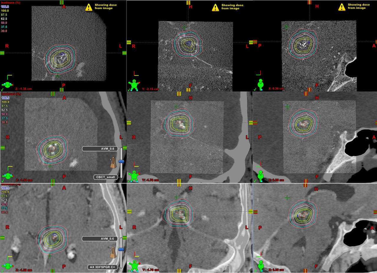
MRI, CT and VasoCT data were transferred to a workstation with radiation therapy planning software (Eclipse 8, Varian Medical Systems, Palo Alto, California, USA) and co-registered using anatomical landmarks. Brain AVM nidus was delineated on VasoCT data by an experienced neurointerventional radiologist. Due to image co-registration, radiation planning could be directly adopted into MRI and CT data (figure 3).

Snapshots of radiation planning software (Eclipse). Axial (left column), coronal (middle column) and sagittal (right column) slices of VasoCT (top row), VasoCT registered with CT (middle row) and VasoCT registered with MRI data. Delineation of arteriovenous malformation nidus was readily completed in three-dimensional mode on VasoCT data and dose planning was performed accordingly. Due to image registration, treatment planning was directly adopted into CT and MRI data.
Radiosurgery was performed with five arcs in four treatment fractions of 5 Gy on a Trilogy accelerator (Varian Medical Systems) equipped with a 120 multileaf collimator. Patient alignment was established using Varian OBI KV, CBCT image guidance.
Outcome and follow-up
Ten months after the last radiosurgery treatment the patient underwent follow-up catheter angiography. DSA showed complete obliteration of the treated AVM without damage to adjacent brain structures (figure 4).

Anterior-posterior (A) and lateral (B) digital subtraction angiograms and MRI (C) acquired 10 months after last radiosurgery treatment showing complete obliteration of brain arteriovenous malformation and no evidence of off-target injury caused by radiosurgery.
Discussion
Radiosurgery is a safe and reliable treatment option for small AVMs that can result in high obliteration rates.6 In order to prevent unwanted radiation injury, accurate target delineation is essential. Because of its high temporal and spatial resolution, the gold standard technique for diagnostic and follow-up imaging of AVMs is catheter angiography. DSA is particularly helpful to identify early filling of the draining vein. However, translation of this 2D image information into a 3D target is an ill-posed problem, especially in case of a complex nidus configuration. Various techniques have been proposed to perform such a task.7–9 Nonetheless, inferring 3D geometries from biplanar imaging will entail a non-negligible uncertainty.
Recently, investigators have successfully demonstrated that contrast-enhanced CBCT data acquired with an angiographic c-arm system can be used for treatment planning for radiosurgery of AVMs.10 ,11 They showed that CBCT data with soft tissue contrast (ie, using a binned mode) and voxel volume from 0.46 to 0.81 mm3 was suitable for delineation of AVMs. However, the spatial resolution of CBCT obtained with pixel binning is not sufficient for full appreciation of micro-AVMs by itself, and required image registration of CBCT to anterior-posterior and lateral DSA to enable nidus delineation.12 We have shown that CBCT data acquired using a non-binned mode provide sufficient spatial resolution to visualize and delineate a micro-AVM.
Key messages
-
Contrast-enhanced high resolution CBCT (VasoCT) provides a feasible method to perform treatment planning for radiosurgery of arteriovenous malformations that are too small to be visualized with CT angiography, MRA or conventional contrast-enhanced CBCT.
-
The advantage of this approach is that it provides direct target delineation in three dimensions instead of inferring information from planar angiography.
-
This technique is readily adaptable to present workflow in stereotactic radiosurgery.
Footnotes
Republished with permission from BMJ Case Reports Published 14 August 2013; doi:10.1136/bcr-2013-010763
-
Contributors IMJvdB: Study design, data collection and processing, manuscript preparation and approval. MJG: Study conception and design, data collection, manuscript revision and approval. LD: Data collection, processing and analysis, radiation physicist, manuscript preparation and approval. ALK: Data collection, manuscript preparation and approval. DG: Interpretation of data, radiation oncologist, manuscript revision and approval. ASP: Study design, interpretation of data, neurointerventional radiologist, manuscript revision and approval. AKW: Study design, interpretation of data, neurointerventional radiologist, manuscript revision and approval.
-
Competing interests AKW receive research funding from Philips Medical HealthCare. None for other authors.
-
Patient consent Obtained.
-
Ethics approval Ethics approval was provided by the University of Massachusetts Medical School institutional review board.
-
Provenance and peer review Not commissioned; externally peer reviewed.