Article Text

Abstract
Background Wide-necked intracranial aneurysms have been a challenge for endovascular techniques. With the advent of adjunctive devices such as balloons or stents, recanalisation rates have decreased secondary to better packing.
Purpose The purpose of this registry was to evaluate the safety and effectiveness of the new Low-profiled Visualized Intraluminal Support LVIS and LVIS Jr. stents in the treatment of unruptured wide-neck intracranial aneurysms.
Methods The LVIS or LVIS Jr. stent-assisted coil embolisation was performed in 78 patients harbouring 78 intracranial aneurysms. There were 59 aneurysms located in the anterior circulation and 19 in the posterior circulation. Clinical data and 6-month follow-up angiograms are presented.
Results The LVIS and LVIS Jr. stents were successfully delivered to the target aneurysm; however, there were seven cases in which the LVIS/LVIS Jr. stents had suboptimal opening and apposition to the parent vessel wall. The overall technical success for all groups was 91% (71 of 78 stents). There was complete angiographic occlusion in 66 (85%) of 78 cases and residual neck remnants in 12 (15%) cases. All patients had 6-month angiographic follow-up, which demonstrated complete occlusion of the target aneurysm in 64 (82%) cases, residual neck remnants in 5 (6%) cases and there was aneurysm filling in 9 (12%) cases.
Conclusions The LVIS/LVIS Jr. stent system is safe and effective for the treatment of wide-neck intracranial aneurysms, providing suitable support of the coil mass, which allows for a high level of occlusion with low rates of recanalisation and subsequent treatments.
- Aneurysm
- Coil
- Stent
- Intervention
Statistics from Altmetric.com
Introduction
Endovascular coiling therapy is an alternative to open neurosurgical treatment of intracranial aneurysms. Coiling embolisation alone is limited to a specific group of aneurysms in which the ratio between the neck and sac of the aneurysm is favourable, which makes it possible to safely position coils within the sac and finally exclude the aneurysm from the circulation. Stents have become very important devices in the treatment of intracranial aneurysms since 1997 when this technique was first described.1 Despite the presence of new devices such as flow-diverters, coil-assist microstents have their own place within the treatment armament with very good rates of aneurysm occlusion and are used primarily to treat wide-neck or fusiform intracranial aneurysms.2 There are many different stents that have been implanted in patients with acceptable results on morbidity and mortality.3 ,4 We report our experience in 78 unruptured aneurysms in 78 patients with 6-month follow-up treated with a hybrid braided closed-cell stents. Low-profile Visualized Intraluminal Support LVIS/LVIS Jr. (MicroVention—Terumo, Tustin, California, USA) are self-expanding, compliant, closed-cell design that are more flexible and have minimal ovalisation compared with the currently used microstents (figures 1 and 2).
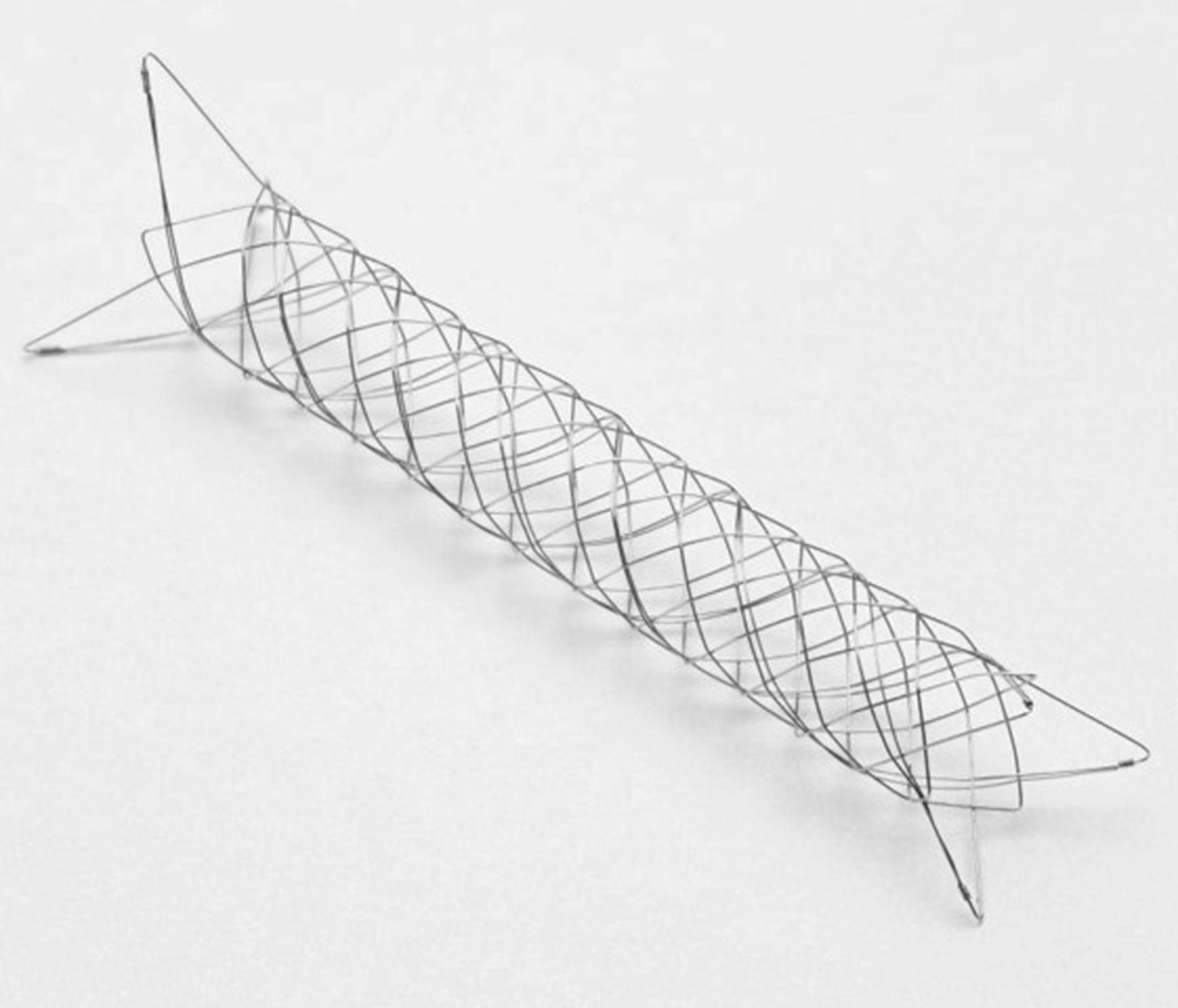
LVIS Jr. stent demonstrating three symmetric radiopaque markers on each flared end and 1.5 mm circular cell size of the stent.

LVIS stent demonstrates four distal radiopaque markers and four proximal paired markers. 1 mm circular cell size of the stent.
The LVIS device is a low-profile self-expanding single wire braid, closed cell construction of nickel-titanium (nitinol) material with compliant cells. They are compatible with microcatheters with a 0.017 inch lumen for LVIS Jr. or with 0.021 inch lumen microcatheters for the LVIS. The LVIS Jr. has three radiopaque tantalum markers on the proximal and distal tines with three tantalum helical strands within the body of the stent. The LVIS has four radiopaque markers on either end with two tantalum threads within the body of the stent. Both devices are well visualised after deployment (figures 3 and 4).
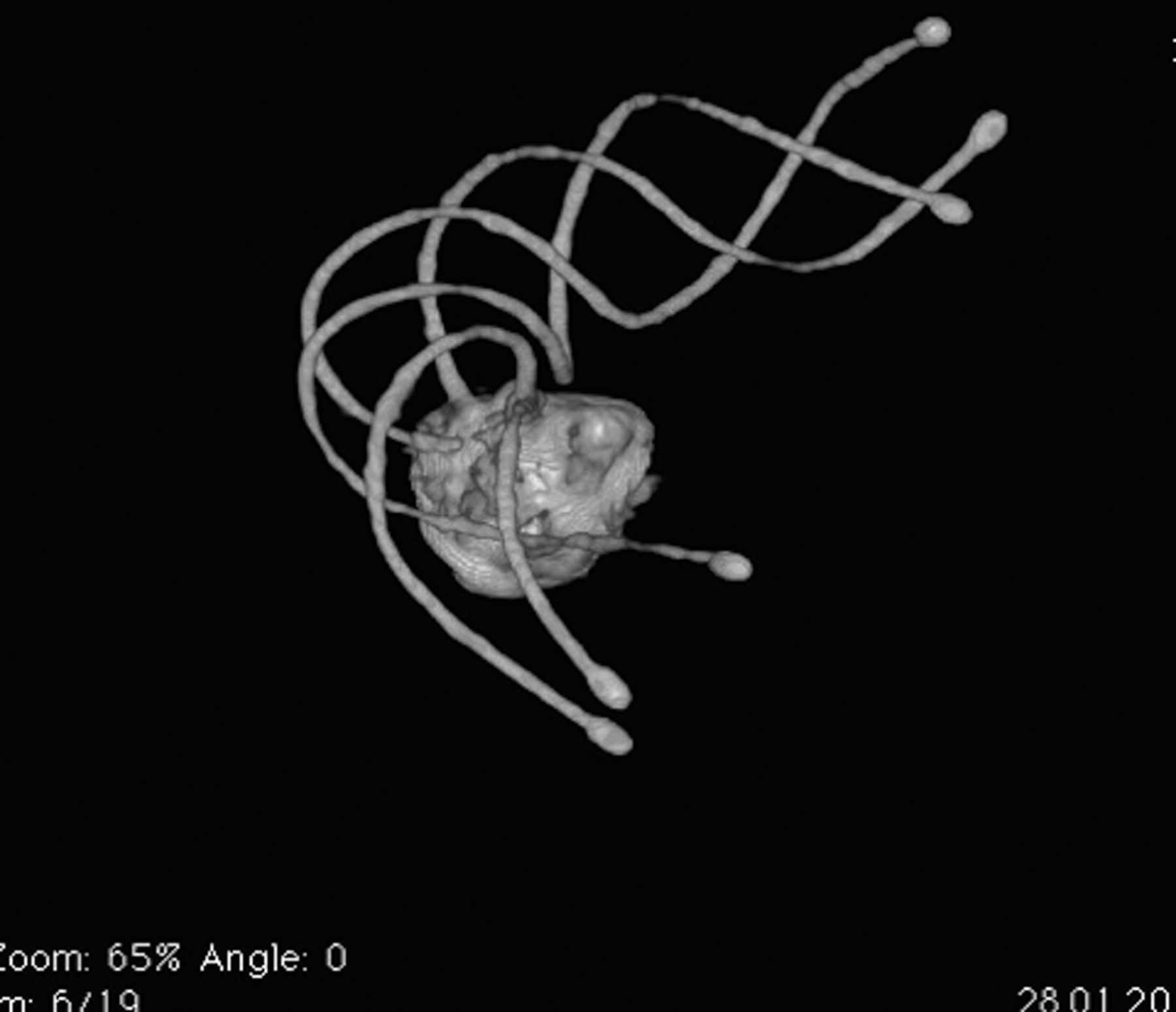
3D reconstruction radiographic image of the LVIS Jr. demonstrating the three radiopaque threads within the body of the stent.
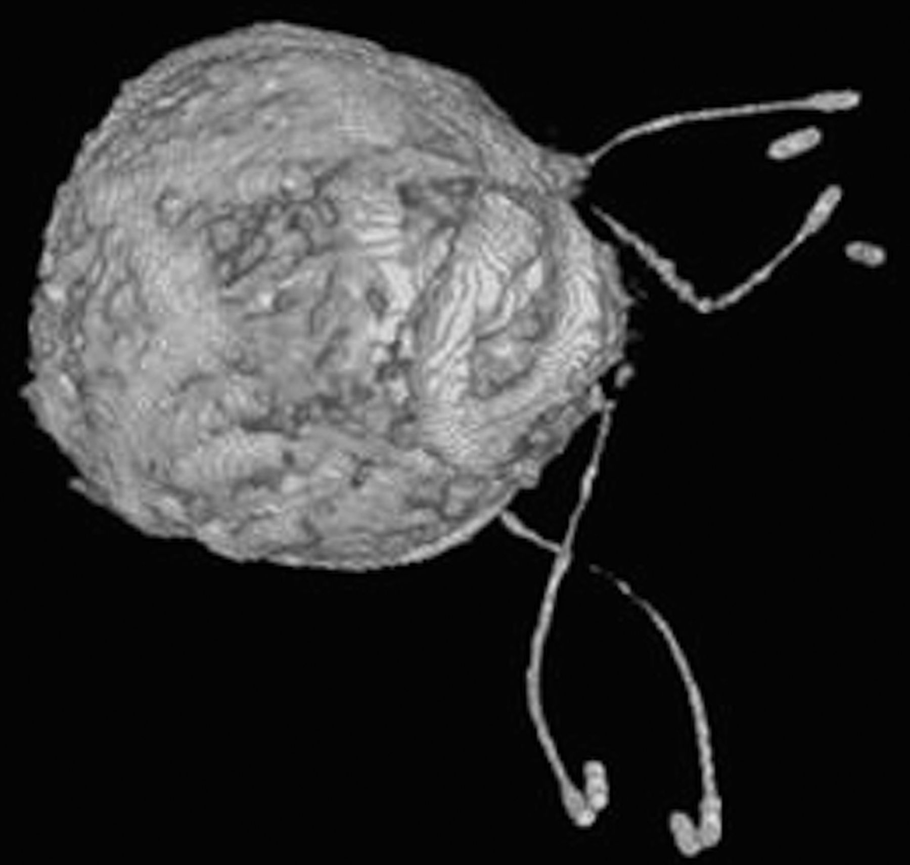
3D reconstruction radiographic image of the LVIS demonstrating the two radiopaque threads within the body of the stent.
Materials and methods
All 78 patients were recruited from neurosurgery or neurology departments at five regional medical centres. The LVIS/LVIS-Jr. stent devices have European Conformity Marking (CE Mark) and are routinely used in each regional institution in the registry. Although this is a relatively new device, it has CE mark and is within the scope of practice at each institution and did not require ethical committee approval for this registry. (The device is not currently approved in the USA but is under an FDA investigational trial.) The registry was carried out with respect to the ethical principles in relation to the participating patients with clinical standards and regulations. The clinical and angiographic data were collected prospectively.
Neurointerventional radiologists and neurosurgeons analysed all possible options including endovascular versus open surgical repair before treatment, and then all patients signed a written consent to have endovascular stent-assisted coiling of their intracranial aneurysms. The individual treating operators at each centre had free will in the choice of stents used for the treatment but was not randomised between the two types. We included only unruptured wide-neck aneurysms, which could not be treated with coiling alone. Assessment of the aneurysms was performed on a radiological workstation based on angiographic reconstructions obtained from CT angiography (CTA) or magnetic resonance angiography (MRA). Clinical examination was performed prior to the procedure, at discharge from the hospital post-treatment and at 6-month follow-up. Angiographic–DSA evaluation was obtained before, after the treatment and then at 6-month follow-up. 3D-DSA was performed to assess opening of the stent struts and body with apposition to the parent vessel arterial wall along the aneurysm neck. Baseline characteristics of the study patients are presented in table 1.
Baseline characteristics of the study patients (n=78)
Interventional procedure and medical therapy
All procedures were performed under general anaesthesia and via the femoral approach using 6F Avanti sheath introducer (Cordis, Bridgewater, New Jersey, USA) and guiding catheter Chaperon 6F (MicroVention, Tustin, California, USA) or Envoy 6F (Cordis, Bridgewater, New Jersey, USA) or Guider Soft (Boston Scientific, Fremont, California, USA), which was positioned in the internal carotid artery (ICA) or basilar artery (BA) depending on aneurysm location. Prior to treatment and deployment of the stents, a bolus of 5000 IU of heparin (Polfa Warszawa, Poland) was administered intravenously in all cases. Acetylsalicylic acid (Polfa Warszawa, Poland) and clopidogrel (Plavix; Bristol-Myers Squibb/Sanofi Pharmaceuticals, New York, USA), each in a dose of 75 mg/daily, were administered 5 days prior to the planned procedure and recommended to be taken for 3 months after discharge and then only acetylsalicylic acid.
3D-DSA was performed to visualise the morphology of the aneurysm sac, aneurysm neck and parent vessel to determine proper working projection for stent placement and endovascular coiling. The Headway 17 microcatheter (0.017 inch inner lumen) (MicroVention, Tustin, California, USA) was used for deployment of the LVIS-Jr. or the Headway 21 (0.021 inch inner lumen) for deployment of the LVIS. A Traxcess 0.014 inch microguidewire (MicroVention, Tustin, California, USA) or Terumo 0.012 inch, double angled (Terumo Medical Corporation, Tokyo, Japan) was placed across the aneurysm neck with the help of 3D roadmap. Before stent deployment, a ‘Jailed’ Echelon 10 (EV3, Plymouth, Minnesota, USA) or Headway 17 microcatheter was placed within the aneurysm sac and used as the coil delivery system. This jailing technique was used in 100% all cases; however, in four cases, additional coiling through the cells of the stents was performed because of lost microcatheter working position within the aneurysm. All stents were deployed according to the standard procedure protocol recommended by the manufacturer. The stent system was flushed, checked for proper opening, introduced into the microcatheter and advanced to the end of microcatheter. The distal end of the LVIS device was placed distal to the target aneurysm with the central portion of the stent placed along the aneurysm neck. Stent selection was made on the basis of vessel diameter and the manufacturer recommendation. If two stents were needed for a single patient in a telescoping method, a proper size stent was deployed first and then a lower profile stent (LVIS Jr.) was chosen to be placed within the previously deployed stent. Using this telescoping technique, the stents were easily accessed and deployed without any friction or difficulty. The unsheath technique for deployment of both stents is by pushing the stent into position and maintaining control of the microcatheter by withdrawing it and unsheathing the stent simultaneously. Care must be taken to observe the free leading tip of the microguidewire and by positioning the distal and proximal ends of the stent at the target location. Proximal and distal radiopaque markers of the stent and helical strands demonstrate full deployment and apposition within the parent vessel of the device.
After stent implantation, coil embolisation was performed using multiple-sized platinum helical coils (MicroVention, Tustin, California, USA). Control angiograms including 3D-DSA was performed in all cases at the end of procedure to demonstrate aneurysm occlusion rate, parent artery and stent patency, stent structure and apposition to the parent vessel wall (figure 5).

Patient presenting with headaches and fatigue underwent coil embolisation of a saccular basilar tip aneurysm. (A) Control angiogram at 24-month follow-up demonstrated basilar aneurysm recanalisation at the junction of right P1 segment. (B) Control angiogram at 6-month follow after the second endovascular treatment demonstrated a continued recanalised segment along the R. P1. (C) XpertCT in coronal reconstruction demonstrates the LVIS stent deployed from the mid portion of the right P1 into the basilar artery. Note is made of the structure of the stent with flared ends and expanded tantalum threads visible. (D) Anteroposterior DSA obtained right after the second LVIS Jr. deployment and coil embolisation.
All centres in this study deployed all versions of the LVIS/LVIS Jr. stents. The implanted stent versions were included in statistical analysis. The LVIS Jr. system version C has approximately 30–50% more radial force than version A and B along with shorter flared ends. The regular LVIS only has two versions, A and C. The changes are exactly the same as LVIS Jr. with more radial force and shorter flared ends. Since version B of LVIS Jr. stent is more similar to version A than to version C, we compared version C with combined versions A+B of stents.
Study end points, definitions and statistical analysis
The primary end points of this study were
Proper delivery of the LVIS/LVIS Jr. stents: As measured by technical success immediately after deployment and embolisation. Technical success of the treatment was defined as full coverage of the aneurysm neck and full stent opening with wall adherence along the length of the device.
Effectiveness: Defined as successful aneurysm treatment with the LVIS/LVIS Jr. stents. According to the three-point Roy–Raymond scale, as complete occlusion, residual neck or residual aneurysm. This was evaluated on the bases of DSA angiographic images immediately postendovascular treatment and after 6-month follow-up.
Parent artery patency and/or neointimal hyperplasia of the treated parent artery measured by DSA images at 6-month follow-up.
Unplanned additional aneurysm treatment within 6-month follow-up period.
Stent position at 6-month follow-up: Evaluation of the stent position and possible migration compared with the immediate post-treatment angiogram.
Safety: Defined as any major stroke or death within 30 days, or major ipsilateral stroke or neurological death within a 6-month follow-up period. Minor strokes defined as no clinical consequences during the initial hospital stay and 6-month follow-up period. A major stroke was defined as a new neurological event that persists for>24 h and results in a ≥4 point increase in the NIHSS score compared with baseline. All patients underwent neurological examinations by a neurosurgeon before treatment and just before their discharge from the hospital and during the 6-month follow-up visit.
Device or procedure related to major or minor adverse events: Major adverse events were defined as those that led to death or life-threatening injury connected with the equipment used and/or the procedure or required hospitalisation with medical or surgical intervention as a result of any given complication. Minor adverse events were all other complications not falling into the major adverse events category.
Statistical analysis
Descriptive data are presented as mean±SD or number and percentage of analysed cases. Mann–Whitney test was used for comparison of quantitative variables between groups. Fisher's exact test was used for the analysis of qualitative data. Multivariate logistic regression models adjusted for patients' age and sex were created to find independent predictors of effectiveness defined as complete aneurysm occlusion immediately after aneurysm embolisation and after 6-month follow-up. OR and 95% CIs were calculated to show the strength of associations. p<0.05 was considered statistically significant. Statistica V.10 software was used for the analysis.
Results
Characteristics of the stent implantation procedure and their outcomes are presented in table 2. In the group of 78 patients, 37 aneurysms were treated with LVIS (version A—25 cases; version C—12 cases) and 41 aneurysms with LVIS Jr. stents (version A—18 cases; version B—7 cases; version C—16 cases).
Characteristics of the stent implantation procedure and the outcomes
All LVIS and LVIS Jr. stents were successfully delivered to the target location; however, in seven cases, the LVIS/LVIS Jr. stents were not fully opened and apposing to the parent vessel wall in the middle segment of the stent (LVIS A—1 case; LVIS Jr. A—2 cases) and distal segment (LVIS A—2 cases; LVIS Jr. A—2 cases). The overall technical success rate for all groups was 91% (71 of 78 stents). The technical success rate for the improved stents (with more radial force) 12 (LVIS C) and 16 (LVIS Jr. C) was 100%; however, for the group of 25 (LVIS A), 18 (LVIS Jr. A) and 7 (LVIS Jr. B) (with less radial force) technical success was only 86% (43 of 50 stents).
Effectiveness of the treatment in this registry is based on the Roy–Raymond classification, which demonstrated complete occlusion of the aneurysms in 66 (85%) of 78 cases (Roy–Raymond grade 1) and residual neck remnants remained in 12 (15%) cases (Roy–Raymond grade 2). It was the responsibility of the treating physician to review and adjudicate the pre-, post- and follow-up images at their own centre. There was no central core lab established for our series. All the patients were discharged in stable condition with no increase in the NIHSS score compared with baseline presentations. After 6-month angiographic follow-up, complete occlusion of the aneurysms appeared in 64 (82%) cases (grade 1), residual neck remnants remained in 5 (6%) cases (grade 2); however, there was recurrent aneurysm filling in 9 (12%) cases (grade 3).
On 6-month follow-up in one case, there was aneurysm thrombosis with occlusion but there was also parent artery occlusion. This is most likely secondary to the patient discontinuing the dual antiplatelet therapy but without clinical consequences. We did not observe any intimal hyperplasia of the treated parent artery along the course of the LVIS stent.
In the nine cases with aneurysm filling at 6-month follow-up, additional coiling was performed and in one of these patients, an additional LVIS Jr. was placed from the P1 segment down into the basilar artery.
The position of all the deployed stents at the 6-month angiographic follow-up remained the same with no proximal or distal migration. All patients underwent neurological examinations by a neurosurgeon or neurologist before treatment as a baseline and just before their discharge from the hospital and then during the 6-month follow-up. No major stroke or death (0%) within 30 days or major ipsilateral stroke or death within the 6-month follow-up period (NIHSS score compared with baseline). No major adverse events occurred (0%). A transient ischemic attack (TIA) episode of neurologic dysfunction with sudden weakness or numbness occurred in 2 (2.6%) cases but resolved within a few hours of onset. In three cases (3.8%), minor adverse event of groin hematomas were observed, but did not require any surgical intervention.
The LVIS Jr. compared with the LVIS stents were more frequently used for more distal aneurysms (83% vs 11%, p<0.00001) and located at arterial bifurcations (73% vs 16%, p<0.00001). The size of aneurysms treated with LVIS stents was significantly larger than of those treated with LVIS Jr. (10.2±5.4 vs 7.8±5.7 mm, p=0.0085).
The rate of incomplete deployment was similar for LVIS and for LVIS Jr. stents (11% vs 7%, p=0.70). There were no cases of incomplete deployment for 12 (LVIS C) and 16 (LVIS Jr. C) stents; however, this difference between the relationships with the 50 older (versions A and B) stents totalled seven cases with incomplete deployments were observed, which was significant (0% vs 14%, p=0.045).
Initial complete occlusion was associated with lower aneurysm size (7.8±4.3 mm in cases with complete occlusion vs 15.0±8.2 mm in cases with residual neck remnants p=0.00045), but similar association with neck size did not reach significance differences (p=0.068).
Multivariate logistic regression analysis showed that larger aneurysm size and implantation of older stent generations ‘A’ or ‘B’ were significant independent predictors of lower chance of initial complete occlusion (table 3). Another multivariate model showed that larger aneurysm size and aneurysm parent artery other than ICA was associated with a lower chance of 6-month complete occlusion (table 4).
Multivariate logistic regression model for initial complete occlusion as the dependent variable
Multivariate logistic regression model for 6-month complete occlusion as the dependent variable
Discussion
Wide-neck, complex or dissecting aneurysms remain an ongoing challenge and usually require scaffolding or bridging of the neck by stents, combined with aneurysm coils or flow-diverters to reconstruct the parent artery.2 ,3 The aim of the endovascular treatment is to completely occlude the aneurysm by exclusion from the intracranial circulation and to prevent possible recanalisation for protection against rupture and re-rupture.
In some cases (about 5–14.5%), it is impossible to complete embolisation of the aneurysm with coils alone because of different anatomical factors.5–9 The large or wide-neck aneurysms usually have lower rates of successful occlusion and higher rates of recanalisation.7–9
Since 1997, when Higashida published the first paper about the use of stents in brain arteries to prevent protrusion of the coils from the aneurysm sac, different companies launched several types of intracranial stents.1 ,3 ,4 Different types of stents are currently available (braided or laser cut) to use with the coils for support at the neck of the aneurysms.3 Some are poorly visualised, and some allow coils to protrude into the parent artery because of not a fully apposed structure of the stent to the side of the artery or too large mesh cells in the stent.4 ,10–14
In this study, we report our results of the treatment of 78 wide-neck aneurysms with the LVIS and LVIS Jr. stents from multiple medical centres. The main finding our study was the high effectiveness and safety for both stents—LVIS and LVIS Jr.—with no morbidity or mortality at the 6-month clinical follow-up. The LVIS are stent devices intended for use with embolic coils for the treatment of intracranial neurovascular diseases. LVIS/LVIS Jr. are self-expanding single nitinol wire stents, braided with flared ends. This compliant closed-cell device is designed to allow simultaneous deployment and retrieval by a single operator. The LVIS/LVIS Jr. systems have working layer coverage marked with radiopaque helix to provide fluoroscopic visibility, distal and proximal markers on its single-layer ends. The implantation/deployment relies on push and pull technique, but the whole system can be reconstrained up to 80% of its deployment. The proximal and distal flared ends splay apart, showing that the proximal and distal ends of the device are open. The helical radiopaque strands, within the body of the stent, also spread into a helix configuration, with alternating wall apposition on fluoroscopy.
There are a couple of differences between both stents and their variations (A, B, C). LVIS Jr. has a lower profile, which helps to reach more distal and tortuous vessels, in a lower profile microcatheter, but also has 50% larger cell size (1.5 mm) versus LVIS device cell (1.0 mm) for easier catheterisation through the stent struts. LVIS Jr. is compatible with smaller 0.017-inch (0.43 mm ID) microcatheters, which allows reaching distal and tortuous vessels. In case of LVIS, we have to use 0.021-inch microcatheters. In our study, the LVIS stent was easier in recatheterisation through the stent struts compared with LVIS Jr. In ICA cases, when using the jailing technique, we observed greater protection across the aneurysm neck with the LVIS compared with other currently available coil-assist stents, which we also use in our institutions.
We implanted three versions of LVIS Jr. (A, B, C) and two versions of LVIS (A and C) that are currently available on the market. LVIS Jr. B is exactly the same as LVIS Jr. A, but with approximately 10–20% more radial force. LVIS Jr. C has 30–50% more radial force than LVIS Jr. B and shorter flared ends. The regular LVIS only has two versions A and C. The changes are exactly the same as LVIS Jr. with more radial force and shorter flared ends. All of these new modifications to both devices have increased the radial force to produce a 100% technical success rate in later versions. There were seven cases where the LVIS and LVIS Jr. (generation A) stent did not fully open without good parent vessel apposition, which we did not observe with the improved LVIS Jr. (generation B and C) and LVIS (C) stents. Multivariate analysis showed that implantation of version C of LVIS or LVIS Jr. stent is an independent factor associated with higher initial complete occlusion rate compared with implantation of previous versions A or B with less radial force. All available devices now have higher radial force to increase parent vessel wall apposition.
No large series of LVIS/LVIS Jr. reports have been published so far. There is only one publication demonstrating technical and clinical success of implantation in three cases of LVIS stent published.15 In this paper, despite it being a very small group, the conclusion is that the LVIS device offered some promise as a stent-assisted coil device and it might be advantageous over currently available stents.
High safety profile and flexibility of the LVIS/LVIS Jr. stent system offers increased advantages as compared with other stent systems.4 ,7–9
According to our previous experience using other coil-assist devices (Leo+/baby Leo, Balt, Montmorency, France/Enterprise, Codman and Shurtleff Inc., Raynham, Massachusetts, USA/Neuroform 3, Boston Scientific, Fremont California, USA/Solitaire, EV3, Plymouth, Minnesota, USA), the behaviour of the LVIS/LVIS Jr. stent is more favourable. The LVIS system is also electropolished and theoretically may produce less intimal changes; however, in one report,1 ,6 without this process, it may provoke more thromboembolic events.
Additionally, there is better visualisation of the LVIS/LVIS Jr. with improved wall apposition, less ovalisation and less stent bending10 ,12 ,17 than with the other devices. It is more flexible and tracks better through the microcatheter. Also, there was a report18 stating more neointimal hyperplasia in 5–6% of patients, which was not observed in our series or other LVIS/LVIS Jr. publications. The Solitaire stent is a fully retrievable stent, and therefore it is now mostly used for stroke treatment.19 However, catheterisation through the deployed mesh of the Solitaire stent back into the aneurysm sac was easier.20 ,21
When we looked at other clinical studies of the different stents, we tried to compare them in terms of technical deployment success rates: Enterprise (93.9%), Leo+ (88%) Solitaire AB (87%) Neuroform (80.8%) but without complete vessel wall apposition evaluation.20 ,22 ,23
Symptomatic thromboembolic events were more frequently reported with the Leo+ 12% and Enterprise 8.7%, then Solitaire AB 1.6% or Neuroform 1.4%.20 ,22 ,23
The Enterprise had a higher rate of immediate aneurysm occlusion 87.3% compared with Neuroform 73.0%, Leo + 65% and Solitaire AB 42.2%.20 ,22 ,23
In our study, the LVIS/LVIS Jr. demonstrated 82% complete aneurysm occlusion at 6-month follow-up with only 3% of cases with TIA episode rate, but all resolved within a few hours and back to baseline, which is comparable with other stents, or even better.
Conclusions
Although we only have mid-term follow-up in this series of patients, the LVIS/LVIS Jr. stent system is safe and effective for the treatment of wide-neck intracranial aneurysms. It provides a high level of occlusion with a low rate of subsequent retreatment rates. It has good parent vessel wall apposition especially in curved or tortuous vessels and provides adequate support of the coil mass within the aneurysm sac. Clinical outcomes are favourable, especially for the version ‘C’.
References
Footnotes
Contributors All authors met all four criteria for authorship: substantial contributions to the conception or design of the work; or the acquisition, analysis or interpretation of data for the work; drafting the work or revising it critically for important intellectual content; final approval of the version to be published; agreement to be accountable for all aspects of the work in ensuring that questions related to the accuracy or integrity of any part of the work are appropriately investigated and resolved.
Competing interests none.
Provenance and peer review Not commissioned; externally peer reviewed.