Article Text

Abstract
Background Recently, an in vitro cerebrovascular occlusion model of the intracranial circulation was developed for testing thrombectomy devices. The Cover accessory (Lazarus Effect; Campbell, California, USA) is a novel nitinol braided mesh device that surrounds the stent retrieval device and thrombus during the retrieval process to help prevent clot fragmentation and embolization.
Methods Using the in vitro model, after introducing fresh clot into the middle cerebral artery, we compared rates of target vessel recanalization and embolization in new territories (areas in which clot had not been introduced) achieved with the Solitaire Flow Restoration (FR) stent retriever (Covidien, Irvine, California) in conjunction with the use of a conventional guide catheter (control group), a balloon guide catheter (BGC group), and the Cover device (Cover group).
Results In a total of 51 thrombectomy experiments (20 in the control group, 20 in the BGC group, and 11 in the Cover group), successful recanalization (Thrombolysis in Cerebral Infarction 2b–3) was achieved more frequently in the Cover group than in the control group or in the BGC group (p=0.047 and p=0.020, respectively). Embolization of new (previously unaffected) territories occurred in five (25%) experiments from the control group and in three (15%) experiments from the BGC group, whereas no embolization of new territories was seen with Cover device assisted thrombectomy.
Conclusions Application of the Cover device in this experimental model resulted in higher successful recanalization rates, no embolic events, and was more effective than use of the conventional guide catheter or BGC.
- Stroke
- Stent
- Thrombectomy
Statistics from Altmetric.com
Introduction
The Solitaire With the Intention For Thrombectomy (SWIFT) and Thrombectomy Revascularization of Large Vessel Occlusions in Acute ischemic stroke (TREVO 2) trials demonstrated the superiority of stent retrievers over the early generation Merci retriever (Concentric Medical/Stryker Neurovascular, Kalamazoo, Michigan, USA) in achieving successful recanalization and favorable clinical outcomes, prompting stent retrievers to assume the primary role in the endovascular treatment of acute stroke.1 ,2 Thrombus breakdown and embolization can occur during a thrombectomy attempt in as many as 2–17% of cases, resulting in increased perioperative morbidity and mortality, and indicating the need for further research with the goal of improving the efficacy and safety of mechanical thrombectomy.3–9
In vitro experiments using a middle cerebral artery (MCA) occlusion model demonstrated a reduction in the risk of distal embolization with the use of a balloon guide catheter (BGC) instead of a conventional guide catheter during thrombectomy.10 Data obtained from patients with acute stroke who were treated with stent retriever thrombectomy also showed an association between BGC use and higher recanalization rates but failed to confirm the laboratory findings of reduced risk of distal embolization or embolization of new territories with the use of a BGC.9
In the present study, we evaluated a novel Cover ‘distal’ embolic protection device (Lazarus Effect; Campbell, California, USA) by comparing its efficacy for stentriever thrombectomy with ‘proximal’ protection using a BGC. A previously developed in vitro cerebrovascular occlusion model of the intracranial circulation was used for these investigations.11–13
Methods
Device description and use
The Cover is a nitinol braided mesh device that surrounds the stent retrieval device and its captured thrombus during retrieval to help prevent clot fragmentation and embolization. The device recently received CE Mark approval in Europe and is used as an accessory during thrombectomy as follows. First, the stent retrieval device (Solitaire Flow Restoration (FR) stent retrievers (Covidien, Irvine, California, USA) were used in our experiments) is delivered and unsheathed to capture the thrombus. Next, the stent retriever delivery microcatheter is removed. The Cover device is loaded and delivered over the 0.014 inch stent retriever pusher wire via the ‘rapid exchange’ port until it is positioned at the proximal end of the stent retriever (see online supplementary video 1). An intermediate catheter can be used to facilitate the delivery process in cases with tortuous anatomy of the intracranial vasculature. Withdrawal of the stent retriever causes the mesh of the Cover device to invert, wrapping around the stent retriever and protecting the clot from fragmentation (see figure 1 and online supplementary videos 2 and 3).
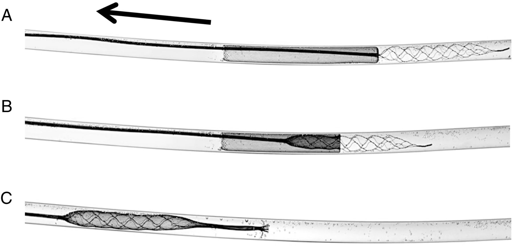
Illustrations depicting (A) initial delivery phase of the Cover device, (B) partial inverted state, and (C) fully inverted state, in which the Cover device is completely wrapped around the stent retriever. In this series of illustrations, the Trevo stent retriever (Stryker Neurovascular, Kalamazoo, Michigan, USA) is shown, demonstrating that the Cover device is compatible with a variety of stent retrievers. The Trevo stent retriever was not tested in our experiments. The arrow shows the direction of withdrawal of the stent retriever pusher wire.
Model description
The design and manufacturing of the in vitro model of the intracranial circulation used for our experiments has been previously described in detail.11 ,12 The model closely resembles the human intracranial circulation and consists of the MCA branches (M1–M4 segments), bilateral A1 anterior cerebral artery segments connected to a single anterior cerebral artery, and a single posterior communicating artery (right side), thus allowing near complete circle of Willis circulation. Fresh clots were prepared, cut into 10 mm length pieces, and introduced into the flow loop through a 9 F sheath, as previously described.12 The clots were navigated into the M1 or M2 MCA segment via the anterograde flow of the circulating fluid.
Digital subtraction angiography was performed using a biplane angiographic suite with 8 inch×8 inch flat panel detectors (Toshiba Infinix-i; Toshiba, Tochigi, Japan). A high resolution region of interest imaging detector, the microangiographic fluoroscope–charge coupled device (previously described in detail14), was utilized to precisely visualize the deployment and retrieval of the Cover device under high magnification.
Thrombectomy procedures
For this study, stent retriever thrombectomy was performed using a conventional guide catheter (control group), using a BGC (BGC group), and in combination with the Cover device (Cover group).
After placing the clot into the target vessel and performing angiography to confirm the occlusion site (ie, M1 or M2 MCA segment), a 0.021 inch microcatheter was delivered over a 0.014 inch wire to cross the clot under fluoroscopy and roadmapping guidance. Microinjection through the microcatheter was performed to document the extent of occlusion distally. The Solitaire FR stent retriever was delivered using the 0.021 inch microcatheter, deployed, left in place for 5 min, and then retrieved back into the guide catheter.
The control group consisted of thrombectomy experiments performed using a conventional guide catheter. For this purpose, we used a 6 F Cook shuttle (Cook Medical, Bloomington, Indiana, USA), which was placed into the segment corresponding to the cervical internal carotid artery (ICA).
For experiments testing the BGC approach, thrombectomy was performed with an 8 F BGC (Merci (Concentric Medical-Stryker Neurovascular) or Cello (Covidien)). The balloon was inflated prior to withdrawal of the stent retriever, and manual aspiration was applied using a 20 mL syringe.
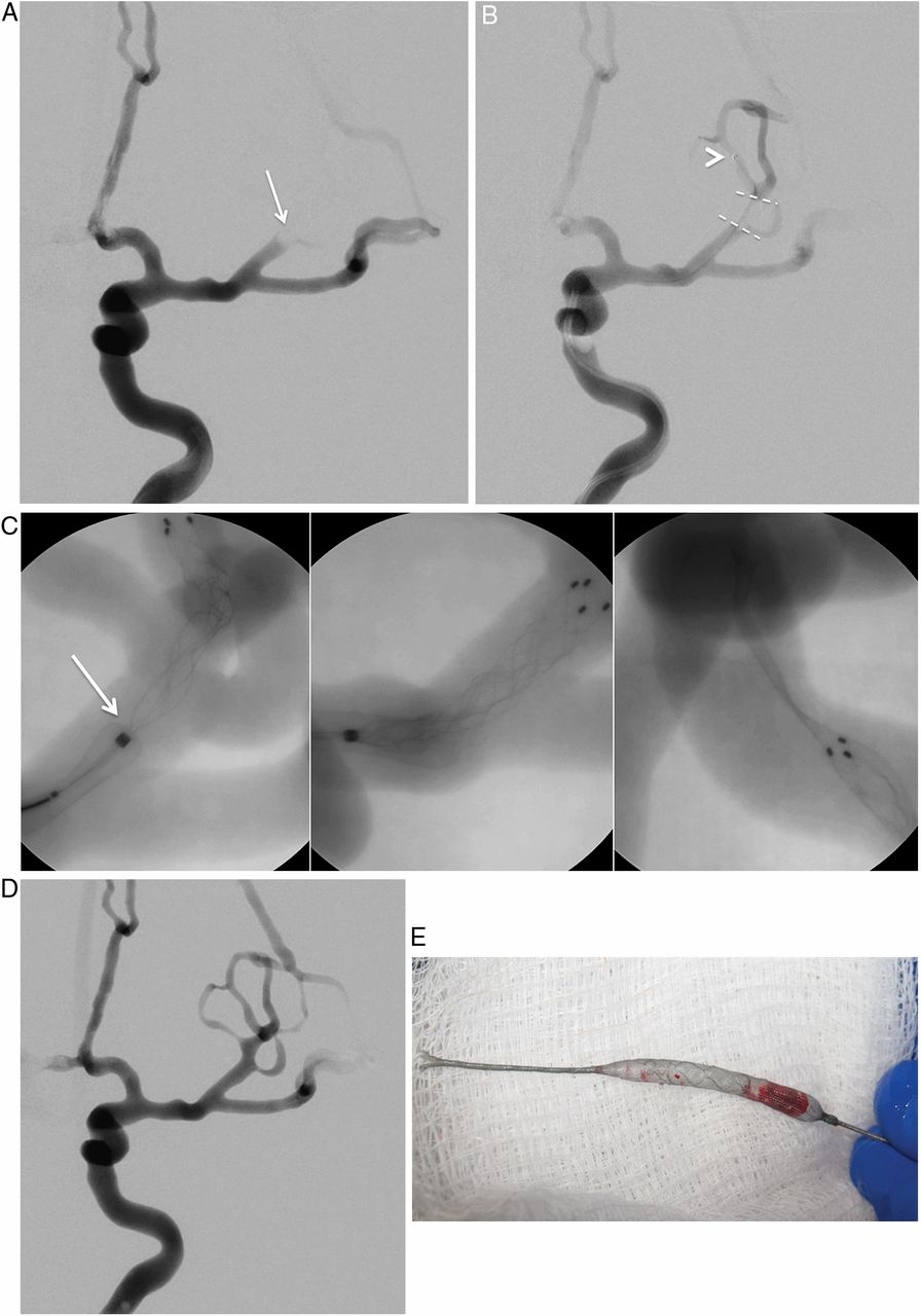
In experiments with the Cover device, we used the 6 F Cook shuttle. An intermediate catheter was utilized (0.057 inch distal access catheter, Concentric Medical-Stryker Neurovascular) to facilitate delivery of the Cover device. No aspiration was applied through the intermediate catheter or the guide catheter while thrombectomy was attempted. An example of Cover assisted thrombectomy is shown in figure 2.

Application of the Cover device (Lazarus Effect; Campbell, California, USA) during stent retriever thrombectomy. (A) Baseline angiogram, anteroposterior (AP) view, left internal carotid artery injection showing occlusion of the superior trunk of the middle cerebral artery (MCA) (arrow). (B) Simultaneous combined injection through the guide catheter and the microcatheter (arrowhead) delivered beyond the clot, demonstrating a filling defect, which indicates the location and length of the clot (broken lines). (C) Microangiographic fluoroscopy images showing gradual withdrawal of the Solitaire Flow Restoration (FR) stent retriever (Covidien, Irvine, California, USA), causing the Cover device to wrap around the Solitaire FR, preventing the clot (not seen) from fragmentation. Arrow points to the proximal end of the Solitaire FR. (D) Final angiogram following successful thrombectomy, AP view, showing robust filling of the superior trunk of the MCA, corresponding to the final recanalization grade of Thrombolysis in Cerebral Infarction 3. (E) Photograph showing the Cover device wrapped around the Solitaire FR stent retriever, helping to protect the clot from defragmentation.
A single thrombectomy attempt was allowed in each experiment. After deployment of the Solitaire FR, visual inspection and fluoroscopy were performed to confirm satisfactory placement of the stent retriever within the clot. Experiments in which Solitaire FR deployment missed the clot (deployment either proximal or distal to the clot position) were aborted and not included in the analysis. While performing thrombectomy, continuous visualization and high definition video camera recording of the clot and the stentriever were performed to capture and document the location of any clot breakdown. Primary outcomes included the degree of recanalization (based on the Thrombolysis in Cerebral Infarction (TICI) grading scale15) and the occurrence of emboli in a new territory (ie, an area in which clot was not introduced; previously unaffected territory). Successful recanalization was defined as a TICI score of 2b–3. A p value of <0.5 was considered statistically significant.
Results
A total of 51 thrombectomy experiments were conducted: 20 in the control group, 20 in the BGC group, and 11 in the Cover group. Successful delivery and inversion of the Cover device around the stent retriever was achieved in all cases. Successful recanalization (TICI 2b–3) was achieved in 50% of control cases, 45% of BGC cases, and 91% of Cover device cases (table 1).
Radiographic outcomes of thrombectomy experiments
The occurrence of successful recanalization (TICI 2b–3) was similar between the control and BGC groups (in 10 of 20 vs 9 of 20 experiments, respectively; p=1.0). TICI 2b–3 was achieved more frequently in the Cover group than in the control group (in 10 of 11 vs 10 of 20 experiments, respectively; p=0.047), or in the BGC group (in 10 of 11 vs 9 of 20 experiments; p=0.020).
Visual inspection of the model during withdrawal of the stentriever allowed us to document the anatomical location of clot loss when it occurred (table 2). Embolization of new territories occurred in five (25%) experiments from the control group and in three (15%) experiments from the BGC group. No embolization of new territories was seen with the use of the Cover device.
Location of clot loss during thrombectomy experiments
Discussion
Distal embolization occurs in up to 17% of thrombectomy cases, and embolization of new, previously unaffected, territories has been reported in 2–11% of stroke cases treated with stentriever devices.4 ,5 ,8 ,9 Such embolization phenomena are associated with the occurrence of distal infarcts, worsening of neurological recovery, added morbidity, and increased mortality.6–8 It is possible that the actual rates of embolic complications are under recognized because angiographic outcomes from many of the reported studies are reviewed by local investigators and not adjudicated by a central core laboratory, which can result in overestimation of recanalization outcome.2
Our in vitro results demonstrate that the use of the Cover device was more effective than the use of the BGC with respect to reducing the number of embolic events, thus leading to higher recanalization rates. Moreover, adjunctive use of the Cover device does not prolong the duration of the interventional procedure. The stent retriever, once unsheathed at the area of the clot, is typically left in place for several minutes to allow improved engagement of the thrombus within its struts and restoration of blood flow to previously deprived brain (creating a ‘temporary endovascular bypass’). Concurrently, the Cover device is loaded and delivered. In addition, the ‘rapid exchange’ design of the Cover device allows the operator to use it with various types of stent retrievers available on the market.
Our experiments showed that a certain amount of initial withdrawal of the stent retriever was necessary to fully invert the Cover device before the clot could be fully protected. During that initial step, clot loss can still occur. This was observed in 2 of 11 thrombectomy attempts using the Cover device (table 2), all of which occurred within the M1 MCA segment.
Our data show that the majority of clot defragmentation occurs within the proximal MCA segment or at the ICA terminus, and the BGC is only partially effective in preventing clot loss at those locations. Jahan16 used a swine vessel occlusion model and demonstrated that, contrary to our results, no clot fragmentation occurred during deployment or retrieval of the stent retriever until the clot reached the tip of the balloon catheter. One possible explanation for this discrepancy is that the swine arterial vasculature is not as tortuous and has a more uniform diameter, allowing less chance for clot loss from the stent retriever struts.
The limitations of our study include testing the efficacy of the Cover device with only one type of stent retriever (Solitaire FR) and lack of histopathological data to investigate whether the Cover device can be protective or, on the contrary, create endothelial damage during device retraction. Our findings should be further tested in an animal thrombectomy model. In clinical practice, some neurointerventionists perform stent retriever thrombectomy under continuous aspiration via an intermediate catheter. We did not test the value of combining the Cover device with continuous aspiration, which we are planning to address in our future experiments. Although the intermediate catheter was used to facilitate delivery of the Cover device, the catheter was not advanced beyond the cervical segments of the ICA, and no aspiration was applied through the catheter during the thrombectomy attempt. Therefore, it is unlikely that the use of the intermediate catheter for delivery purposes affected the outcomes of our experiments.
Conclusion
On the basis of our in vitro stroke model experiments, the Cover device is successful in achieving higher rates of successful recanalization and reducing the number of embolic events, and is more effective than the use of the conventional guide catheter or the BGC.
Acknowledgments
The authors thank Stephen Rudin PhD for guidance, Liza Pope BS for clot preparation, Paul H Dressel BFA for illustration preparation, and Debra J Zimmer for editorial assistance.
References
Supplementary materials
Supplementary Data
This web only file has been produced by the BMJ Publishing Group from an electronic file supplied by the author(s) and has not been edited for content.
Files in this Data Supplement:
- Data supplement 1 - Online video 1
- Data supplement 2 - Online video 2
- Data supplement 3 - Online video 3
Footnotes
Contributors Conception and design: MM, JM, and AHS. Data acquisition: all authors. Data analysis and interpretation: MM and AHS. Drafting the manuscript: MM. Critically revising the manuscript and final approval of the manuscript: all authors.
Funding Devices were provided by Lazarus Effect, Covidien, and Stryker Neurovascular. All data collection, analysis, and interpretation were performed by the authors, independent of company input or interpretation.
Competing interests CNI: grants from the National Institutes of Health (2RO1EB002873; for partial support of student and faculty) and Toshiba (equipment) in conjunction with the present study. JM: consultant—Lazarus Effect, Reverse, Pulsar, Edge Therapeutics, and Medina; investor—Blockade Medical and Medina; advisory board—Codman Neurovascular Mokin; educational grant—Toshiba. AHS: research grants—National Institutes of Health (co-investigator: NINDS 1RO1NS064592-01A1; NIBIB 5RO1EB002873-07), University at Buffalo (Research Development Award), none related to the present study; financial interests—Hotspur, Intratech Medical, StimSox, Valor Medical, Blockade Medical, and Lazarus Effect; consultant—Codman and Shurtleff Inc, Concentric Medical, ev3/Covidien Vascular Therapies, GuidePoint Global Consulting, Penumbra, Stryker Pulsar Vascular, MicroVention, Lazarus Effect, and Blockade Medical; speakers’ bureau—Codman and Shurtleff Inc; National Steering Committee member—Penumbra Inc's 3D Separator Trial, Covidien's SWIFT PRIME trial, and MicroVention's FRED trial; advisory boards—Codman and Shurtleff and Covidien Neurovascular; honoraria—Abbott Vascular, Codman and Shurtleff, and Penumbra Inc.
Ethics approval Collection of clinical data for the model design was approved by the University at Buffalo institutional review board (project 567513-1).
Provenance and peer review Not commissioned; externally peer reviewed.
Data sharing statement Additional data may be available on a per request basis directed to the corresponding author.