Article Text

Abstract
Preclinical studies are important in helping practitioners and device developers improve techniques and tools for endovascular treatment of intracranial aneurysms. Thus an understanding of the major animal models used in such studies is important. The New Zealand rabbit elastase induced arterial aneurysm of the common carotid artery is one of the most commonly used models in testing the safety and efficacy of new endovascular devices. In this review we discuss: (1) the various techniques used to create the aneurysm, (2) complications of aneurysm creation, (3) natural history of the arterial aneurysm, (4) histopathologic and hemodynamic features of the aneurysm, (5) devices tested using this model, and (6) weaknesses of the model. We demonstrate how preclinical studies using this model are applied in the treatment of intracranial aneurysms in humans. The model has similar hemodynamic, morphological, and histologic characteristics to human aneurysms, and demonstrates similar healing responses to coiling as human aneurysms. Despite these strengths, however, the model does have many weaknesses, including the fact that the model does not emulate the complex inflammatory processes affecting growing and ruptured aneurysms. Furthermore, the extracranial location of the model affects its ability to be used in preclinical safety assessments of new devices. We conclude that the rabbit elastase model has characteristics that make it a simple and effective model for preclinical studies on the endovascular treatment of intracranial aneurysms, but further work is needed to develop aneurysm models that simulate the histopathologic and morphologic characteristics of growing and ruptured aneurysms.
- Aneurysm
- Device
- Intervention
- Subarachnoid
- Technique
Statistics from Altmetric.com
Introduction
Intracranial aneurysms are increasingly treated with endovascular therapies. A number of devices have been developed to improve the safety and efficacy of endovascular intracranial embolization, including coils, modified coils, stents, glues, intraluminal flow diverters, and intra-saccular flow diverters. A number of preclinical trials based on animal studies have been performed testing the safety and efficacy of these devices prior to use in humans. Such trials have provided valuable information regarding potential pitfalls of newer devices in the treatment of aneurysms as well as information regarding the mechanism of action of newer devices.
Because of the importance of preclinical studies in helping practitioners and device developers improve the tools used in endovascular treatment of intracranial aneurysms, an understanding of the major animal models used in such studies is important. Currently, the New Zealand rabbit elastase induced arterial aneurysm of the common carotid artery is one of the most commonly used models in testing the safety and efficacy of new endovascular devices. In this review we discuss: (1) the various techniques used to create the aneurysm, (2) complications of aneurysm creation, (3) natural history of the arterial aneurysm, (4) histopathologic and hemodynamic features of the aneurysm, and (5) devices that have been tested using this model. Our primary focus is to demonstrate how preclinical studies using this model have been applied in the treatment of intracranial aneurysms in human patients.
Aneurysm creation
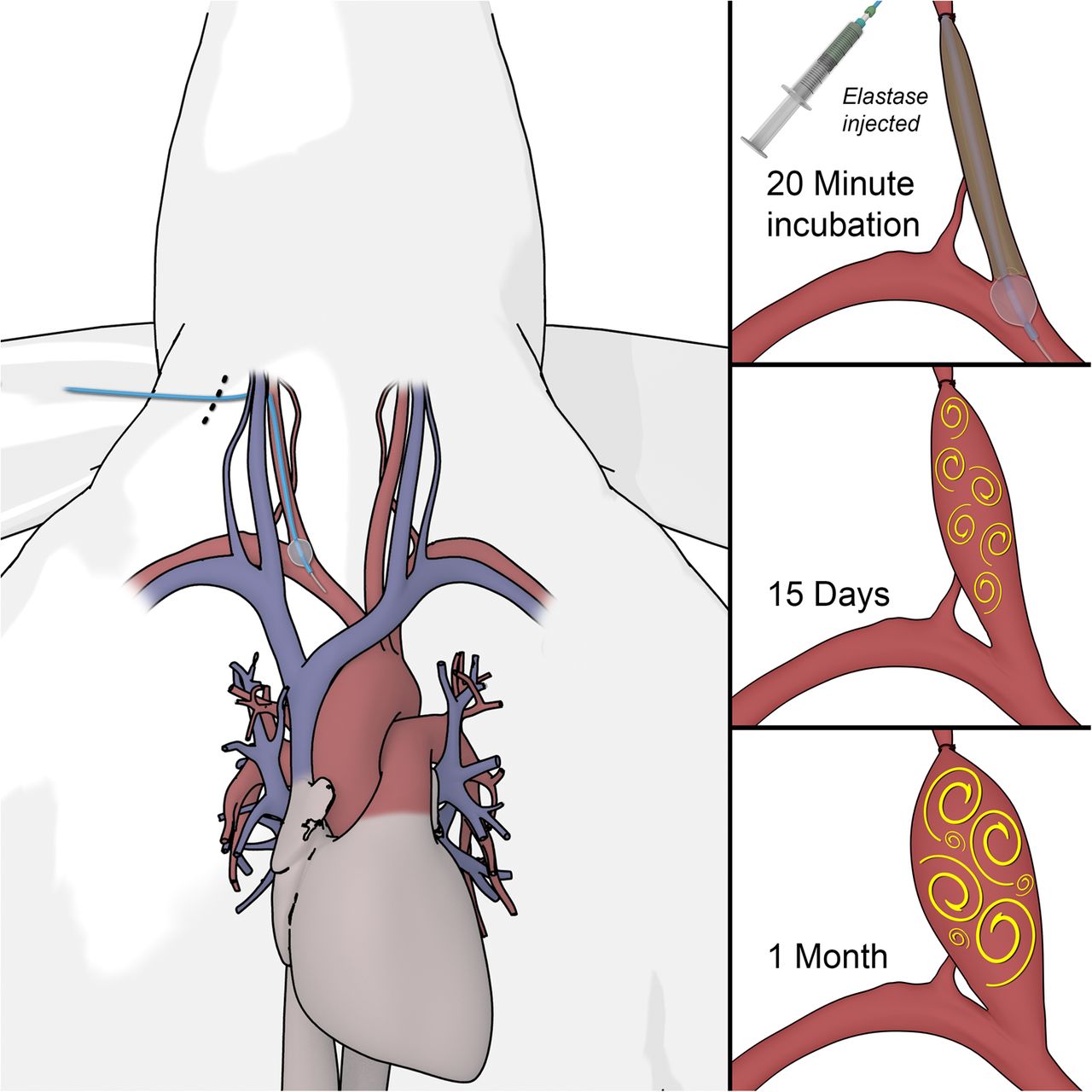
The New Zealand white rabbit elastase induced arterial aneurysm is created through a combination of open and endovascular techniques. The classic techniques used to create the arterial aneurysm have been previously described.1 Following administration of anesthesia, the right carotid sheath and the right common carotid artery (RCCA) are surgically exposed. A 1–2 mm beveled arteriotomy is created in the RCCA and a 5 F vascular sheath is inserted retrograde into the mid portion of the RCCA. The origin of the RCCA is identified with a small injection of iodinated contrast. Then, using fluoroscopic guidance, a 3 F balloon catheter is advanced through the sheath to the RCCA origin and inflated with iodinated contrast material. Following confirmation of occlusion of the RCCA origin with another small injection of contrast material, 100 U of elastase are incubated within the lumen of the proximal RCCA for 20 min. After this, the balloon is deflated and the catheter system is removed. The vessel is then ligated in its midportion and the skin is closed. Two weeks following the treatment, the saccular aneurysm is formed. Figure 1 demonstrates the steps used in the creation of the aneurysm model.

Creation of the rabbit elastase aneurysm model. This cartoon illustrates the operative process used to create the rabbit elastase aneurysm model. Following exposure of the right carotid sheath and creation of a small arteriotomy, a sheath is introduced into the right common carotid artery. A balloon is introduced through the sheath and placed at the origin of the right common carotid artery and inflated. The right common carotid artery is ligated. Following this, elastase is introduced into the artery and allowed to incubate for 20 min. The sheath and balloon are then withdrawn and the superior aspect of the aneurysm is cinched off. The aneurysm continues to grow and then generally stabilizes at 1 month due to a combination of elastin digestion and hemodynamic forces.
Modifications to the model
A number of investigators have proposed modifications in creating the elastase arterial aneurysm model. The occurrence of collateral vessel branches between the RCCA and arteries supplying the trachea could lead to washout of elastase to the trachea, resulting in hemorrhagic necrosis. Krings et al2 proposed the following modifications to the abovementioned model: (1) injection of elastase by the region of the aneurysm neck rather than the mid RCCA, (2) rule out elastase leakage to arteries supplying the trachea using angiography, and (3) lowering the dose of elastase to 20 U (instead of 100 U).
Hoh et al3 demonstrated that by applying an aneurysm clip to the RCCA origin rather than a balloon catheter, elastase could be kept within the RCCA during incubation with consistent results.
Ding et al proposed a modified aneurysm creation technique in which the entirety of the RCCA, including its origin at its junction with the subclavian and brachiocephalic arteries, was completely exposed and isolated. This allowed for assessment of the presence of branch vessels which could be ligated to avoid complications from flow of elastase to the trachea and allow for increased concentrations of elastase in the aneurysm sac.4
Other modifications, such as adjusting the position of the ligation, adjusting the position of the inflated balloon, injuring the aneurysm neck, and using the left common carotid artery instead of the RCCA, have been proposed as techniques to alter the volume, neck size, and configuration of the aneurysm, respectively.5–8
Complications of aneurysm creation
Creation of aneurysms using the rabbit elastase model approach is not without complications. One of the most commonly reported complications is tracheal necrosis following application of elastase to the RCCA. This complication occurs due to the origin of the tracheoesophageal artery arising from the RCCA, leading to elastase deposition in the trachea.9 ,10 One series of over 700 procedures in rabbits demonstrated that the aneurysm creation process is associated with an 8.4% risk of mortality related to factors such as anesthesia, failure to thrive, self mutilation (by biting at sutures), and stroke.11 Other complications that have been reported are creation of thoracic aortic aneurysms, laryngeal paralysis due to damage of the recurrent laryngeal nerve, tracheitis–laryngitis due to intubation, pneumonia, and vessel thrombosis.12–14
Aneurysm natural history, morphology, and histology
Histopathologic studies of aneurysms created using the rabbit elastase model demonstrate many similarities to aneurysms in humans. In the elastase model, the elastic lamina within the aneurysm wall is markedly attenuated secondary to elastase deposition. This feature simulates the characteristics of human aneurysms where the internal elastic lamina is substantially diminished. The aneurysm wall of the rabbit elastase model has a high concentration of smooth muscle cells, similar to human aneurysms. The endothelium of the aneurysm remains intact, also simulating features found in human aneurysms.1 ,3 ,15 Other models which use a venous pouch fail to simulate the histological characteristics of human aneurysms. The use of sutures in the venous pouch model at the aneurysm neck and dome results in an extensive healing and fibrotic response about the sutures, thus limiting the applicability of these types of models in assessing post-embolization healing.16–18
Another advantage of the rabbit elastase model is its favorable natural history. When left untreated, these aneurysms remain patent for long periods of time.19–21 One study followed 20 elastase induced saccular aneurysm models for 24 months and found no cases of spontaneous thrombosis and no changes in aneurysm geometry over this time period.22 This long term stability is important as it simulates the characteristics of most unruptured intracranial aneurysms in humans. One disadvantage of this stability is that it is difficult to simulate the characteristics of growing and ruptured aneurysms.
The long term patency of saccular RCCA aneurysms in the rabbit elastase model differs substantially from the spontaneous healing seen in the rabbit elastase abdominal aortic aneurysm model. This is thought to be due to differences in the hemodynamic stresses in the side wall type abdominal aortic aneurysm and bifurcation type RCCA aneurysm. The side wall abdominal aortic aneurysm model is exposed to lower shear stresses than the RCCA aneurysm, thus resulting in a higher propensity to spontaneously thrombose and heal.23 ,24 The swine model of intracranial aneurysms, another popular model, also has short term aneurysm patency.16 ,25 Spontaneous healing of these aneurysms is thought to be due to the robust neointimal proliferation response seen in pigs following endothelial injury. Such a robust reaction is not appreciated in rabbit models.16 ,26
Another important factor in designing intracranial aneurysm models is ensuring that the preclinical model has similar morphological characteristics to human aneurysms. The average diameter and height of the rabbit elastase aneurysm model is 4.5 and 7.5 mm, respectively.1 The distribution of sizes seen in creation of this model are similar to the distribution of sizes seen in human aneurysms.27 In addition, the diameter of the parent artery is similar to that of intracranial arteries (approximately 4 mm).27 Modifications can also be made to increase the size of the aneurysm. Ding et al28 demonstrated that by creating an RCCA right jugular arteriovenous fistula, larger aneurysms, measuring approximately 15 mm in maximum dimension, can be formed. As mentioned above, changing the position of the ligation can be used to change aneurysm size; and neck size can be adjusted by changing the position of the balloon at the RCCA origin.6 ,7 In general, the morphology and size of the aneurysm can be designed to model the type of aneurysm that is desired with consistent and predictable results.4 ,5 ,29 ,30
ANEURYSM HEMODYNAMICS
Because hemodynamic factors play a large role in aneurysm formation, growth, and recanalization following endovascular treatment, it is important for a preclinical model to simulate the hemodynamic characteristics of human models. In a study of 51 elastase induced RCCA aneurysms, Zeng et al found that the range in values of pressure, wall shear stress, and oscillatory shear index were all within the ranges seen in human aneurysms. The number of recirculation regions seen within the aneurysms was also similar to that seen in human aneurysms.31 ,32
The hemodynamic similarities between the rabbit model and humans is due to similarities in size and vessel curvature of the rabbit aneurysms. Other animal models, such as the venous pouch side wall model found in canines, fail to simulate the flow seen in human aneurysms, as human aneurysms are rarely present along the side walls of long straight vessels. Because of the curvature of the parent vessel of the RCCA aneurysm in rabbits, there is substantial inertia driven flow in the aneurysm. This contrasts with the shear driven flow seen in side wall aneurysms.33 Because most human aneurysms are located at bifurcation points or vessel curvatures, these are subject to high rates of inertia driven flow, contributing to aneurysm growth and recanalization.
Device testing
A number of preclinical studies have been performed using the rabbit elastase model. Such studies have been performed to assess the safety and efficacy of aneurysm treatment as well as the healing response following aneurysm embolization. In general, studies examining the efficacy of these devices include a combination of imaging and histopathologic studies to determine aneurysm occlusion rates and mechanism of healing. A variety of imaging modalities have been tested to evaluate the angiographic occlusion rates, including conventional angiography, intravenous digital subtraction angiography, CT angiography, phase contrast MR angiography (MRA), time of flight MRA, and contrast enhanced MRA.34–39 All of these techniques have been shown to be effective, but conventional angiography remains the gold standard for evaluation of aneurysm patency following coil embolization, stenting, and flow diverter treatment.35 ,40 For histologic characterization, an ordinal histologic scale combines data on recanalization/coil compaction, neck healing, and dome healing using both gross and microscopic images.41
Bare platinum coils have been the most extensively tested devices in the rabbit elastase model. Histopathologic and angiographic studies have demonstrated that bare platinum coils are highly effective in obliterating the aneurysm in both humans and rabbits.42 Interestingly, the healing response of the rabbit model closely resembles that found in humans. Rabbit studies of bare platinum coil embolization demonstrate early thrombus formation within the aneurysm sac within days of embolization. This is followed by infiltration of the aneurysm dome by fibroblasts and inflammatory cells up to 4 weeks following embolization. Fibroblasts deposit a loosely packed extracellular matrix of fibrin and collagen in the aneurysm dome.42 The tissue response in the aneurysm dome stops at approximately 4 weeks post-embolization due to apoptosis of the fibroblasts and inflammatory cells. After 4 weeks, the aneurysm dome is filled with acellular loose connective tissue, similar to what is seen in humans.43 Long term studies of the aneurysm neck of the rabbit aneurysm demonstrate a thin hypocellular layer of tissue with a sparse number of endothelial cells.43 This is also similar to the response seen in humans and contrasts with the responses seen in pigs where a thick layer of neointima forms.25
Modified coils, such as the Matrix and HydroCoil, have also been tested in the rabbit elastase model and have demonstrated similar angiographic results to humans.44 One series embolized 33 rabbit aneurysms with either Matrix coils, HydroCoils, or bare platinum coils and found that HydroCoils were associated with lower rates of coil compaction, higher rates of angiographic occlusion, and improved long term occlusion rates compared with Matrix and bare platinum coils.45 In addition, Matrix coils were not associated with improved aneurysm occlusion and recanalization rates compared with bare platinum. These results are similar to those seen in the HELPS trial which demonstrated higher rates of aneurysm recanalization in bare platinum compared with HydroCoils46 and the Matrix and Platinum Science (MAPS) trial which demonstrated similar aneurysm occlusion and recanalization rates between Matrix and bare platinum trials.47 Cruise et al48 tested HydroCoils in rabbit elastase aneurysms and found high rates of long term occlusion and a twofold increase in volumetric filling of the aneurysm sac, similar to findings in human studies. These findings have been corroborated by other preclinical and clinical studies.44 ,49
This model has also been used in preclinical studies of aneurysm stenting and stent assisted coiling. In a study of 10 rabbits, Hans et al50 applied porous stents with and without detachable coils and found high rates of aneurysm occlusion using both techniques. Krings et al51 tested stent assisted coiling and found that porous stents resulted in higher rates of aneurysm recanalization than covered stents. In addition, Krings et al52 demonstrated comparable rates of in-stent stenosis with those seen in human aneurysms treated with stent assisted coiling. As mentioned above, the curvature of the parent vessel of the rabbit elastase aneurysm allows for accurate characterization of intra-aneurysm flow dynamics both before and after treatment. The hemodynamics of the stent coiled and stented rabbit aneurysm closely resemble those seen in models of human aneurysms.33 This is likely the reason behind the similar rates of aneurysm occlusion seen in humans and rabbit aneurysms treated with stenting and stent coiling.
Many animal models, such as the canine side wall aneurysm model and the rabbit model, were instrumental in the development and testing of flow diverters.53–55 The rabbit model was extensively used in the development and testing of flow diverter devices created with modern braiding technology. Because flow diverters reconstruct the parent vessel, they can be used in a wide variety of aneurysm sizes and morphologies. Initial testing of flow diverter devices were focused on determining the optimum porosities and pore densities of flow diverters. Sadasivan et al40 found that a medium porosity performed better than high and low porosity devices in limiting intra-aneurysmal flow. In a later study, Sadasivan et al56 found that increasing pore density was the most critical factor in modulating the efficacy of flow diverters. Such work paved the way for the Pipeline Embolization Device (PED), a braided bimetallic endoluminal implant. Initial studies in the rabbit model found complete and near complete occlusion rates of 53% and 35%, respectively, similar to rates seen in human studies.57 One of the concerns regarding this device was the patency of branch vessels covered by the struts. Studies in the rabbit model have demonstrated high rates (up to 100%) of branch artery patency, similar to findings in clinical studies.58 In addition, rates of parent artery compromise from neointimal hyperplasia are low.57 ,59 Mechanisms of healing following flow diverter treatment have also been studied using rabbits. Initial studies suggest that endothelial progenitor cell migration to the flow diverters is essential to neointima formation and re-endothelialization of the aneurysm neck following treatment.60
Intra-aneurysmal flow diverters are the newest devices to make the jump from bench to bedside using the rabbit elastase model. In a study using 24 elastase induced aneurysms in rabbits, Ding et al studied the efficacy of the Woven EndoBridge occlusion device. These authors found high rates of complete and near complete occlusion. Aneurysms progressively occluded with increasing follow-up. Similar to coil embolization, histopathologic studies demonstrated loose connective tissue and organized thrombus in the aneurysm dome.61 These results have been corroborated in other animal studies as well as in human studies.39 Current small case series have demonstrated that the Woven EndoBridge device has a lot of promise for the treatment of wide neck bifurcation aneurysms.62
A number of preclinical devices have been tested in the rabbit model that have yet to be tested in humans. Due to the poor healing and fibrotic response seen in both rabbit and human saccular aneurysms following coil embolization, Kallmes et al tested a collagen based coil in the rabbit model and found that collagen based coils had a marked cellular response and dense matrix deposition with high rates of progressive occlusion. This contrasts with the low rates of progressive thrombosis and loose matrix deposition seen with bare platinum coils. Other coil modifications that have been tested include polyvinyl alcohol coils covered in basic fibroblast growth factor. These coils have been found to stimulate aneurysm healing in the aneurysm dome and neck compared with bare platinum coils.63 The Luna aneurysm embolization system is another intra-saccular flow diverter similar to the Woven EndoBridge system that has been tested in rabbit models and shown to be effective.64 But to date, there are no reports of the use of this device in humans. Polyurethane covered stent grafts have also been tested in the rabbit elastase model with high rates of aneurysm occlusion and low rates of in-stent stenosis but these have not been used in humans. Other devices that have been shown to be effective in rabbits, but not yet tested in humans, include asymmetric vascular stents, nanofiber covered stents, variable porosity flow diverters, biodegradable flow diverters, and magnetic microparticles.65–71
Other advantages
The rabbit elastase model has a number of other advantages. For example, the saccular aneurysm model can be used as a training tool for neurointerventionalists. Because of the ability to ‘design’ both easily treatable and difficult to treat aneurysms, this model can be used to train practitioners in the treatment of a wide variety of aneurysm morphologies.72 Studies using the rabbit elastase model have demonstrated that using the rabbit model helps in the development of proficiency with endovascular techniques and device deployment among neurointerventional trainees.73 It is important to note however that access systems are typically limited to 7 F systems and therefore commonly used setups with long sheaths are not possible. In addition, training multiple physicians on a single model is complicated by the limited amount of contrast and fluids that can be delivered to the animal, high rates of anesthesia related mortality, and the fact that each femoral artery can only be accessed once due to the fact that the femoral artery is generally ligated after each intervention.
Pitfalls
Histologic pitfalls
As with any disease model, the rabbit elastase model is not the perfect representative of the intracranial aneurysm disease process. As mentioned above, the rabbit elastase model serves as a model only for non-growing, stable, unruptured aneurysms. Thus histologic and molecular studies of this model cannot be applied to growing or ruptured aneurysms as these aneurysms are more biologically complex with high concentrations of inflammatory cells, something that is not seen in the wall of the untreated rabbit elastase model.74 ,75 For example, in a study of 24 unruptured and 42 ruptured aneurysms, Frosen et al75 found that ruptured aneurysms were more likely to have macrophage and T cell infiltration, smooth muscle cell proliferation, apoptosis, luminal thrombosis, and de-endothelialization. The structure of the aneurysm wall in the rabbit elastase model is generally homogeneous and does not include regions of atherosclerotic thickening or regions of extremely thin walls, such as those seen in both ruptured and unruptured human aneurysms.75 Prior studies in humans have demonstrated that the aneurysm wall of human aneurysms is in no way homogeneous in nature, as aneurysms are known to develop atherosclerosis and have areas of mural tearing.75 ,76 Because these aneurysms are essentially a stump of the internal carotid artery, all created with generally similar techniques, there is very little variation in the structure and biology of aneurysms between individual rabbits. However, in humans, we see a wide variability in the size, structure, vulnerability, and geometry of unruptured aneurysms.75 ,77
Device safety and efficacy
While the rabbit model has been extensively used in the development and testing of various endovascular devices, it is by no means a perfect model for assessment of device efficacy and safety. As described above, the rabbit elastase model does not emulate the biologic conditions of unstable growing or ruptured aneurysms. As such, it is possible that devices or treatments that may help in improving outcomes of ruptured or growing aneurysms may not prove of benefit in the thick walled, stable, unruptured aneurysms of the rabbit elastase model. Conversely, it is also possible that devices that are deemed effective in treating the unruptured aneurysms of the rabbit model may not be effective in treating ruptured or unstable growing aneurysms. In addition, in some cases, device efficacy studies can yield different results in the rabbit model compared with human studies. For example, in a study of large neck bifurcation aneurysms treated with the Woven EndoBridge device, Cognard et al78 found that more than 70% of aneurysms had worsening of aneurysm filling on follow-up angiography compared with a rate of just 13% in preclinical rabbit studies.61 Lastly, the extracranial location of these aneurysms adjacent to the thoracic cavity exposes devices to stresses due to the respiratory and cardiac cycles that they would not be exposed to in the brain, which could impact the translatability of efficacy results from preclinical to clinical studies.
Aside from concerns regarding device efficacy, device safety is difficult to assess due to the extracranial location of the aneurysms and the thick aneurysm wall. Because the aneurysm wall is thicker than that seen in most human histologic studies, certain complications, such as intraoperative aneurysm wall perforation, are exceedingly rare in the treatment of rabbit aneurysms. Furthermore, due to the extracranial location, it is impossible to assess the potential intracranial complications that could potentially result from placement of different coils or flow diverters. For example, there have been several reports of hydrocephalus possibly due to a chemical meningitis among patients treated with HydroCoils.79 However, preclinical studies on treatment of extracranially located aneurysms in the rabbit model could not explain or predict this complication. Likewise, with the PED, post treatment intraparenchymal and subarachnoid hemorrhages occurred in 2.4% and 0.6% of patients, respectively, in the IntrePED registry. The exact cause of these complications is unknown but, again, preclinical studies in the rabbit model could not have predicted this complication given the extracranial location.80 Likewise, in PED patients, there have been higher rates of perforator infarcts and thromboembolic complications than would be expected from preclinical rabbit studies.80 ,81
Conclusions
In conclusion, the rabbit elastase model has characteristics that make it a simple and effective model for preclinical studies on the endovascular treatment of intracranial aneurysms. The model has a similar histologic appearance to human aneurysms and demonstrates a similar healing response to endovascular coiling as human aneurysms. The morphology and hemodynamic characteristics of these aneurysms are similar to human intracranial aneurysms. In addition, many preclinical studies of new devices, such as flow diverters and modified coils, in rabbit aneurysms have yielded similar results in the treatment of human aneurysms. Despite these strengths however, the model does have many weaknesses, including the fact that the model does not emulate the complex inflammatory processes affecting growing and ruptured aneurysms. Furthermore, the extracranial location of the model affects its ability to be used in preclinical safety assessments of new devices. Further work is needed to develop aneurysm models that simulate the histopathologic and morphologic characteristics of growing and ruptured aneurysms.
References
Footnotes
Contributors YHD, WB, RK, and DFK participated in drafting the article and revising it critically for important intellectual content. The authors made substantial contributions to conception and design, acquisition of the data, and analysis and interpretation of the data. All authors provided final approval of the version to be published.
Funding This study was funded in part by NIH grant NS076491.
Competing interests WB: grants/grants pending from Brain Aneurysm Foundation. DFK: consultancy for ev3,* Medtronic,* and Codman*; grants/grants pending from ev3,* MicroVention,* Sequent,* and Codman*; payment for lectures (including service on speakers bureaus) from MicroVention*; royalties from UVA Patent Foundation*; payment for development of educational presentations from ev3*; and travel/accommodations/meeting expenses unrelated to the activities listed from MicroVention.* *Money paid to the institution.
Provenance and peer review Not commissioned; externally peer reviewed.