Article Text

Abstract
The follow-up and indications for retreatment of intracranial aneurysms treated endovascularly are still a matter of debate. We report the case of a patient with a ruptured aneurysm who was treated twice with coils and regularly followed up with MRI/MR angiography which showed a neck remnant that finally rebled.
- Aneurysm
Statistics from Altmetric.com
Background
This case report raises several questions regarding the treatment and follow-up of intracranial aneurysms treated by an endovascular approach, including when and how to retreat recanalized aneurysms, how neck remnants evolve, and the best imaging follow-up for neck remnants.
Case presentation
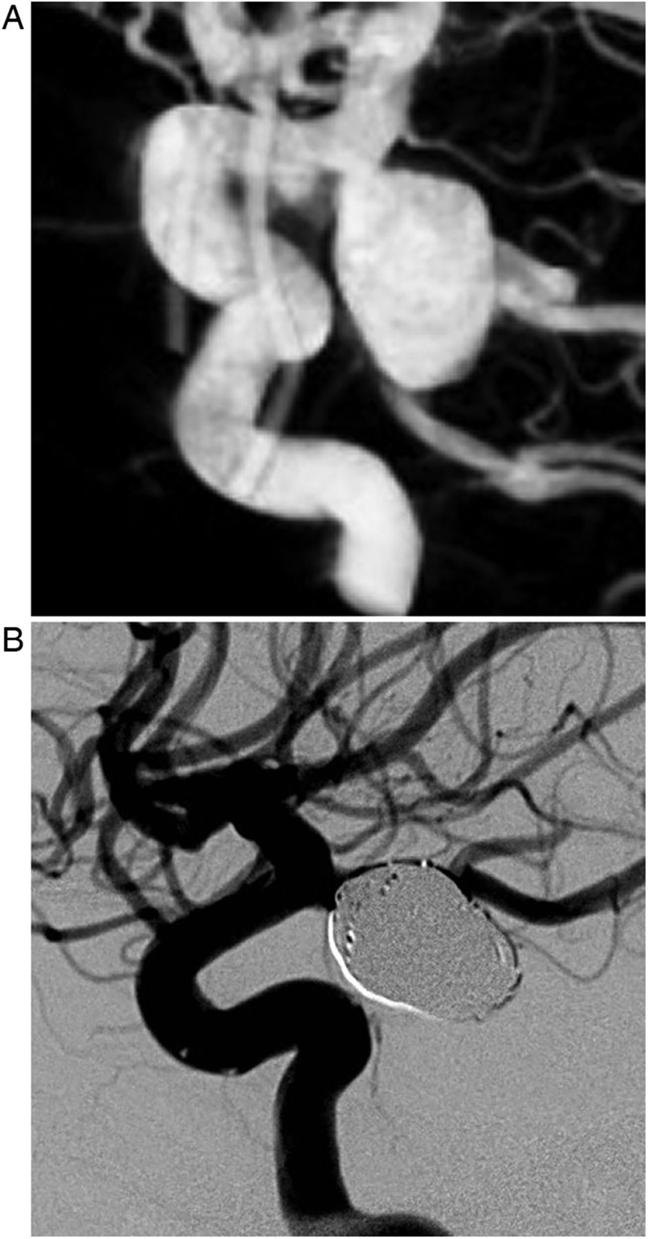
In October 2006 a patient in the fifth decade presented with subarachnoid hemorrhage due to rupture of a right non-thrombosed internal carotid artery aneurysm measuring 20×12 mm (figure 1A). The patient was treated with coils and immediate post-procedural digital subtraction angiography (DSA) showed complete aneurysm occlusion (figure 1B). MRI with MR angiography (MRA) performed 4 months after treatment showed stable total aneurysm occlusion. Twelve months after the initial treatment, MRI/MRA showed aneurysm recanalization (12×8 mm) confirmed by DSA (figure 2A) which was retreated with coils 15 months after initial treatment with complete aneurysm occlusion following the procedure (figure 2B). MRI/MRA performed 3 months after retreatment showed complete aneurysm occlusion. One year after retreatment, MRI/MRA and DSA showed a neck remnant that was also stable at MRI/MRA performed 2 and 3 years after retreatment (figure 3A, B). The next MRI/MRA was scheduled for 2015 (9 years after the initial treatment and 7 years after retreatment). In September 2014 the patient presented with a sudden coma due to a massive subarachnoid and intraventricular hemorrhage (WFNS grade 5, Fisher grade 4) (figure 4A). DSA showed a large aneurysm recanalization (14×7 mm, figure 4B) which was treated with coiling, but the patient died a few days later from intracranial hypertension.

Right internal carotid artery (ICA) aneurysm before and after the first treatment. (A) Three-dimensional conventional angiography before treatment shows a right ICA aneurysm measuring 20×12 mm. (B) Lateral view of conventional angiogram after treatment shows complete occlusion of the aneurysm (Montreal grade A).
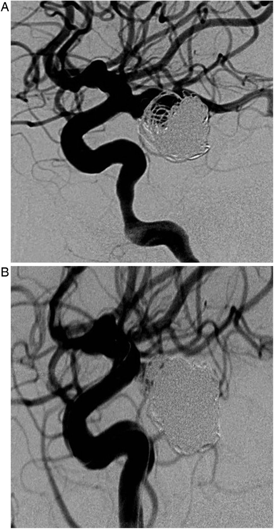
Aneurysm recanalization of the right internal carotid artery before and after the second treatment. (A) Digital subtraction angiography (DSA) lateral projection before the second treatment shows a neck remnant of the previously coiled aneurysm (Montreal grade B). (B) DSA lateral projection after the second treatment shows complete occlusion of the coiled aneurysm.

Stable neck remnant of the right internal carotid artery (ICA). Three-dimensional time-of-flight MR angiography maximal intensity projection performed (A) 2 years and (B) 3 years after retreatment shows a stable neck remnant of the right ICA.

Ruptured aneurysm recanalization of the right internal carotid artery with subarachnoid and intraventricular hemorrhage. (A) Axial maximal projection reconstruction CT shows a massive subarachnoid and intraventricular hemorrhage due to aneurysm rupture. (B) Digital subtraction angiography lateral projection shows a large ruptured aneurysm recanalization of the right internal carotid artery.
Discussion
Endovascular treatment of ruptured intracranial aneurysms is increasingly performed with satisfactory clinical and angiographic results.1 Nevertheless, aneurysm or neck reopening occurs in approximately 20% of patients, necessitating retreatment in about half of them.2 There is no consensus or recommendations regarding retreatment after aneurysm coiling.3 Indications for retreatment have to take into account the risks of retreatment and the risk of bleeding/rebleeding. Recent long-term analysis of the International Subarachnoid Aneurysm Trial (ISAT) shows that the risk of rebleeding of coiled aneurysms is low.4 The Cerebral Aneurysm Rerupture After Treatment (CARAT) study showed that the risk of early rebleeding after treatment of ruptured aneurysms was strongly related to aneurysm occlusion status.5 In their recent literature review, Tsurumi et al6 confirmed that delayed aneurysm rupture after coiling occurs in the majority of cases when there is an aneurysm remnant, but is also possible in cases of neck remnant. The risk is also higher in patients with large and giant aneurysms, as in our case.
Evolving remnants and aneurysm remnants are usually retreated because they are considered to have a relatively high risk of rerupture.4 In our case, the remnant after the second treatment was classified as a neck remnant and was not evolving. However, it should be noted that the differentiation between neck and aneurysm remnants is not always easy, as illustrated by the moderate interobserver reproducibility of the Montreal scale. To make a decision regarding retreatment, other factors must be taken into account such as age and risk factors (eg, smoking and elevated blood pressure). The modalities of retreatment are also relatively unclear. Coiling is probably the first choice for retreatment, but adjunctive devices are also useful for avoiding further growth of the aneurysm (eg, singular flow diverters).7 Clipping is also considered a viable option. In our case, initial retreatment would now probably consist of coiling with a flow diverter, but this option was not available in 2007.
This case also illustrates the fact that aneurysm growth is not a regular process. Until recently, aneurysms and remnants were considered to have a relatively linear growth. However, experience shows that aneurysms and remnants can remain stable for a long time and then have a rapid or sudden growth. Statistical models have also confirmed that the aneurysm growth process does not have a constant time-independent rate but is irregular and discontinuous, which results in periods with and without aneurysm growth and with high and low risks of rupture.8 This phenomenon was perfectly illustrated in our case with a long-term stable neck remnant followed by the rupture of a probably rapidly growing aneurysm. However, the voluminous aneurysm pouch observed at the time of rebleeding was probably not exclusively the result of aneurysm regrowth, but also partially a pseudoaneurysm created by the rupture itself.
The unpredictable evolution of aneurysms makes it difficult to plan effective follow-up strategies. Because of the low rate of late rebleeding, it is debatable whether long-term follow-up imaging should be performed. In addition, there is no consensus regarding which modality of imaging follow-up should be used. DSA is the gold standard for follow-up but has some disadvantages including rare neurological complications and radiation exposure. MRA (three-dimensional time-of-flight) is an appropriate technique for the follow-up of coiled aneurysms with 3 T, but has some limitations for aneurysms treated with intravascular devices.9 ,10 CT and CT angiography have not been well evaluated for this purpose. Whatever the technique used, it is noteworthy that it gives mostly morphological information including whether the aneurysm is completely occluded and whether there is a neck or aneurysm remnant. There is actually no imaging tool that evaluates the scarring process at the level of the neck, and further work is needed in this field. Regarding the timing, early imaging follow-up is usually performed 3–6 months after treatment, with further follow-up at 12 or 18 months. Later follow-up varies from one center to another and must be adapted to the aneurysm occlusion status at 12/18 months. The case presented here shows that follow-up does not afford complete protection against rebleeding.
Key messages
Late aneurysm rerupture is rare, especially in cases with a stable neck remnant.
Large studies should be performed to evaluate the risk factors for aneurysm recanalization, the relation between recanalization and rerupture, and the potential value of imaging follow-up in terms of reduction of the morbidity/mortality rates.
Further studies have to be performed to assess the potential value of imaging follow-up in terms of reduction of the morbidity/mortality rates.
Footnotes
Republished with permission from BMJ Case Reports Published 16 April 2015; doi:10.1136/bcr-2014-011601
Contributors AB and LP collected and analyzed the data and wrote the manuscript.
Competing interests None.
Patient consent Obtained.
Ethics approval Ethical approval was obtained from Reims IRB.
Provenance and peer review Not commissioned; externally peer reviewed.