Article Text

Abstract
Introduction The advancement of technology has allowed the development of new catheters that may provide safe intracranial navigation.
Objective To report our first experience with the direct aspiration first pass technique in small arteries as the primary method for recanalization with the Penumbra 3MAX cerebral reperfusion catheter.
Methods A retrospective case series analysis study of patients with acute ischemic stroke endovascularly treated with the direct aspiration technique using the 3MAX reperfusion catheter in our hospital in the past year.
Results We treated six patients in our hospital for acute ischemic stroke using the 3MAX aspiration catheter as first choice. The patients had a median National Institutes of Health Strokes Scale (NIHSS) score of 12 (range 10–17) at admission, with occlusions of an M2 segment of a middle cerebral artery (MCA) treated through an anterior communicating artery, pericallosal artery, P2 artery, and M2-MCA and M3-MCA arteries. Recanalization (TICI 2b–3) was achieved in all cases and no complications occurred. It was not necessary to combine treatment with a stent retriever in any of the patients. All the patients showed early neurological improvement. The median NIHSS score at discharge was 1 (0–3) and 5/6 (83%) patients had a modified Rankin Scale score 0–2 at discharge.
Conclusions Our initial experience suggests that treatment of distal cerebrovascular occlusions with the 3MAX catheter is feasible. We achieved complete recanalization in all cases without unexpected complications while obtaining good clinical results. However, larger studies are necessary to establish its benefits and its safety.
- Thrombectomy
- Stroke
- Catheter
- Technique
Statistics from Altmetric.com
Introduction
Ischemic stroke is a devastating illness that continues to be one of the leading causes of disability and death. Efficient revascularization of large cerebral artery occlusions has been correlated with better prognosis in selected patients with acute ischemic stroke.1–4
In 1995, the National Institute of Neurological Disorders and Stroke (NINDS) study5 showed the benefits of intravenous recombinant tissue plasminogen activator treatment in cases of acute ischemic stroke; however, the rate of recanalization by this method is limited.6 Endovascular treatment of acute ischemic stroke has evolved greatly in recent years. The first approach to intra-arterial treatment of large cerebral vessel occlusion was local thrombolytic drug treatment. These drugs were administered through a microcatheter, a technique initially validated by the Prolyse in Acute Cerebral Thromboembolism study (PROACT II).7 Afterwards, various mechanical extraction systems that allowed a more rapid and effective recanalization of the artery8–10 were developed. The recent publication of different randomized studies11–15 has finally shown the benefits of endovascular treatment for acute ischemic stroke; in these studies most patients were treated with stent retrievers.
Since these studies, the advancement of technology has allowed the development of new, larger-caliber thrombectomy catheters that may provide safe intracranial navigation. Recent articles16 ,17 have assessed the ADAPT technique (a direct aspiration first pass technique for stroke thrombectomy) as an effective and safe thrombectomy technique in large cerebral vessels, using state-of-the-art aspiration catheters.
The occlusion of smaller arteries, such as the M2 or M3 segments of the middle cerebral artery (MCA), pericallosal artery, or the posterior cerebral artery, sometimes causes ischemic strokes with great clinical impact on the patient. The benefits of thrombectomy of small arteries must be carefully considered in relation to the risks and one should take into account, among other things, the diameter of the artery occluded, the elongations before the arterial occlusion and, particularly, the clinical benefit that can be obtained by the recanalization based on the eloquence of the dependent territory of the artery occluded. ACE and ACE 64 catheters have a distal outer diameter of 5.4 and 5.8F, respectively, and may be too large for the calibre of those smaller arteries. The purpose of this study is to describe our first experience with a direct aspiration first pass technique using catheters, similar but smaller in size (3.8F), in these more distal arteries as the primary method for recanalization.
Materials and methods
We conducted a retrospective case series analysis study of the patients with acute ischemic stroke treated endovascularly with the 3MAX reperfusion catheter as first intention at Hospital Universitario Donostia, San Sebastian. In each case, demographic and clinical characteristics of the patients and data on the procedures performed in each case were collected.
Device description
The body of the 3MAX reperfusion catheter is made of nitinol and coated with Tecoflex polymer. It has a distal outer diameter of 3.8F (1.27 mm), with a distal internal diameter of 0.035 in, a proximal internal diameter of 0.043 in, and a length of 153 cm. The ADAPT aspiration technique is performed using the Penumbra aspiration pump, with the associated consumables. In this technique, the ACE is deployed first, followed by the 3MAX to create a seamless procedure. At the same time, the ACE provides support for further advancement of the 3MAX.
Patient selection
Inclusion criteria were anterior or posterior circulation acute cerebral occlusion, regardless of age, with National Institutes of Health Strokes Scale (NIHSS) score ≥5. When not contraindicated, in stroke <4.5 h of evolution, all patients received intravenous thrombolysis before following the guidelines of the Spanish Society of Neurology.18
Exclusion criteria included the presence of intracranial hemorrhage or established cerebral infarction according to the Alberta Stroke Program Early CT Score (ASPECTS ≤7).19
A neurologist evaluated the NIHSS and modified Rankin Scale score at admission, at discharge, and after 3 months.
Imaging evaluation
At admission and after a clinical evaluation by a neurologist, all patients underwent a baseline CT scan, supra-aortic and cerebral CT angiography, and cerebral perfusion CT. A CT control was performed 24 h after surgery to rule out intracranial hemorrhage.
Endovascular procedure
All procedures were performed under general anesthesia, except for the last patient where the procedure was carried out under sedation. After femoral puncture, the presence of arterial occlusion previously displayed in the CT angiogram was initially confirmed by placing a long sheath (Penumbra Neuron Max 088) at the origin of the internal carotid artery in the case of anterior circulation occlusions or in the vertebral artery V2 segment for posterior circulation occlusions. Through this long sheath the internal carotid or basilar artery was distally catheterized with a 5.4F/5.75F catheter (Penumbra ACE/ACE 64) over a 3.8F reperfusion catheter (Penumbra 3MAX reperfusion catheter) and a 0.014 microwire (MicroVention Traxcess). Under road map assistance, the Penumbra 3MAX catheter, guided by this microwire, was advanced and positioned to gently touch the proximal part of the clot. Then the microwire was removed and the 3MAX catheter was connected to the Penumbra aspiration pump for at least 2 min. Then both reperfusion catheters were slowly removed under aspiration (3MAX catheter with the aspiration pump and the ACE/ACE 64 with a 60 mL syringe), maintaining the long sheath in position and aspirating the fluid with a 60 mL syringe through the side port.
Recanalization was considered successful upon achieving a Thrombolysis in Cerebral Infarction (TICI) 2b–3 score.20 If aspiration failed, the ACE/ACE 64 catheter was quickly put in position and another aspiration thrombectomy or stent retriever attempt was performed, at the discretion of the operator. The time to recanalization was defined as the time from the femoral puncture until achieving, at least, a TICI 2b score. Vasospasm after thrombectomy was considered present if >50% stenosis was noted on follow-up angiography.
Early neurological improvement was defined as an improvement of ≥8 points on the NIHSS score (compared with baseline) 24 h after treatment.
Results
Between April 2014 and March 2015, six patients with acute ischemic stroke have been treated in our hospital (Hospital Universitario Donostia, San Sebastian) with direct aspiration as first intention using the 3MAX reperfusion catheter, as described above. All patients had primary distal occlusions. The median age of the patients was 60 years (range 46–76). The baseline characteristics of the patients and the radiological and clinical outcome are shown in table 1.
Treatment variables and outcomes
The first patient, middle aged, came to our institution’s emergency department with a 90 min evolution left hemiplegia. CT angiography showed an occlusion at the origin of the right internal carotid artery associated with complete occlusion at the origin of the superior branch of the right MCA. A fibrinolytic agent was administered intravenously, but this did not improve symptoms. Catheterization of the contralateral internal carotid was performed with a long introducer followed by the ACE reperfusion catheter. Then, an anterior communicating artery of good calibre was observed and a 3MAX catheter was passed through it, and positioned adjacent to the right MCA thromboembolism. After the first aspiration attempt, a complete reopening (TICI 3) of the right MCA and its branches was seen. The puncture–recanalization time was 65 min.
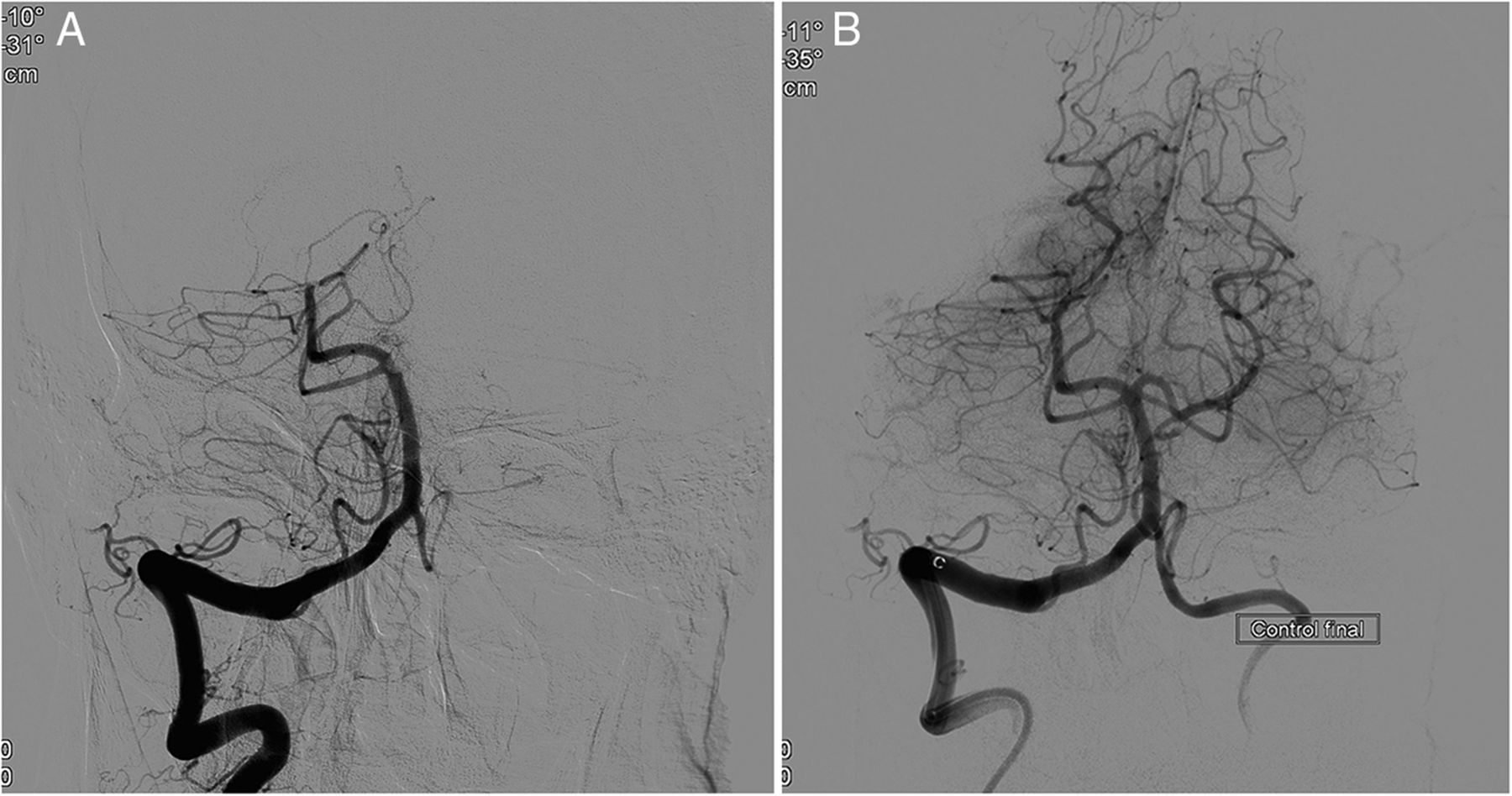
The second patient, also middle aged, came to the emergency department with an 80 min evolution left hemiplegia. CT angiography showed an occlusion in a superior branch-M2 segment of the right MCA and a concurrent occlusion of the right pericallosal artery distal to the origin of the callosomarginal artery. A fibrinolytic agent was administered intravenously, but this was not effective. Endovascular treatment (figure 1) was started, with effective aspiration (TICI 3) on the first attempt of the M2-MCA occlusion using the ACE. Then the right pericallosal artery was catheterized up to the occlusion using the 3MAX. When removing the 3MAX under aspiration, we approached the ACE, which was positioned at the origin of the anterior cerebral artery, and then removed both catheters together and aspirated through them to try to minimize the possibility of MCA embolism. Aspiration was effective (TICI 3) at the first attempt. The puncture–recanalization time for the MCA was 32 min and 44 min for the pericallosal artery.

(A) Lateral view showing right-M2 and distal right-anterior cerebral artery occlusions. (B) Lateral angiogram after treatment.
The third patient, elderly, had been admitted 2 weeks earlier at another hospital owing to splenic infarcts. He was transferred from the other hospital with 3 h clinical signs of upper and lower left limb paresis, dysarthria, and decreased level of consciousness. CT angiography showed an occlusion of the basilar artery top that completely occluded the origin of the left posterior cerebral and superior cerebellar arteries and a concurrent occlusion of the right posterior cerebral artery at the origin of the P2 segment, with significant mismatch seen in this artery’s territory. Owing to the patient’s recent clinical history, intravenous fibrinolytic therapy was not considered as a possible treatment. Endovascular treatment (figure 2) was started with thrombus aspiration of the clot of the basilar artery with an ACE 64 catheter, which was effective at the first attempt. Subsequently, the origin of the right posterior cerebral artery was selectively catheterized with this ACE 64 catheter and this artery was distally microcatheterized up to the occlusion in the right P2 segment with the 3MAX. Aspiration was performed from both catheters, achieving complete recanalization (TICI 3) of the artery at the first attempt. Puncture–recanalization time of the basilar artery was 28 min and 39 min for the posterior cerebral artery.

(A) Angiogram showing basilar and right-P2 occlusions. (B) Angiogram after treatment.
The fourth patient, elderly, with a history of Killip IV anterior myocardial infarction with contained cardiac rupture 3 weeks earlier, was admitted to the emergency department with symptoms of aphasia and right hemiparesis on awakening. Infarction was not established by CT, but occlusion of an M2 segment top branch of the left MCA was confirmed by CT angiography and a significant mismatch was seen in this branch’s territory. Owing to the patient’s recent clinical history, intravenous treatment was not considered a possible choice. After selective catheterization of the left MCA with the ACE, a microcatheterization of the M2 branch was conducted with the 3MAX. The 3MAX was removed at the first attempt, but not the ACE, which remained in the MCA. The distal migration of the thrombus in the M2 branch was shown at the angiographic check-up subsequently performed. The artery was catheterized again with the 3MAX up to the occlusion, followed by aspiration; then, both catheters, the 3MAX and the ACE were removed. Complete recanalization (TICI 3) of the artery was then achieved. The puncture–recanalization time was 40 min. The clot was located on the 3MAX catheter tip.
The fifth patient, elderly, was transferred to our hospital from another institution owing to 3.5 h clinical signs of global aphasia and right upper limb hemiparesis. CT angiography showed an occlusion in a M2 inferior branch of the left MCA. Owing to the patient´s treatment with acenocumarol, intravenous therapy was not considered and the patient was transferred directly to the angiosuite. After selective catheterization with the ACE catheter of the left MCA, microcatheterization of the occluded branch was conducted with a 3MAX, followed by aspiration of the clot with this catheter. Then, removing the 3MAX and the ACE catheter, complete recanalization (TICI 3) of the artery was achieved. The puncture–recanalization time was 25 min.
The last patient, middle aged, was admitted on awakening with left hemiparesis. A small infarction was established by CT but with a significant mismatch seen in this branch’s territory on perfusion CT. The occlusion of an M3 segment top branch of the right MCA was confirmed by CT angiography. A fibrinolytic agent was administered intravenously, but this was not effective. After selective catheterization of the right MCA with the ACE, a microcatheterization of the M3 branch was conducted with the 3MAX, followed by aspiration of the clot with this catheter. Then, after removing the 3MAX and the ACE catheters, complete recanalization (TICI 2b) of the artery was achieved. The puncture–recanalization time was 20 min.
No technical complications or vasospasm occurred in any of the cases. Neither bleeding nor any other complications were observed in the CT control performed 24 h after the intervention.
Discussion
Distal cerebrovascular occlusions may have an important clinical impact,2 but the benefits of the thrombectomy of small arteries must be carefully considered in relation to the risks. A case series review of the Trevo 3×20 device for the treatment of distal intracranial occlusions has just been published,21 emphasising the importance of this matter. In our case series, all patients had a high NIHSS score (median 12, range 10–17) and had either not responded to intravenous fibrinolytic therapy or this treatment was contraindicated. We think it is important to emphasise that this procedure is not carried out merely to recanalize an artery but to achieve a clinical benefit.
The technical development of new cerebral reperfusion catheters, with better navigability, more flexibility, and less traumatic tips, allows the catheterization of more distal occlusions and the aspiration of these as first intention endovascular treatment.10 ,11 The 3MAX catheter, commonly used to support catheterization with the ACE/ACE 64 reperfusion catheters, may also be an effective catheter for the recanalization of more distal arteries.
In our initial series, we achieved recanalization (TICI 2b–3) in all six cases, with 5/6 on the first attempt. It was not necessary to combine treatment with a stent retriever. In none of the cases was it necessary to pass the clot with a microwire, thus minimizing the chances of distal embolization.
We believe that the ACE/ACE 64 catheter and long sheath associated aspiration is necessary to decrease the chances of thrombus disaggregation in distal emboli. Furthermore, selective catheterization with the ACE/ACE 64 catheter of the proximal segment of the artery to be treated can minimize the chance of stroke in new territories and increase the safety of the procedure. In one of the cases, the ACE catheter was not removed during the first aspiration attempt and distal embolism occurred in the same branch where the initial occlusion was located. On the next attempt, the ACE was removed together with the 3MAX and the thrombus was effectively aspirated. The thrombus was on the tip of the 3MAX and it was probably released on contact with the ACE catheter tip during the first aspiration attempt.
The M2-MCA segment has a parent vessel with a mean diameter of 2 mm.22 ,23 In the cases we have described above with an M2 occlusion, the diameter of the parent vessel was smaller, so the 3MAX was the preferred catheter, but in other cases it might be advantageous to use a 4MAX (internal diameter 0.041 in and 1.42 mm outer diameter) and/or ACE, as we used in the second patient with a larger M2.
All six patients showed early neurological improvement, with a median NIHSS at discharge of 1. No complications occurred during the procedures, even though there were occlusions in distal arteries.
Conclusion
Our initial experience suggests that treatment of distal cerebrovascular occlusions with the 3MAX catheter is feasible. We achieved complete recanalization in all cases without unexpected complications while obtaining good clinical results. However, larger studies are necessary to establish the benefits and safety of the procedure.
References
Footnotes
Twitter Follow PEDRO NAVIA at @pnavia
Collaborators G Arenaza, VG Montoya, E Garmendia.
Contributors All authors made a substantial, direct, and intellectual contribution to the work.
Competing interests None declared.
Ethics approval Local institutional review board (Comité de Ética Asistencial del Hospital Universitario Donostia).
Provenance and peer review Not commissioned; externally peer reviewed.