Article Text

Abstract
Purpose To evaluate the clinical and anatomical results of treatment of intracranial aneurysms (IA) with the Woven EndoBridge (WEB) device, with emphasis on mid term and long term follow-up.
Methods Between November 2010 and November 2015, we retrospectively identified, in our prospectively maintained database, all patients treated by WEB device placement for an IA at three institutions. Clinical charts, procedural data, and angiographic results were reviewed.
Results 48 patients with 49 IAs were identified. There were 35 women and 13 men with a mean age of 57 years (range 35–76 years). All IA were wide necked. Mean aneurysm size was 8.6 mm. There were 44 unruptured IA and 5 ruptured IA. During endovascular treatment (EVT), adjunctive devices were used in 22.4% of procedures. A good clinical outcome (modified Rankin Scale score ≤2) was achieved in 44/48 patients (92%). There was no mortality. Mean follow-up was 25 months (range 3–72 months; median 24 months). Between mid term and long term follow-up, occlusion was stable in 19/23 IA (82.6%), improved in 2/23 IA (8.7%), and worsened in 2/23 IA (8.7%). Retreatment was performed in 8/49 IA (16.3%). At the latest available follow-up, there were 34/47 (72.3%) complete occlusions and 13/47 (27.7%) neck remnants.
Conclusions Our study suggests that EVT of IA with the WEB device provides adequate and stable long term occlusion.
- Aneurysm
- Device
Statistics from Altmetric.com
Introduction
Endovascular treatment (EVT) is now recognized as the first therapeutic option for ruptured and unruptured intracranial aneurysms (IA).1–5 Among several technical developments, intrasaccular flow disruption has contributed towards extending EVT indications to wide necked bifurcation IA that remain challenging to treat, even with bridging neck devices such as balloons and stents. Moreover, these IA are associated with a higher rate of recanalization.6 The first available flow disrupter device is the Woven EndoBridge (WEB) (Sequent Medical, Aliso Viejo, California, USA), a self-expandable, detachable, intrasaccular device made of one or two woven nitinol cages.7 Since the first clinical experience reported in 2012,8 the WEB dual layer (WEB-DL, figure 1A) has been replaced by a single layer device (WEB-SL, figure 1B), and a second device with a spherical shape has also been developed (WEB-SLS, figure 1C).9 Early series have reported good clinical and anatomical outcomes in unruptured8 ,10–14 and ruptured IA.9 ,15 ,16 However, few data are available on mid term12 ,17 ,18 and long term follow-up,19 which raise questions about long term stability of neck remnants related to potential device compression.17
The purpose of this work was to report the clinical and anatomical results of our 6 years of experience in IA treatment with WEB devices, with emphasis on long term follow-up.
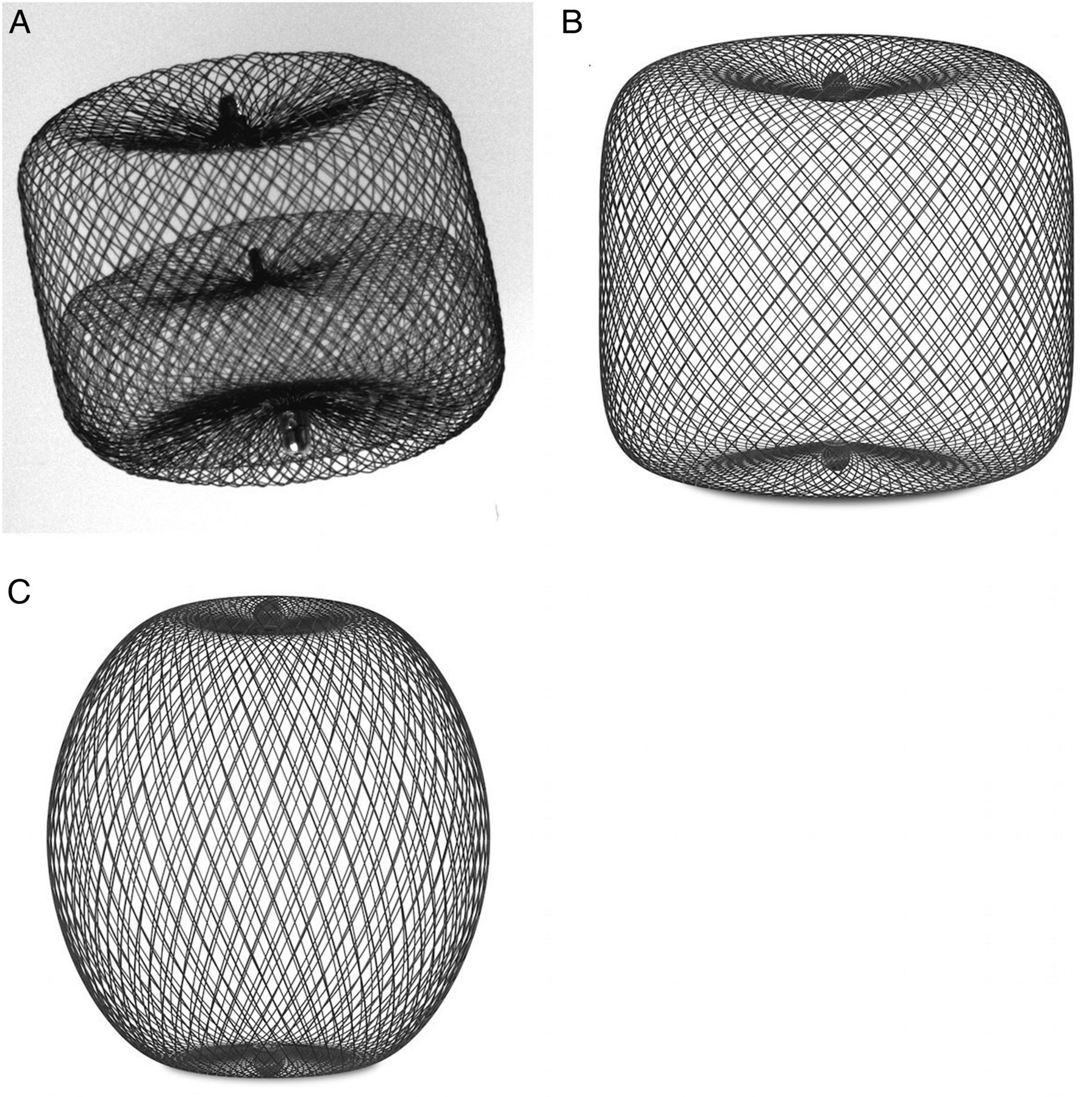
Evolution of the Woven EndoBridge (WEB) devices. (A) WEB dual layer device (WEB-DL). (B) WEB single layer device (WEB-SL). (C) WEB single layer spherical device (WEB-SLS).
Materials and methods
Population
The study protocol was approved by the local ethics committee.
We identified, in our prospectively maintained database, all patients in whom EVT of an IA with a WEB device was performed between November 2010 and November 2015 in three centers. All patients underwent pre-therapeutic DSA, including anteroposterior and lateral views of both carotid and vertebral arteries. Then, three-dimensional rotational angiography was obtained on the artery bearing the target IA to precisely depict aneurysm morphology. IA location and size were recorded, as well as neck size.
Endovascular procedures
The indication and modality of IA treatment were based on multidisciplinary discussion for all patients. All EVT were performed under general anesthesia and systemic heparinization. The adequacy of anticoagulation was monitored by repeated measurements of the activated clotting time (ACT). A baseline ACT value was obtained prior to femoral puncture. Then a 5000 IU bolus infusion of heparin was administered, followed by a continuous drip (1500–2500 IU/hour), with the purpose of doubling the baseline ACT value. At the end of the procedure, systemic heparinization was maintained for 24 hours in most patients. Arterial access was obtained with a coaxial system, including a 6 F long sheath placed in the lower cervical vessels, a 6 F guiding catheter placed in the cervical internal carotid artery (ICA) or vertebral artery, and a microcatheter used to access the aneurysm and deliver the device. The first procedures were performed with a DAC 038 (Stryker Neurovascular, Kalamazoo, Michigan, USA) as the delivery catheter to obtain sufficient support because of the stiffness of early devices and the poor support of its initial pusher. The pusher was replaced after a few months based on our early evaluation, which allowed delivery of the device with conventional 0.027 inch microcatheters. Since December 2012, the dedicated VIA microcatheters (0.021 inch and 0.027 inch; Sequent Medical) were available and used for all procedures. All endovascular procedures were performed by three senior interventional neuroradiologists with 5–15 years of experience.
Immediate outcome
Clinical outcome
Procedural and early post-procedural (within 48 hours) complications were recorded. Clinical course was recorded by review of clinical charts following the modified Rankin Scale (mRS).20 Worsening of symptoms or death were evaluated by a vascular neurologist and/or an interventional neuroradiologist.
Anatomical outcome
Aneurysm occlusion was evaluated on immediate post-treatment DSA using a 3 gradescale:12 grade I, complete occlusion; grade II, almost complete occlusion—neck remnant; and grade III, incomplete occlusion—aneurysm remnant.
Patient follow-up
Clinical follow-up
We recorded delayed complications based on clinical chart review. The latest clinical follow-up was obtained at the same time as the latest imaging follow-up and evaluated using mRS.
Imaging follow-up
Our imaging follow-up protocol consisted of DSA at 3, 6, and 12 months. Contemporarily MR angiography (MRA) at 12 months was performed and used as a baseline comparative non-invasive technique. Then, follow-up was performed by MRA, including time of flight and contrast enhanced MRA, every 2 years for unruptured IA and yearly for ruptured IA. Imaging follow-up duration was defined as the delay between EVT and the latest imaging examination.
Imaging analysis
Aneurysm occlusion was evaluated by two senior neuroradiologists with 5 and 15 years of experience, respectively, reaching consensus. The latest MRA was compared with immediate post-EVT DSA.
The same grading scale12 was used for immediate and follow-up imaging examinations.
Results
Patient and aneurysm population
Overall, we identified 48 patients with 49 IA. There were 35 women and 13 men with a mean age of 57 years (range 35–76 years). Mean aneurysm diameter was 8.6 mm (range 4–22 mm) and mean neck diameter was 4.9 mm (range 2–10 mm). There were 41 unruptured incidental IA, 3 IA recurrences after EVT with coils, and 5 ruptured IA. We found 29/49 IA located on the middle cerebral artery (MCA) (59%), 7/49 on the basilar tip (14%), 5/49 on the ICA (10%), 5/49 on the anterior communicating artery (10%), 2/49 on the posteroinferior cerebellar artery (4%), and 1/49 on the vertebral artery (2%).
Treatment modalities
Procedures and WEB devices
Fifty-two procedures were performed: 50 were successful in treating 47 IA (success rate 96%). Failure occurred in two cases: one device was retrieved because of a major protrusion in the parent artery and one device could not be implanted because of an acute angle between the parent artery and the aneurysmal neck. These two cases were excluded for anatomical evaluation.
A single device was implanted in all but two procedures in which two devices were needed because the largest available device (11 mm) was too small to completely occlude the sac.
Forty-five IA were treated in one procedure whereas two IA required several procedures. One right MCA recanalized IA required a second WEB placement because of a significant remnant after the first procedure. One previously ruptured and recanalized giant partially thrombosed anterior communicating artery aneurysm required three procedures because of persistent growth.
Overall, 52 WEB were implanted, including 33 WEB-DL, 15 WEB-SL, and 4 WEB-SLS.
Additional devices
An additional device was used in 11/52 procedures (22.4%). There were 10 stent placements: 1 was planned prior to EVT and 9 were decided during the procedure because of a WEB protrusion (n=7) or clot formation (n=2). In one patient, additional coiling was performed because of a significant IA remnant after WEB placement.
Clinical, anatomical, and treatment data are summarized in table 1.
Summary of clinical, anatomical, and treatment data
Technical issues, complications, and clinical outcome
Unexpected procedural events occurred in 7/52 procedures (14%), including 3 thromboembolic complications, 1 IA rupture, 2 cervical artery dissections, and 1 femoral artery dissection.
Thromboembolic events, consisting of clot formation at the neck, were observed in 3/52 procedures (6%). Two were immediately treated by IV administration of abciximab and stent placement with no further consequences. In the third patient with a ruptured IA, abciximab was not given to avoid hemorrhagic complications. A subsequent occlusion of a temporal M2 branch occurred resulting in temporo-parietal ischemia with right hemiparesis and dysphasia (mRS score of 4). The patient gradually recovered with residual slight hemiparesis at follow-up (mRS score of 2).
In one patient, acute rupture of the dome occurred during placement of the WEB device. Complete IA occlusion was obtained with coils and glue preserving parent artery. Subsequent symptomatic vasospasm resulted in a hemiplegia (mRS score of 3) that remained stable at follow-up.
Two cervical artery dissections occurred. One vertebral artery dissection was identified during the procedure and treated by stenting with no clinical consequences. One ICA dissection was diagnosed by DSA 24 hours after EVT because the patient showed a mild left hemiparesis. Stenting was performed immediately with good anatomical results. Rolandic and capsular ischemic lesions were identified at follow-up CT and MR. The patient had left upper limb paresis (mRS 2) at follow-up.
Finally, there was one femoral artery dissection requiring surgical bypass with no further clinical consequences.
Among 48 patients, 40 (83%) had a normal (n=38; mRS=0) or unchanged (n=2) neurological examination at discharge. Four patients (8.3%) presented neurological deterioration due to symptomatic vasospasm in the setting of aneurysmal subarachnoid hemorrhage. Three showed a good clinical evolution at follow-up (mRS score of 0, n=2; mRS score of 1, n=1). The last patient retained left paresis (mRS 3).
Overall clinical outcome results at the last clinical follow-up (range 3–72 months, mean 25 months, median 24 months) were as follows: mRS score of 0 in 38/48 patients (79.2%), mRS score of 1 in 3/48 patients (6.2%), mRS score of 2 in 3/48 patients (6.2%), mRS score of 3 in 2/48 patients (4.2%), and mRS score of 6 in 2/48 patients (4.2%). Two patients died from unrelated reasons during follow-up. Overall permanent neurological morbidity and mortality rates were 3/48 (6.2%) and 0/48 (0%), respectively. A good clinical outcome (mRS ≤2) was achieved in 44/48 patients (92%).
Retreatment
Eight IA had to be retreated (1 retreatment for 6 IA, 2 retreatments for 1 IA, and 3 retreatments for 1 IA). Retreatments consisted of stent assisted coiling (n=5), WEB placement (n=3), coiling (n=2), and flow diverter stent placement (n=1).
Short term anatomic outcome
Because the goal of our study was to report our daily practice experience with WEB treatments, we included patients with additional treatments in the anatomical results. Short term anatomical results were available for 45/47 IA (94%) (range 1–6 months, mean 4.8 months, median 6 months) and were graded as follows: grade I in 31/45 IA (68.9%), including 1 retreatment, grade II in 12/45 IA (26.7%), and grade III in 2/45 IA (4.4%). Adequate occlusion (grade I and grade II) was observed in 43/45 IA (95.6%).
Mid term anatomic outcome
Mid term anatomical results (12 months) were available for 35/47 IA (74.5%) and were graded as follows: grade I in 26/35 IA (74.3%), including 2 retreatments, grade II in 7/35 IA (20%), including 2 retreatments, and grade III in 2/35 IA (5.7%), including 1 retreatment. Adequate occlusion was observed in 33/35 IA (94.3%), including 5 retreatments.
Evolution between short term and mid term follow-up
In 31/35 IA (88.6%), occlusion was stable. In 2/35 IA (5.7%), 1 with immediate occlusion graded II and 1 graded III, occlusion was improved and graded I, both after retreatment. In 2/35 IA (5.7%), occlusion was worsened (1 immediate grade I worsened to grade II and 1 immediate grade II worsened to grade III).
Long term anatomic outcome
Long term anatomical results (range 24–72 months, mean 39 months, median 36 months) were available for 25/47 IA (53.2%) and were graded as follows: grade I in 18/25 IA (72%), including 2 retreatments, and grade II in 7/25 IA (28%), including 2 retreatments. Adequate occlusion was observed in 25/25 IA, including 4 retreatments.
Evolution between mid term and long term follow-up
In 19/23 IA (82.6%), occlusion was stable (figure 2). In 2/23 IA (8.7%), both with immediate occlusion graded III, occlusion was improved and graded I after retreatment. In 2/23 IA (8.7%), both with immediate occlusion graded I, occlusion was worsened, including 1 retreated IA.

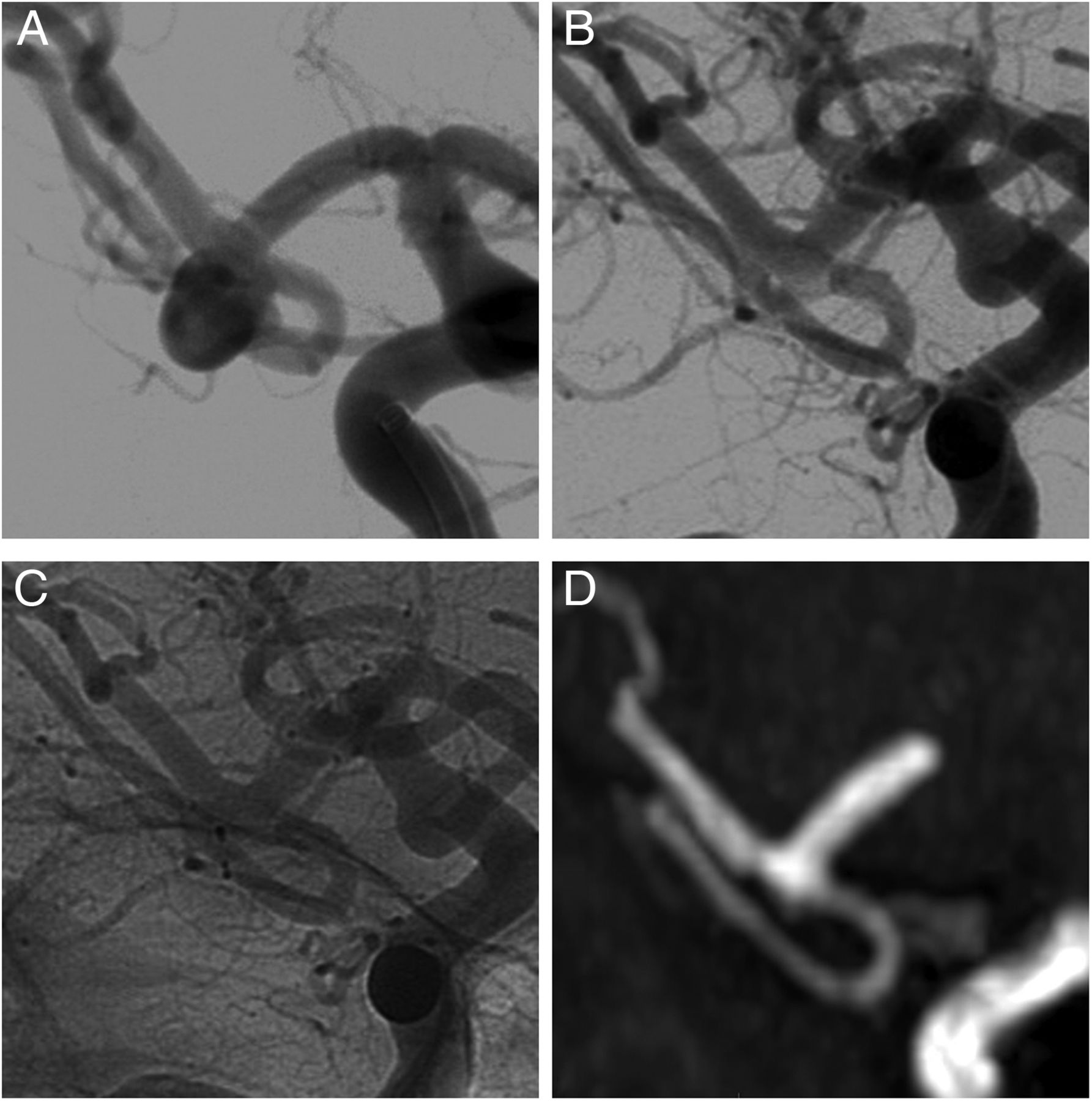
Long term stability of complete occlusion of an unruptured wide necked middle cerebral artery bifurcation aneurysm treated with a Woven EndoBridge dual layer (WEB-DL) device. (A) Pretreatment DSA working view. (B, C) Immediate post-treatment DSA showing complete occlusion (B, subtracted image; C, unsubtracted image). (D) Five year follow-up time of flight MR angiography showing complete occlusion of the aneurysm.
Overall, occlusion at the latest available follow-up (range 3–72 months, mean 25 months, median 24 months) was rated as grade I in 34/47 IA (72.3%) and grade II in 13/47 IA (27.7%).
Discussion
This study confirms the safety and efficacy of EVT of IA with the WEB device. Moreover, it showed that it provides stable occlusion on mid term and long term follow-up in a subgroup of complex IA that are technically challenging to treat and prone to recanalize.
Endovascular procedures
As reported in recent systematic reviews,21 ,22 the technical success rate was high in our series (96%) despite the fact that our experience included the very early learning curve because our main center was one of the first three centers to use the WEB device worldwide. Successive device and dedicated microcatheter improvements23 allowed us to get easier access to the IA and to obtain better control of device deployment. These improvements have progressively widened the range of IA amenable to flow disruption and increased the technical success rate.
Adjunctive devices—mostly stents—were used in 22.4% of IA, which was slightly higher than in a recent systematic review.21 This was not associated with an increased complication rate in our series. Again, the need to use these adjunctive techniques may partially be explained by our early experience. Indeed, some of the protrusions, clot formations, or incomplete occlusions requiring additional treatment may be related to the learning curve.
Intraprocedural thromboembolic complications occurred in 3/52 procedures. Only one had clinical consequences because no abciximab was administered in the setting of subarachnoid hemorrhage, as discussed above. This rate of thromboembolic events (5.8%) and their management in unruptured IA is comparable with that of coiling.24
Finally, one perforation occurred (2%) in an unruptured MCA IA. Although the rupture rate with the WEB was low21 and similar to that of coiling,25 a very good understanding of IA anatomy is mandatory to reduce this risk as much as possible. Indeed, the device needs to be advanced a few millimeters before it expands and gets a wider wall apposition. During this first step, the distal part might cause perforation if it is oriented towards the wall as the device itself remains proximally stiffer. A biplane angio suite might also help to ensure adequate orientation of the device towards the sac.
Clinical outcome
Immediate neurological morbidity in this series was 8.3%, which was higher than reported with conventional coiling. This may partially be related to our population of complex wide necked and some ruptured IA. Moreover, as mentioned above, our experience included the very early learning curve. With growing experience, sizing was more accurate, stiffness of the WEB devices decreased, and more specific catheters were available.
As previously reported,22 we observed good clinical outcomes at follow-up in most patients (92%). We reported no mortality and a permanent morbidity of 6.2%, which are satisfying results considering the population of complex and some ruptured IA. We observed no delayed IA rupture or complications related to EVT.
Anatomical outcome
The rate of immediate adequate occlusion in our series was high (95.6%) despite the use of antiplatelet therapy in the case of stent placement. In their systematic review, Asnafi et al22 reported a rate of immediate adequate occlusion of 59%. This may be explained by the fact that we used adjunctive devices in almost 25% of our cases.
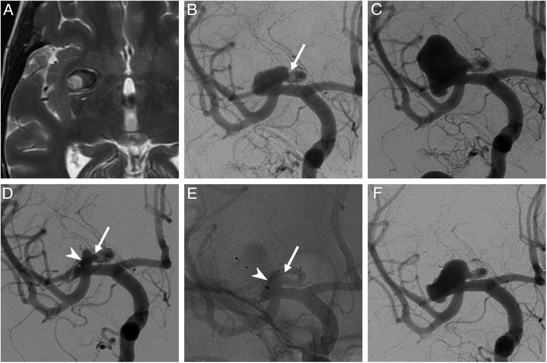
The rate of retreatments in our series (16%) was higher than reported in the long term series of Pierot et al (11.5%). This may be explained by the fact that we initially included very large and partially thrombosed IA. It has been reported26 and confirmed in our experience that the device may progressively migrate in the clot and/or the aneurysm resulting in incomplete occlusion (figure 3). Inadequate sizing in the early experience may also explain some of these retreatments as significant remnants were left to avoid device protrusion. Thus we believe that with growing experience, optimal device and IA selection may reduce the rate of retreatment.

Regrowth of a large partially thrombosed right middle cerebral artery bifurcation aneurysm after treatment with a Woven EndoBridge dual layer (WEB-DL) device. (A) Axial T2 weighted MR image showing a partially thrombosed aneurysm. (B) DSA (frontal view) showing the circulating portion of this wide necked aneurysm with a branch arising from the sac (arrow). (C) DSA (frontal view) showing a partial recanalization of the previously thrombosed portion. (D, E) Immediate occlusion after treatment with a WEB-DL device. Subtracted (D) and unsubtracted views showing a small neck remnant (arrowhead) and the preserved branch (arrow). (F) Follow-up at 3 months showing recanalization of the aneurysm caused by migration and compaction of the device.
We decided to include retreated IA in the anatomical follow-up because this corresponds to our daily practice with the WEB device. Our results showed that IA occlusion was stable and adequate at long term follow-up in this population of wide neck bifurcation IA that are prone to recanalization with other endovascular techniques.27 These findings confirm those of previous mid term12 ,17 and long term series.19 Moreover, it confirms that the small proximal recess related to the shape of the device should not be considered as a remnant and is stable over time.12 This long term anatomical stability suggests that flow disruption may be considered as a firstline option for these complex IA because they compare favorably with surgical results.28
The endovascular alternatives for these wide neck bifurcation IA are the PulseRider, the barrel stent, and the pCONus devices. However, their use is limited for ruptured IA because antiplatelet therapy is required. Moreover, we have fewer clinical data on these devices to date.29–31
Limitations
This study had several limitations. Firstly, it was a retrospective work based on a relatively small population. Patients and follow-up durations were heterogeneous and many adjunctive devices were used which makes extrapolation of our results difficult.
Conclusion
This study showed that flow disruption represents a safe and efficient endovascular approach to treat complex ruptured and unruptured wide neck bifurcation IA. It may be used as a standalone technique or in conjunction with other endovascular techniques. Moreover, our results confirm that the WEB device provided a high rate of adequate occlusion that was stable over time.
References
Footnotes
Contributors BM, AG, DB, and BL: substantial contributions to the conception or design of the work, or the acquisition, analysis, or interpretation of data for the work; drafting the work or revising it critically for important intellectual content; final approval of the version to be published;and agreement to be accountable for all aspects of the work in ensuring that questions related to the accuracy or integrity of any part of the work are appropriately investigated and resolved.
Competing interests None declared.
Ethics approval The study was approved by the ethics committee of Erasme University Hospital.
Provenance and peer review Not commissioned; externally peer reviewed.