Article Text

Abstract
Objectives To develop an in vitro model for studying the biological effect of complex-flow stress on endothelial cells in three-dimensional (3D) patient-specific vascular geometry.
Materials and methods A vessel replica was fabricated with polydimethylsiloxanes using 3D printing technology from vascular image data acquired by rotational angiography. The vascular model was coated with fibronectin and immersed in a tube filled with a cell suspension of endothelium, and then cultured while being slowly rotated in three dimensions. Culture medium with viscosity was perfused in the circulation with the endothelialized vascular model. A computational fluid dynamics (CFD) study was conducted using perfusion conditions used in the flow experiment. The morphology of endothelial cells was observed under a confocal microscope.
Results The CFD study showed low wall shear stress and circulating flow in the apex of the basilar tip aneurysm, with linear flow in the parent artery. Confocal imaging demonstrated that the inner surface of the vascular model was evenly covered with monolayer endothelial cells. After 24 h of flow circulation, endothelial cells in the parent artery exhibited a spindle shape and aligned with the flow direction. In contrast, endothelial cells in the aneurysmal apex were irregular in shape and size.
Conclusions A geometrically realistic intracranial aneurysm model with live endothelial lining was successfully developed. This in vitro model enables a new research approach combining study of the biological impact of complex flow on endothelial cells with CFD analysis and patient information, including the presence of aneurysmal growth or rupture.
- aneurysm
- blood flow
- vessel wall
- technology
Statistics from Altmetric.com
Introduction
Subarachnoid hemorrhages from rupture of intracranial aneurysms account for 5–10% of all cases of stroke being fatal in about 50% of cases, and occur more often than with other forms of strokes.1 Non-invasive vascular imaging techniques, such as magnetic resonance angiography, have detected unruptured aneurysms with high sensitivity.2 The treatment needs microsurgical clipping or endovascular techniques, both of which involve some procedural risks.3 4 As intracranial aneurysms are relatively common, an in-depth understanding of their natural history is imperative for a comprehensive management of patients.
The growth or rupture of intracranial aneurysms is thought to be a pathological consequence of complex arterial wall remodeling caused by an interaction of biological and hemodynamic factors. Endothelial cells in the artery physiologically respond to blood flow, especially flow direction and wall shear stress (WSS). Image-based computational fluid dynamics (CFD) modeling allows simulation of complex flow in intracranial aneurysms and has led to the identification of connections between hemodynamics and the increased likelihood of their growth and rupture.5 6 However, both high and low aneurysmal WSS have been separately correlated with intracranial aneurysm growth and rupture. This controversy might stem from limitations of CFD, such as inconsistent parameter definitions, calculation assumptions including outflow boundary conditions, rigid walls and Newtonian properties, or lack of biological information.7 To make CFD more reliable for rupture or growth prediction, the biologic mechanisms underlying growth and rupture and their interaction with hemodynamics should be elucidated.
The biologic findings obtained from experiments using a flow chamber or cone and plate have been used to interpret the relationship between the change of WSS and the tendency toward growth or rupture.8 9 However, these in vitro flow experimental systems are too simple to reflect the physiologic conditions in complex three-dimensional (3D) geometry. Recently, a 3D microfluidic aneurysm model has been reported.10 However, the patient-specific geometry cannot be replicated in the model and the validity of biological outcomes obtained during microfluidic experimentation is still being questioned because of uncertainty about the effects of culturing in low media volume.11 To use a CFD study as a predicting tool for growth or rupture from a patient’s image, the interaction between each hemodynamic parameter and the biologic response on the vascular wall needs to be understood.
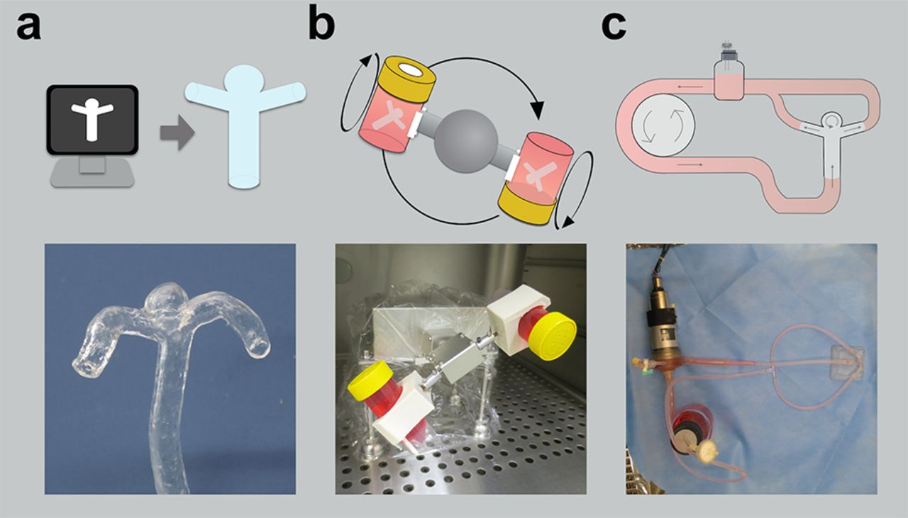
Therefore, we have developed a new in vitro model to study the biophysical cellular interactions in intracranial aneurysms (figure 1). In this model, endothelial cells are attached to the lumen of a patient-specific aneurysm model using 3D rotational equipment. Then, the endothelial cells are loaded with hemodynamic conditions, which can be measured with particle imaging velocimetry or can be simulated in the CFD calculation. After the flow stress, gene expression in endothelial cells can be examined by molecular biology methods such as immunocytochemistry. This new in vitro model is unique in that the flow condition used in the CFD study can be replicated in the realistic geometry, which can combine flow dynamics, biology, and patient information, including the presence of aneurysmal growth or rupture. If CFD parameters can be correlated with biologic changes, rupture prediction by CFD may become more achievable. In this study, we examined the flow effect on endothelial morphology in the patient-specific aneurysmal model.

Process of an in vitro patient-specific vessel research model. (A) The PDMS vessel is fabricated using 3D printing technology from vascular image data acquired by CT angiography, MR angiography, or 3D rotational angiography. (B) The vascular model in the tube filled with a cell suspension of endothelium is slowly rotated in a CO2 incubator in three dimensions by a rotator with two axes. (C) The endothelialized vascular model is connected to an experimental system consisting of silicone tubing, a centrifugal pump and a reservoir. Culture medium with viscosity by dextran is perfused in the circuit in a CO2 incubator during the flow culture. PDMS, polydimethylsiloxanes.
Methods
Vascular model
The study, including use of patient image data, was approved by the institutional review board, which waived the requirement for patients’ informed consent. In this study, a basilar tip aneurysm (dome width 5.0 mm, dome height 5.1 mm, and neck width 3.8 mm) was used as our research model because of its simplicity and its lack of influence on flow from other vessels. Digital Imaging and Communication in Medicine (DICOM) data acquired from 3D rotational angiography were exported and loaded onto 3D computer-aided design software (Amira 5.4.5, VSG, Burlington, Massachusetts, USA). The small artery not required for simulation was removed. The 3D vessel image was converted to a stereolithography format (.stl). Patient-specific hollow models were created as previously reported.12 Briefly, vascular molds were fabricated using a 3D printer (UP! Plus 2, OPT, Tokyo, Japan). The vascular molds made of acrylonitrile butadiene styrene (ABS) were smoothed by immersion in ABS solvent (eSolve, Kaneko Chemical, Saitama, Japan) to remove the stair-like layers on the surface of printed objects. After drying, each vascular mold was coated with degassed polydimethylsiloxanes (PDMS; Dow Corning Corp, Midland, Michigan, USA) three times and then cured at 60°C overnight. The wall thickness ranged between about 430 and 650 µm. The ABS mold and cured PDMS was immersed in acetone for 4–12 hours to dissolve and remove the ABS (figure 1A). After washing three times with acetone, the vascular replicas were dried.
3D rotating cell culture
To enhance cell adhesion to PDMS, the vessel replicas were immersed in 10% (3-aminopropyl)trimethoxysilane (Sigma-Aldrich, St Louis, Missouri, USA) in 100% ethanol overnight and cross-linked with 0.1 mM sulfosuccinimidyl-6-(4′-azido-2′-nitrophenylamino)-hexanoate (sulfo-SANPAH, Sigma-Aldrich) in water with the aid of UV activation (10 min x two treatments; 365 nm, 36 W, at a distance of 5 cm). After washing with phosphate-buffered saline (PBS), the vascular replicas were coated with fibronectin in PBS at 40 µg/mL. The vascular models were again washed with PBS and then exposed to UV light for sterilization before cell seeding.
Bovine carotid artery endothelial cells (BCAECs; JRCB cell bank, Osaka, Japan) were cultured in growth medium composed of Eagle’s minimum essential medium with 10% fetal bovine serum, penicillin and streptomycin, and used between passages 16 and 20. The prepared vascular replicas were transferred into a tube filled with a cell suspension of BCAECs (105 cells/mL). The tube was covered with a cap with a filter for sterile gas exchange and rotated in a CO2 incubator at a rate of 1 revolution per 4 min in three dimensions by a rotator with two axes for 48 hours (figure 1B).
Perfusion culture
Eagle’s minimum essential medium containing 5% dextran (molecular weight 200,000, Wako Pure Chemical, Osaka, Japan) was used for the study. The viscosity of the solution was 3.9 cPa, as measured by a rotating-cone, plate-type viscometer, BIORHEOLIZER (Tokimec, Osaka, Japan) at 37°C. The endothelialized vascular replica was connected to an experimental system consisting of silicone tubing, a centrifugal pump, and a reservoir with a filter for gas exchange (figure 1C). Culture medium with the appropriate viscosity was perfused in the circuit with steady flow at the rate of 120 mL/min by a centrifugal six-blade impeller-type pump driven by a small DC servomotor13 after overnight culture at the rate of 20 mL/min. The entire experimental system was placed in a CO2 incubator during the continuous flow culture.
CFD analysis
Commercial software (SCRYU/Tetra for Windows version 12, Software Cradle Co, Osaka, Japan) was used for meshing and fluid simulation. The computational mesh comprised layered prismatic elements near the wall and interior tetrahedral elements. The mesh size was determined based on the mesh-independence convergence study and resulted in 5.44×10⁵ mesh elements. The Navier–Stokes equations were solved to simulate fluid flow on the mesh. The culture medium was assumed to be an incompressible Newtonian fluid. A measured viscosity of 3.9 cPa and specific gravity of 1.05 kg/cm3 and a constant laminar flow at a speed of 120 mL/min were applied to the calculation. The walls of the model were assumed to be rigid with a non-slip boundary condition, and zero pressure was set at the outlet. The wall shear stress distribution, streamlines, and flow velocity were calculated and visualized for the model.
Immunocytochemistry/confocal microscopy
After flow stimulation for 24 hours, the vascular replica with endothelial cells was washed with PBS and fixed in 2% paraformaldehyde for 10 min. After washing three times with PBS, parts of the aneurysm and the parent artery were cut out into pieces of about 4 mm. Cells on the PDMS pieces were permeabilized by 0.5% Triton X-100 in PBS for 5 min. Cells were stained with rhodamine-phalloidin (Cytoskeleton, Inc, Denver, Colorado, USA) for 30 min at room temperature. Cells were washed once with PBS and mounted using Prolong gold antifade reagent with 4',6-diamidino-2-phenylindole (Invitrogen). Cells were observed under an Olympus FluoView 1000 confocal microscope (Olympus Co, Tokyo, Japan) with a 20x objective. To observe the endothelial cells on the curved PDMS surface, z stacks of typically 20 10 µm were taken to about a 200 µm thickness. Confocal z-stack images were used to process into 2D maximum intensity projection images using Olympus FluoView software version 4.2a. The minor/major axis ratio was determined according to the method described previously.14 The length of the long (major) and the short (minor) axes of each cell were measured using the Fit Ellipse tool in ImageJ software by tracing the ellipse which fits the outline of cell. The individual cell area was calculated by manually outlining the boundary of each cell.
Statistical analysis
Data are expressed as the mean±SD obtained from at least 50 cells in projected images of the parent artery or aneurysm tip from four independent experiments. Student’s t-test was used to determine statistical significance (p<0.05) between before and after perfusion culture.
Results
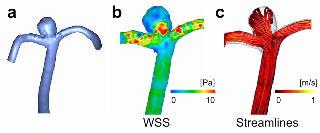
We simulated the WSS and streamline by CFD techniques using the same .stl data as that in used in the 3D printing (figure 2A-C). The average WSS of the aneurysm was 1.2 Pa, which was lower than that of the parent artery 4.2 Pa (figure 2B) A streamline study showed that the flow was linear along the parent artery at the rate of 0.41 m/s, whereas a slow circulating flow at the rate of 0.19 m/s was observed near the aneurysmal apex (figure 2C).

Analysis of computational fluid dynamics. (A) 3D image of the basilar tip aneurysm. (B) WSS distribution. (C) Streamlines coloured with the magnitude of velocity.
This hemodynamic analysis showed contrasting results in the parent artery and aneurysm. We next investigated the endothelial cells of the parent artery and the aneurysmal apex in a vascular replica. Before performing the perfusion culture, we examined the effect of the 3D rotational culture of the endothelial cells on the complex PDMS shape. To achieve this, we observed cytoskeletal F-actin stained with rhodamine-phalloidin in endothelial cells by confocal microscopy after rotational culture without flow stimulation. Confocal imaging showed that the inner surface of the PDMS vascular model was evenly covered with monolayer endothelial cells (figure 3A). Most of the cells were spindle-shaped, but were randomly oriented in various directions. After 24 hours of flow circulation, the endothelial cells in the parent artery became longer and larger in the longitudinal direction and aligned with their long axis parallel with the flow direction (figure 3B, D, E) In contrast, the endothelial cells in the aneurysm had irregular shapes, and most of the cells lost their spindle shape (figure 3C, D). In addition, the size variation in the aneurysm increased after flow stimulation (figure 3E). Collectively, these data suggest that endothelial cells alter the morphology dependent on the flow stress in complex-shaped PDMS models of macroscopic size.

Morphological change of the endothelial cells by flow stress under confocal microscopy. Representative 2D projection images are shown. (A) Static. (B) Parent artery after flow stimulation. (C) Aneurysmal apex after flow stimulation. (d) Minor/major axes ratio. (E) Cell area of endothelial cells before and after perfusion culture. F-actin cytoskeleton (red, phalloidin) and cell nuclei (blue, 4’,6-diamidino-2-phenylindole) are fluorescently stained. The scale bar is 100 µm. *p<0.05; **p<0.001.
Discussion
In this study, we report on a patient-specific in vitro vascular model with an endothelial lining to study the biological effect of flow stress on endothelial cells in a realistic geometry. The biological information in endothelial cells can be combined with CFD data, which can bridge the gap between biology and flow dynamics. Our macroscale vascular model with endothelial lining was achieved by overcoming two hurdles: the step-like layers of 3D printing and the gravitational settling of cells.
Although there are several methods for 3D printing, most of the techniques print objects layer by layer into the designed object, which yields stair-like steps on the surface. If the vascular replica is created using the model as a negative mold without removing these step-like layers, a micro-turbulent flow might occur owing to the steps and flow stress will not properly affect the endothelial cells cultured on the steps. Some reports have demonstrated polishing methods of the surface of ABS vascular models.15 16 However, they fabricated normal vessels without aneurysms and did not evaluate the conformational change after smoothing. Recently, we reported a chemical smoothing method to remove the step-like layers with minimal change to the aneurysmal conformation.12 By applying that method to this model, it became possible to enable endothelial cells to adhere to the smoothed lumen of the aneurysmal replica.
Moreover, the 3D rotating machine with two axes allowed us to avoid cellular settling to the bottom by gravity. Although culture methods of endothelial cells in tubular silicone have been reported,13 17 18 these methods used single-axis rotation and cannot provide evenly coated endothelium in a complex-shaped intracranial vessel or aneurysm. In this study, confocal study showed that the endothelium was successfully attached onto the complex-shaped vascular lumen after the 3D rotational culture with the use of two-axes for 24 hours.
A confocal microscopic study demonstrated that the shape of the endothelial cells was modified by flow stress (figure 3). We previous reported that WSS and flow rate in the aneurysms at the peak of the cardiac cycle were found to differ in magnitude between different locations and a basilar tip aneurysm showed a lower WSS and flow rate, which is similar to the CFD result in this study.19 After the flow stimulation, the endothelial cells in the parent artery elongated and aligned with the longitudinal direction of the flow (figure 3B) and cells in the aneurysm lost their spindle shape under low wall shear or slow circulatory flow (figure 3C). The morphological findings are consistent with the previous studies which used simple flow chamber or cone and plate apparatus.20–22 Because endothelial cells are known to alter their morphology and align along the direction of the laminar fluid shear stress through activation of the Rho family small GTPase23 and upregulation of an intercellular adhesion molecule,24 the findings in this study indicate that in our model flow stress can adequately alter intracellular signaling and induce remodeling of the actin cytoskeleton in the endothelial cells. Moreover, an early histopathological study from a human aneurysmal sample showed that the endothelial cells lining the luminal surface are irregularly arranged,25 which is consistent with our results. Overall, these data support our proposed method functioning as an in vitro model of intracranial aneurysm for studying the impact of complex flow stress on endothelial cells.
Its basic trait as an in vitro flow model provides the flexibility to perform various experiments, such as gene modification, changes in the properties of the circulating fluid, changes in the perfusion conditions, and addition of blood cells to the circuit. In addition, this model provides unique insight into the biophysical interactions that occur in the patient-specific 3D geometry, and can add CFD analysis and clinical information, such as a patient’s time course and place of growth or rupture.
On the other hand, this research model has limitations because it is an in vitro model. The model seems physiologically poorer than animal models as it does not adequately replicate the cellular microenvironment. Therefore, a drug study or migration assay might result in conflicting data. While in vitro models are incomplete substitutes for examining the human disease process, they generally provide a considerable number of advantages compared with the in vivo animal disease models.
In conclusion, this model provides new insights into the relationship between the biological effects of complex flow in intracranial aneurysms and CFD research. Further studies will be required to evaluate the relation between endothelial gene expression and location of an aneurysm or patient history such as aneurysmal growth or rupture. Importantly, hemodynamics are implicated in intracranial aneurysms and also in the pathogenesis of aortic aneurysms, coronary artery disease and arteriovenous malformation. Therefore, this in vitro model can potentially be applied to these important vascular diseases.
Acknowledgments
We also thank Dr Joji Ando, Dr Kimiko Yamamoto, Dr Shin-ichiro Sugiyama and Dr Masaaki Shojima for useful and valuable suggestions.
References
Footnotes
Contributors NK: concept, design and development of the study, acquisition and analysis of the data, writing of the article. TM, EW: development of the study. KN: acquisition of the data. ST, KK: critical review of the article.
Funding KAKENHI (Grants-in Aid for Scientific Research) from Japan Society for the Promotion of Science (JSPS), grant no. 15K19978 to NK and Jichi Medical University Young Investigator Award, 2015 to NK.
Competing interests None declared.
Patient consent The study, including use of patient image data, was approved by the institutional review board, which waived the requirement for patients’ informed consent.
Ethics approval Institutional review board.
Provenance and peer review Not commissioned; externally peer reviewed.
Data sharing statement Any technical detail concerning the creation of the model and the results are available from the corresponding author.