Article Text

Abstract
Background and purpose Despite significant technical advances, recanalization rates after endovascular therapy of ruptured intracranial aneurysms (IAs) remain a clinical challenge. A histopathological hallmark of ruptured human IA walls is mural cell loss. Mural smooth muscle cells (SMCs) are known to promote intraluminal healing in thrombosed experimental aneurysms. In this rat model we assess the natural history and healing process after coil embolization in SMC-rich and decellularized aneurysms.
Methods Saccular aneurysms were created by end-to-side anastomosis of an arterial graft from the descending thoracic aorta of a syngeneic donor rat to the infrarenal abdominal aorta of recipient male Wistar rats. Untreated arterial grafts were immediately transplanted, whereas aneurysms with loss of mural cells were chemically decellularized before implantation. Aneurysms underwent coil implantation during aneurysm anastomosis. Animals were randomly assigned either to the non-decellularized or decellularized group and underwent macroscopic and histological analyses on days 3, 7, 21, or 90 post-coil implantation.
Results A total of 55 rats underwent macroscopic and histologic analysis. After coil embolization, aneurysms with SMC-rich walls showed a linear course of thrombosis and neointima formation whereas decellularized aneurysms showed marked inflammatory wall degeneration with increased recanalization rates 21 days (p=0.002) and 90 days (p=0.037) later. The SMCs showed the ability to actively migrate into the intra-aneurysmal thrombus and participate in thrombus organization.
Conclusions Coil embolization of aneurysms with highly degenerated walls is prone to further wall degeneration, increased inflammation, and recanalization compared with aneurysms with vital SMC-rich walls.
- aneurysm
- coil
- vessel wall
- inflammation
Statistics from Altmetric.com
Introduction
Since the introduction of detachable coils for the treatment of intracranial aneurysms (IAs) in 1991 by Guido Guglielmi,1 2 this technique has evolved in many neurosurgical centers as the most widely used therapy for IAs. Although it represents a less invasive approach than other modalities for many types of aneurysms, concerns regarding the durability of coil treatment have been raised. Angiographic recurrences have been detected in up to 20–30% of coiled aneurysms, with higher rates of recurrences in rupture cases.3–5 The course of an IA after coil embolization depends largely on a biological response in which the intraluminal thrombus transforms into fibrotic scar tissue, thus guaranteeing long-lasting exclusion of the aneurysm from circulation.6
This organization of the intraluminal thrombus and subsequent neointima formation appears to depend on the physiology and health of the aneurysmal wall.7–13 The walls of many IAs, especially ruptured ones, are characterized by loss of mural smooth muscle cells (SMCs), fibroblasts, and endothelial cells,14–16 which may explain the findings of impaired healing and recurrence after embolization.13
The concept that loss of healthy SMCs from the IA wall can impair the healing response after embolization and thus predispose to recanalization has not been studied before in a controlled experimental setting. We therefore investigated the effects of aneurysm wall cellularity related to the natural history and recurrence rates after coil embolization of saccular aneurysms in a rat model.
Materials and methods
Study design, animals, and anesthesia
Following an in vivo pilot series with six animals to test this protocol, we randomly assigned 57 male Wistar rats (Janvier, Saint-Berthevin Cedex, France) weighing 430±50 g to either the vital (non-decellularized) or decellularized group. Healing of the aneurysms was rated at follow-up days 3, 7, 21, or 90 postoperatively. At 21-day follow-up, the migration of aneurysm mural cells was assessed with two transgenic green fluorescence protein (GFP) mutant and two GFP wild type male Lewis rats (inbred, initially kindly provided by Professor Eiji Kobayashi, Center for Development of Advanced Medical Technology, Jichi Medical University, Tochigi, Japan). Figure 1 depicts the study design, dropout rates, and follow-up scheme.

Flowchart of the study design and follow-up modalities. Operated animals were randomly assigned to either the control or experimental group—that is, vital (non-decellularized) or decellularized aneurysm wall groups. Animals included were male Wistar rats, except for male rats that were either transgenic green fluorescence protein (GFP) mutant or wild type Lewis rats.
The animals were housed in animal facilities at room temperatures of 22–24°C and 12 hours light/dark cycle. They had free access to tap water and pellet diet and were cared for in accordance with institutional guidelines. Rats were anesthetized by weight-adjusted intra-abdominal injection of medetomidine hydrochloride (0.5 mg/kg) and ketamine hydrochloride (50 mg/kg). Postoperative analgesia was performed by subcutaneous injections of buprenorphine (0.3 mg/kg). The experiments were approved by the Committee for Animal Welfare at the University of Bern, Switzerland (BE 103/13) and strictly followed the recommendations for good laboratory practice and guidelines for reporting animal research.
Aneurysm model, surgical technique, and graft harvesting
According to the Helsinki rat microsurgical sidewall aneurysm model,17 the aneurysms were created under the operating microscope (Zeiss, OPMI pico, Germany) by end-to-side anastomosis of an arterial graft from the descending thoracic aorta of a syngeneic donor rat to the infrarenal abdominal aorta of a recipient male Wistar rat. Vital (non-decellularized) arterial grafts were immediately transplanted, whereas aneurysms with loss of mural cells were decellularized by sodium dodecyl sulfate before implantation.11 Coil implantation was performed with a predefined coil volume of 2 cm of 360° 3 mm coils (Target Detachable Coils 360, Stryker, USA) during aneurysm anastomosis (see online supplementary video I).7
Supplementary video
The operated animals were then randomly allocated to either the vital aneurysm cohort or decellularized aneurysm cohort.18 Both the Wistar rats and GFP transgenic Lewis rats were operated in the same fashion. To assess the migration capability of aneurysm wall cells into the thrombus, a GFP mutant arterial graft was transplanted in a GFP wild type recipient rat. To assess the impact of a possible cell migration from the carrying vessel into the intra-aneurysmal thrombus, an arterial graft of a GFP wild type Lewis rat was decellularized and transplanted into a GFP mutant recipient rat.
All surgical procedures were video recorded (HXR-MC1P, Sony, Japan). Aneurysm dimensions were measured and photographed before and after implantation (including endoluminal assessment of the aneurysm orifice). Surgical characteristics including total operating time, vessel clamping time, anastomosis suturing time, and graft ischemia time were summarized (see online supplementary table I).
Supplemental material
At follow-up the animals underwent laparotomy and microsurgical harvesting of the aneurysm. With harvesting of the aneurysm and its carrying vessel, the posterior wall of the fragmented abdominal aorta was opened and the aneurysm ostium was photographed for neointima evaluation. We rated the macroscopic neointima maturity on a 6-point scale in a blinded manner (see online supplementary table II).12 Perfusion fixation of the tissue was performed with 4% paraformaldehyde.
Supplemental material
Aneurysm volume, neointima evaluation, and histology
Aneurysm volume was calculated at baseline (day of surgery) and follow-up using the cylinder formula (V = π × width/2 × depth/2 × length).19 Coil volume was determined using the cylinder formula, while considering the coil helix outer diameter according to the manufacturer's information as volume = (π (outer diameter)2 × length)/4. Aneurysm percent packing volume was calculated by dividing the volume of the coils delivered by the volume of the aneurysm. An online aneurysm volume calculator supported the measurements (http://www.angiocalc.com/percent_volume.php).
All aneurysm samples were stored in formaldehyde until they were embedded in paraffin blocks. The blocks were then sectioned in half along the longitudinal axis with a diamond saw (cut-grinder charly, Walter Messner GmbH, Germany), and the coils were carefully removed with a 26G hollow needle and microforceps under the operating microscope.20 Aneurysms were re-embedded and cut into 2 µm sections for hematoxylin-eosin (HE), monoclonal anti-α muscle actin (α-SMA), and Masson-Goldner’s (MT) staining. The probes for immunohistological assessment of GFP were boiled in citrate buffer (pH 6.0; 2.04 g tri-Na-Citrat-Dihydrat, 1 L distilled water, 500 μL hydrochloric acid) for 4 min at 350 W, then defrosted for a further 4 min. In the next step, the primary antibody (rabbit anti-GFP antibody, 1:250, Cell Signalling) was added and slides were incubated at 4°C overnight. Counterstaining with the secondary red fluorescent antibody (Alexa Anti-Rabbit 594, 1:250) as well as with 4′,6-diamidino-2-phenylindole (DAPI) (DAPI-Methanol, Roche Diagnostics AG, Basel, Switzerland) to identify GFP mutant cells was then performed. Slides were assessed by fluorescence microscopy with an exposure time of 50 ms for DAPI and 90–130 ms for TXRED. Distribution of GFP mutant cells was recorded in regions showing positive staining for both GFP and DAPI. Negative controls were performed in GFP wild type and GFP mutant muscle cells from hind limbs (see online supplementary material 1, the online supplementary figures 1 and 2).
Supplemental material
Supplemental material
GFP staining: Representative microphotographs showing distinct autofluorescence in hind limb muscle tissue of a green fluorescence protein (GFP) mutant rat. Sections were investigated using the green (A) and red emission filter (B) by means of an epifluorescence microscope. Note the strong green signal when excited with light in the blue range (A) while only background fluorescence is observed when excited with light in the green range. Scale bar: 100µm.Supplemental material
Immunofluorescence GFP staining: Representative microphotographs illustrating immunofluorescence staining for GFP in sections from hind limb muscle tissue of GFP mutant (A) and wild type rats (B). Sections were investigated using the red filter (A, B) by means of an epifluorescence microscope. Note the specific immunofluorescence in the GFP mutant rats (A) while only background fluorescence is observed in wild type animals (B). Scale bar: 100µm.Stained slides were evaluated by two observers (JR and EN) under a light microscope (Olympus BX51, Olympus AG, Switzerland) equipped with a digital camera (Olympus DP72, Olympus AG, Switzerland). Histological slides were rated for changes affecting the aneurysm wall as well as intra-aneurysmal thrombus using a previously defined scoring method (see online supplementary table III)12 by reviewers blinded to the treatment group. Post-processing was performed using Adobe Photoshop 6 (V13.0, Adobe Systems) and Adobe Illustrator CC (V17.0, Adobe System).
Supplemental material
Statistical analysis
Scores for histological characteristics and macroscopic neointima formation of vital and decellularized aneurysms were compared with the Mann–Whitney U test. Non-parametric values are expressed as median (IQR). Differences in aneurysm volumes and percent packing volumes were determined with the Student's t-test. Parametric values are expressed as mean±SD and 95% CI. A p value of <0.05 was considered statistically significant.
Data analyses and visualizations were performed using Graph Pad Prism statistical software 7 (GraphPad Software, San Diego, California, USA) for Mac.
Results
Aneurysm coiling was successful in all 59 aneurysms with no coil displacements observed during follow-up. Four animals were lost to follow-up. Three animals in the vital group and one animal in the decellularized group died preterm. One animal died with induction of anesthesia, resulting in a total mortality rate of 7% associated with this surgical procedure. Autopsy was performed in all five animals.
Aneurysm volume
Over time, volume measurements showed that coiled aneurysms remained stable in size in both vital and decellularized aneurysms (see online supplementary figure 3). However, a clear trend towards growth versus baseline values was seen on day 7 in the decellularized group (p=0.056), with 2/6 of these aneurysms demonstrating volume increases up to 260% on day 7. This trend became less obvious with advanced maturing aneurysms at later time points on days 21 and 90. Baseline aneurysm volumes were similar between vital and decellularized aneurysms at all time points. There were no differences in aneurysm coil percent packing volume between the groups at baseline: means for vital and decellularized aneurysms were 6.5±1.6% and 6.7±1.4%, respectively (see online supplementary figure 4).
Supplemental material
Aneurysm volume change after coiling: Aneurysm volume of vital and decellularized aneurysms from baseline/day 0 (date of surgery) to final follow-up. No significant changes were detected between vital or decellularized groups, or between baseline and follow-up. However, a clear trend was seen in the decellularized group at follow-up day 7 (p = 0.056). Data are expressed as mean and ± SD.Supplemental material
Aneurysm percent packing volume: Aneurysm percent packing volume in vital and decellularized aneurysms at baseline (date of surgery). No significant changes are detected. Data are expressed as mean and ± SD.Assessment of macroscopic neointima
Significant differences in the macroscopic evaluation of neointima between vital and decellularized aneurysms first became apparent at 21 days (p=0.002) and remained significant at 90 days (p=0.037). As the distribution in vital aneurysms tapered toward a complete and near-complete neointima formation at day 90, the decellularized aneurysms showed the largest diversification—that is, either complete recanalization (4/6 aneurysms) or complete neointima formation (2/6 aneurysms) (figure 2). A case-by-case analysis of neointima evolution is shown at various time points in vital and decellularized aneurysms (see online supplementary figure 5).
Supplemental material
Macroscopic neointima findings in vital and decellularized aneurysms at different follow-up time points.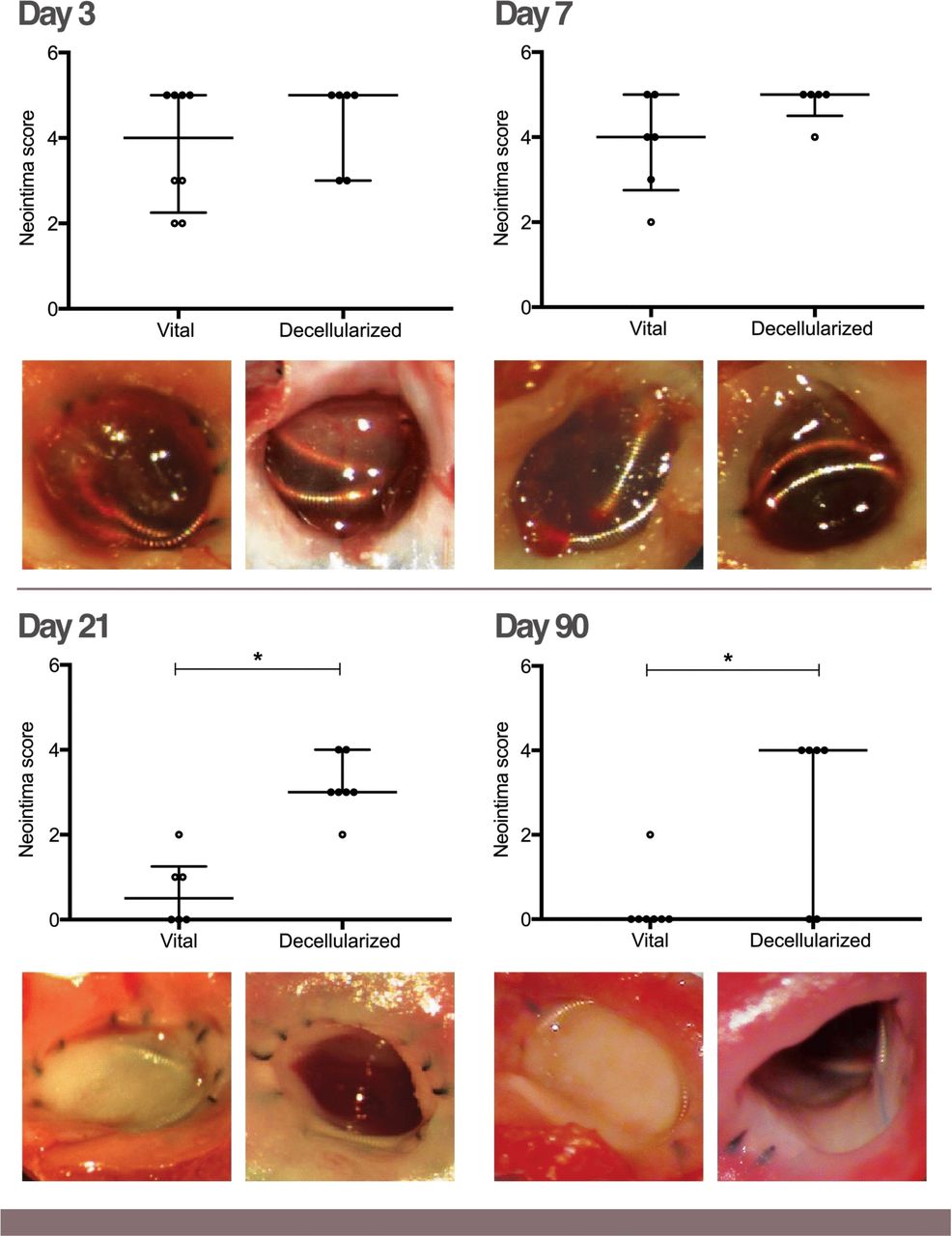
Macroscopic neointima evaluation. Upper row: differences in neointima maturation between vital and decellularized aneurysms at various times. Significant changes between the groups were first detected at day 21 (p=0.002) and remained significant until day 90 (p=0.037). Lower row: example images of aneurysm neointimas between the groups at various times. Corresponding to each follow-up day, the left image represents in each case a vital aneurysm orifice and the right image represents in each case a decellularized aneurysm orifice. Data are presented as median with interquartile range. *Significant differences (p<0.05) between vital and decellularized aneurysms.
Histological changes of aneurysm wall
Compared with vital aneurysm walls, detailed histological analysis of the decellularized aneurysm wall demonstrated increased inflammation (p=0.022), periadventitial inflammation (p=0.007), and fibrosis (p=0.033) (see) with significant changes starting at day 7. Vital aneurysms showed a continuous decline in its wall and periadventitial inflammation over time, with missing or hardly detectable inflammation at day 90. Decellularized aneurysms maintained an elevated inflammation response in this compartment with poor inflammation suppression up to day 90. Both dissection and hematoma of the aneurysm wall were almost exclusively seen in the decellularized group at all time points. Physiologic wall cellularity remained similar in vital aneurysms throughout the observation period, while no aneurysm wall cells could be detected in the decellularized group (see online supplementary figure 6).
Supplemental material
Histological thrombus organization
Detailed histological analysis of the intra-aneurysmal compartment revealed similar levels of neutrophils in the thrombus of vital and decellularized aneurysms on days 3 and 7. The number of neutrophils within the thrombus remained unchanged from day 3 to day 7, declined progressively by day 21, and disappeared at day 90 in vital aneurysms, while it had significantly increased on day 21 (p=0.004) in decellularized aneurysms.
Neointima formation was already more complete and thicker in vital aneurysms than in decellularized aneurysms on day 21 and more clearly differed on day 90 (p=0.001). The α-SMA staining in vital aneurysms showed the tendency of circumferential distribution of α-SMA positive cells along the aneurysm wall or broader spaced coverage along the dome compared with the thrombus organization of decellularized aneurysms. With MT staining, vital aneurysms revealed a denser collagen deposition in the neointima and detectable collagen depositions at the dome by day 21. In contrast, decellularized aneurysms showed insufficient collagen depositions in the neointima and no evidence of collagen at the dome (figure 3).
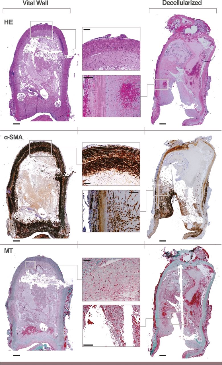
Characteristics of aneurysm healing. Histological characteristics of a vital and decellularized aneurysm at follow-up day 21. The left and right columns represent a histological overview of the aneurysm (x4 magnification, scale bars 200 µm). The middle column highlights specific features of the aneurysms (x10 magnification, scale bars 100 µm or 20x magnification with a scale bar 50 µm). Upper row: histological characteristics in the hematoxylin-eosin (HE) stain; middle row: histological characteristics in the immunofluorescence staining monoclonal anti-α muscle actin (α-SMA); lower row: histological characteristics in the Masson-Goldner’s (MT) stain. HE: The vital aneurysm shows continuously preserved aneurysm wall cellularity with hardly detectable inflammatory cells. The magnification (x10 magnification) demonstrates the cell-rich aneurysm wall at the dome. The decellularized aneurysm shows no aneurysm wall cells and is marked by a periadventitial inflammation, neutrophils within the aneurysm wall, and a lack of thrombus organization as demonstrated in the magnification (x10). α-SMA: The vital aneurysm demonstrates in the overview (x4 magnification) a thick neointima at the orifice and a centripetal distribution of α-SMA positive cells along the aneurysm wall, which also reaches the dome of the aneurysm (x10 magnification). The decellularized aneurysm demonstrates an insufficient neointima formation with obvious recanalization (x4 magnification). The concentration of α-SMA positive cells is higher along the carrier vessel and the orifice and markedly thins out towards the aneurysm dome (x10 magnification). MT: The vital aneurysm shows a thick collagen deposition (green) in the neointima (x4 magnification). Collagen can be detected also in the aneurysm dome (x20 magnification). The decellularized aneurysm shows less collagen deposition in the neointima (x10 magnification) compared with the vital aneurysm.
Cell migration
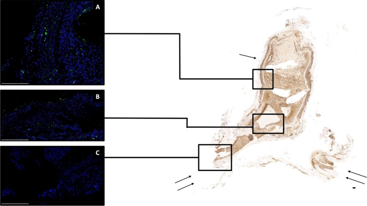
In α-SMA staining, vital (non-decellularized) aneurysms showed infiltration of the luminal thrombus by α-SMA positive cells (figure 3). This infiltration and subsequent thrombus organization and neointima formation started from the distal thrombus adjacent to the aneurysm wall in the vital aneurysms and followed a centripetal progression at consecutive time points. In contrast, thrombus organization and neointima formation was concentrated at the aneurysm orifice and neck in decellularized aneurysms. Experiments with GFP mutant aneurysms implanted in GFP wild type recipients, and vice versa, demonstrated that the cells organizing the luminal thrombus and neointima originated from the aneurysm wall in vital (non-decellularized) aneurysms and originated from the parent artery in decellularized aneurysms (figure 4).
Assessment of aneurysm wall cell migration. Green fluorescence protein (GFP) mutant vital aneurysm (single arrow) is implanted in a GFP wild type carrier vessel (double arrow). Right, image overviews of monoclonal anti-α muscle actin (α-SMA) positive cells (scale bar 150 µm). (A) Magnified area with aneurysm vessel wall, luminal organized thrombus, and artefact from the coil. GFP positive cells (green) are present in the aneurysm wall and also among cells organizing the luminal thrombus. (B) GFP positive cells originating from the aneurysm wall are also found in the neointima (scale bar 100 µm). (C) GFP wild type cells (blue) of the parent artery (scale bar 100 µm).
Discussion
The results of our study show that preservation of aneurysm wall cellularity significantly benefits healing of aneurysms after coil treatment. Thus, loss of mural cells from the aneurysm wall may explain post-embolization recurrence. Early histological changes after coil embolization are characterized by inflammatory responses that affect the aneurysm wall and intra-aneurysmal compartment in aneurysms with a vital (non-decellularized) and decellularized wall. Aneurysms with vital walls demonstrate the ability to attenuate the inflammatory response, organize the thrombus into a mature neointima, and thereby exclude the aneurysm from the circulation. Aneurysms with decellularized walls show a prolonged inflammatory state with the propensity to undergo wall remodeling toward an increased wall vulnerability, insufficient thrombus organization, and failure of neointima formation.
Ruptured intracranial aneurysms following coil embolization have higher rates of recanalization compared with their unruptured counterparts.3–5 Wall degeneration and loss of mural cells are histopathological findings characteristic of ruptured intracranial aneurysms.14 Our results underline the eminent role of preserved aneurysm wall cellularity toward mature neointima formation following coil treatment, whereas aneurysms with a decellularized wall show a significant tendency in macro- and microscopic neointima transformation failure. Aneurysms with preserved cytoarchitecture demonstrated the ability of SMCs to migrate into the thrombus, proliferate, and participate in collagen production and thereby directly contribute to neointima formation.
Intraluminal cell transplantation of myocytes compensated for the inability of decellularized aneurysms to organize the thrombus.12 Similar results have been described with autologous mesenchymal stem cells attached to platinum coils, which significantly improve histological healing.21 Our histopathological findings of aneurysm healing show similarities with human histological studies after Guglielmi detachable coil treatment of intracranial aneurysms—specifically, an initial migration of inflammatory cells into the intra-aneurysmal thrombus is followed by a centripetal invasion of fibroblasts and transformation of the thrombus into scar tissue.22–24 The importance of the aneurysm wall’s cell contribution to aneurysm thrombus organization with neointima formation has been previously demonstrated in animal studies7 and aligns with our observations in GFP transgenic animals in which SMCs from the aneurysm wall migrated into the thrombus and participated in neointima formation.
One major difference in our series comparing groups of aneurysms with vital and decellularized aneurysm walls was the regulation of the inflammatory response after coil treatment. An increased inflammatory reaction following intracranial aneurysm coil embolization appears to be an initial physiologic response to the intraluminal thrombus.25 26 Initially, both vital and decellularized aneurysms had a similar inflammatory reaction after coil treatment that affected the intra-aneurysmal compartment and aneurysm wall. Although vital aneurysms showed the ability to attenuate the initial immunologic reaction, decellularized aneurysms demonstrated throughout the observation period significantly increased numbers of neutrophils in the aneurysm wall and intraluminal thrombus, with elevated prevalence of wall dissection and wall hematoma. This degenerative reverse wall remodeling predisposes the aneurysm to growth and rupture.11 Although a the increase in aneurysm volume was not statistically significant, a clear trend was observed in decellularized coiled aneurysms at the time when the inflammatory reaction peaked. Several aneurysms demonstrated volume increases of up to 260% of their baseline value. One can postulate that the coil mass provided mechanical protection and prevented further growth and ultimately rupture. Furthermore, in our series, the coil treatment of aneurysms may have stabilized the thrombus by influencing intra-aneurysmal flow dynamics and thereby preventing the aneurysm from repeat cycles of thrombolysis and thrombus formation. Both cycles are associated with the maintenance of a pro-inflammatory state27 and destabilization of the aneurysm wall.11 Histopathological assessment of endothelial cells may provide additional information on aneurysm healing, as early endothelial invasion of the intra-aneurysmal thrombus has been associated with recanalization and recurrences after coil embolization of aneurysms.28
Limitations of study
Although the model used in our study does not represent a true intracranial aneurysm such as the model described by Hashimoto et al,29 it permits the assessment of basic concepts of aneurysm wall biology7 11 12 while representing a highly standardized and reproducible aneurysm model.17 Furthermore, the low morbidity/mortality of the model and relatively low costs make it a suitable tool to test and refine embolization devices that will be tested later in other more complicated and expensive models.30–32 Another limitation of the study is the lack of imaging modalities. While some aneurysms show a stepwise thrombosis, other aneurysms demonstrate continually repeating cycles of clot formation, dissolution, and aneurysm recanalization.11 T2* gradient echo sequences on a 7T MRI scan show a significant correlation with the amount of thrombus formation identified histologically in a rat aneurysm model.33 Repeat imaging modalities using this technique would potentially provide additional information on the natural history of aneurysm healing. Furthermore, high frequency optical coherence tomography, a novel intravascular imaging technology, enables precise and accurate measurement of coverage gaps at the neck of aneurysms in vivo.34 There is evidence that uniform aneurysm neck coverage by intrasaccular devices is critical for aneurysm occlusion. This imaging modality would be beneficial in group stratification and potentially exclude recurrent aneurysms due to insufficient aneurysm neck coverage. In our series we used a standardized coil volume in conjunction with a very standardized aneurysm volume, thereby expecting a homogeneous distribution of orifice coverage between the different groups. Furthermore, we included a firm follow-up modality using macroscopic and histopathological assessment of recurrence and aneurysm healing. The follow-up periods in our series assessed the short-term (days 3 and 7) and long-term (days 21 and 90) aneurysm healing following coil embolization.
Conclusions
Experimental coil-treated saccular aneurysms missing mural cells are incapable of organizing an intraluminal thrombus, are marked by a prolonged inflammatory reaction that affects the intraluminal thrombus and aneurysm wall, and thereby leads to aneurysm wall instability with frequent aneurysm recurrences. Aneurysms with preserved mural cells show the ability of myocytes to migrate into the thrombus, attenuate the inflammatory response, organize the thrombus into a mature neointima, and thereby exclude the aneurysm from the circulation and prevent recurrence. online supplementary figure 6
Acknowledgments
We thank Pablo Faundez, Alexander Yang, and Zach Folzenlogen for their technical support, and Majlinda Kalanderi for the illustrations.
References
Footnotes
Contributors All authors made a substantial contribution to the concept and design or analysis and interpretation of data, drafting or critical revision of the manuscript, and final approval of the submitted version of the manuscript.
Funding This work was supported by a research grant from the Research Council of the Kantonsspital Aarau, grant number 1410.000.05.
Competing interests None declared.
Patient consent for publication Not required.
Provenance and peer review Not commissioned; externally peer reviewed.
Data availability statement Data are available upon reasonable request. All data relevant to the study are included in the article or uploaded as supplementary information.