Article Text

Abstract
Background Endovascular internal trapping is an effective procedure for the treatment of acute vertebral artery dissection (VAD). However, the outcomes of reconstructive treatment have not been well established. The aim of our study is to evaluate the long-term clinical and angiographic results of endovascular internal trapping or reconstructive treatment of acute VAD.
Methods Between 2005 and 2013, 26 patients with acute VAD were managed with internal coil trapping (n=10), stent-assisted coiling (n=14), stent only (n=1), and proximal occlusion (n=1). Stent-assisted coiling included the modified stent-assisted semi-jailing technique (n=10), balloon-in-stent technique (n=2), and coiling followed by balloon mounted stent (n=2). Digital subtraction angiography (DSA) was performed in all patients except for three who died during the acute stage.
Results Of 26 patients with VAD, 14 and 12 presented with hemorrhagic and non-hemorrhagic types, respectively. The dominancy of the relevant artery was defined as dominant (n=9), even (n=12), and non-dominant (n=5). Reconstructive treatment was performed in six patients with ruptured VADs which failed balloon test occlusion and nine with non-hemorrhagic VADs. Clinical outcomes were favorable in 22 (84.6%), severe disability occurred in one, and there were three deaths (11.5%). All patients except the three who died had angiographic follow-up at 6–32 months (mean 10.4 months). The angiographic results of nine cases of internal trapping and one of proximal occlusion all showed a stable occlusion state. Among the 15 cases of reconstructive treatment, follow-up DSAs were available for the 13 surviving patients, 10 of which demonstrated stable occlusion of aneurysmal dilation and patent parent artery.
Conclusions This study suggests that internal trapping is a stable and effective treatment for acute VAD. Reconstructive treatment using stent and coils could also be a feasible alternative modality for hemorrhagic type VAD. However, serial DSA follow-up is essential.
- Aneurysm
- Artery
- Dissection
- Intervention
- Technique
Statistics from Altmetric.com
Introduction
Spontaneous vertebral artery dissection (VAD) is a rare pathophysiology of subarachnoid hemorrhage or ischemia, but has recently been recognized more frequently with the improvement in radiologic diagnostic tools.1–3 Intracranial acute VAD may lead to narrowing of the parent artery, occlusion, or pseudoaneurysm formation, resulting in posterior circulation infarction or subarachnoid hemorrhage. In particular, hemorrhagic type VAD has a high rate of early rebleeding with a catastrophic outcome.4 ,5 Thus, aggressive treatment of these complicated lesions is needed. Before the era of endovascular treatment, surgical trapping of a dissecting segment of the vertebral artery (VA) with or without bypass surgery was a curative treatment option; however, this operation has relatively high rates of morbidity and mortality.6 ,7
Endovascular internal trapping of the dissecting segment is currently the most accurate and effective treatment for prevention of rebleeding.8–10 However, sacrifice of the VA is not a realistic option under certain special conditions such as dominant VAD with poor collateral flow, VAD involving the posterior inferior cerebellar artery (PICA), or non-hemorrhagic type acute VAD. Endovascular reconstructive treatment using coils and stents could be a feasible alternative treatment option under these special conditions. A few series have reported a relatively good outcome with reconstructive treatment, but the case size was small and limited to ischemic type VADs or stent application.11–16 In addition, some case reports demonstrated the fatal complication of rebleeding of VAD after stent only therapy.17 ,18
The treatment of acute VAD remains highly individualized and is still controversial. In the current series we report the long-term angiographic and clinical follow-up results of endovascular treatment for acute VAD, internal trapping and reconstructive treatment with coils and stents, particularly the modified stent-assisted semi-jailing technique with or without the stent-within-a stent technique.
Methods
Patients
From September 2005 to January 2014, 26 consecutive patients with acute VAD underwent endovascular treatment at our institution. Owing to its retrospective nature, the current study was approved by the Institutional Review Board. The patients consisted of 13 men and 13 women with a mean age of 53.6 years (range 34–70 years). Brain CT, CT angiography, and digital subtraction angiography (DSA) were performed in all hemorrhagic cases and additional brain MR images were obtained in non-hemorrhagic cases. The clinical severity was assessed using the Hunt–Hess grade in cases of rupture and clinical outcomes were evaluated using the modified Rankin Scale (mRS). All DSAs were analyzed for size, shape, and location of the dissections by three neurointerventionists. Anatomical findings were classified into four types based on their relationship to the PICA: PICA proximal type in 3 patients, PICA involved (PI) in 3 patients, PICA distal in 10 patients, and absent PICA in 10 patients. Dominancy of the involved VA was as follows: dominant in 9 patients, non-dominant in 5 patients, and even in 12 patients. The angiographic features of the dissections were classified into four groups: (1) stenosis-fusiform-stenosis in 5 patients; (2) fusiform-stenosis in 5 patients; (3) stenosis-fusiform in 8 patients; and (4) fusiform in 8 patients.
Ten of the 26 patients underwent internal trapping with coils and one patient underwent proximal artery occlusion. Reconstructive treatment using coils and stents was applied to the remaining 15 patients. Reconstructive treatment included the modified stent-assisted semi-jailing technique (n=10), balloon-in-stent technique (n=2), coiling followed by balloon-mounted stent (n=2), and stent only (n=1). All hemorrhagic type VADs were treated as emergencies within 12 h of arrival at the hospital and all non-hemorrhagic symptomatic VADs were treated within 2 weeks of symptom onset. All procedures were performed under light sedation with propofol and remifentanil in order to maintain continuous neurological monitoring. Demographic and clinical presentation data of the patients with hemorrhagic type VAD and non-hemorrhagic VAD are shown in table 1 and table 2, respectively.
Summary of 14 patients with hemorrhagic type of VAD
Summary of 12 patients with non-hemorrhagic type of acute symptomatic VAD
Treatment criteria
The indication for treatment of acute VAD was all ruptured cases. Internal trapping using detachable coils was the first treatment strategy for all hemorrhagic type VADs, but reconstructive treatment using stents and coils was an alternative treatment option for those with dominant side or PI type VAD. In non-hemorrhagic asymptomatic cases, conservative management with serial image follow-up was the initial treatment option. However, patients with acute symptomatic non-hemorrhagic VAD in whom the fusiform dilation portion was confirmed on DSA were considered candidates for treatment. Headache with or without neck pain was an acute symptom of VAD. Patients with ischemic symptom with stroke confirmed by MR diffusion images were first treated with anticoagulation or antiplatelet therapy. The risks and objectives of the proposed endovascular treatment were explained to all non-hemorrhagic symptomatic patients and their clinical guardians.
Endovascular treatment
The patients took premedication with 5 days of dual antiplatelet therapy consisting of 75 mg clopidogrel and 100 mg aspirin in only non-hemorrhagic cases. The right femoral artery was accessed using a 6 Fr 80 cm long Shuttle sheath (Cook, Bloomington, Indiana, USA). A 6 Fr guiding catheter (Envoy; Cordis, Miami Lakes, Florida, USA) was placed in the distal V2 segment of the VA. After attaining access to the femoral artery, a bolus of 3000 IU heparin was administered intravenously at the beginning of the procedure in cases of non-hemorrhage VADs. However, in cases of ruptured VADs, heparin was administered after delivery of a microcatheter and stent system. We performed a balloon test occlusion (BTO) in selective cases, particularly dominant VAD, immediately after diagnostic angiography in order to determine an alternative option for internal trapping.
Modified stent-assisted semi-jailing with or without stent-within-a-stent technique
We first delivered a Neuroform stent using a 300 cm long exchange microwire over the dissecting portion and then selected the aneurysmal portion using an angled tip microcatheter. The stent was deployed approximately one-third and some coils were placed loosely and circumferentially at the aneurysmal dilation. We focused on the sharpest and most dilated portion of the VAD in order to increase the packing density. When repeated angiography showed slight distal flow compromise, we deployed the stent completely. Additional stent application for placement of unrecognized coil loops between the mesh of two stents was optional. Thus, all VADs treated using this technique had a fusiform sharp dilation portion. The procedure is illustrated in figure 1A–F.
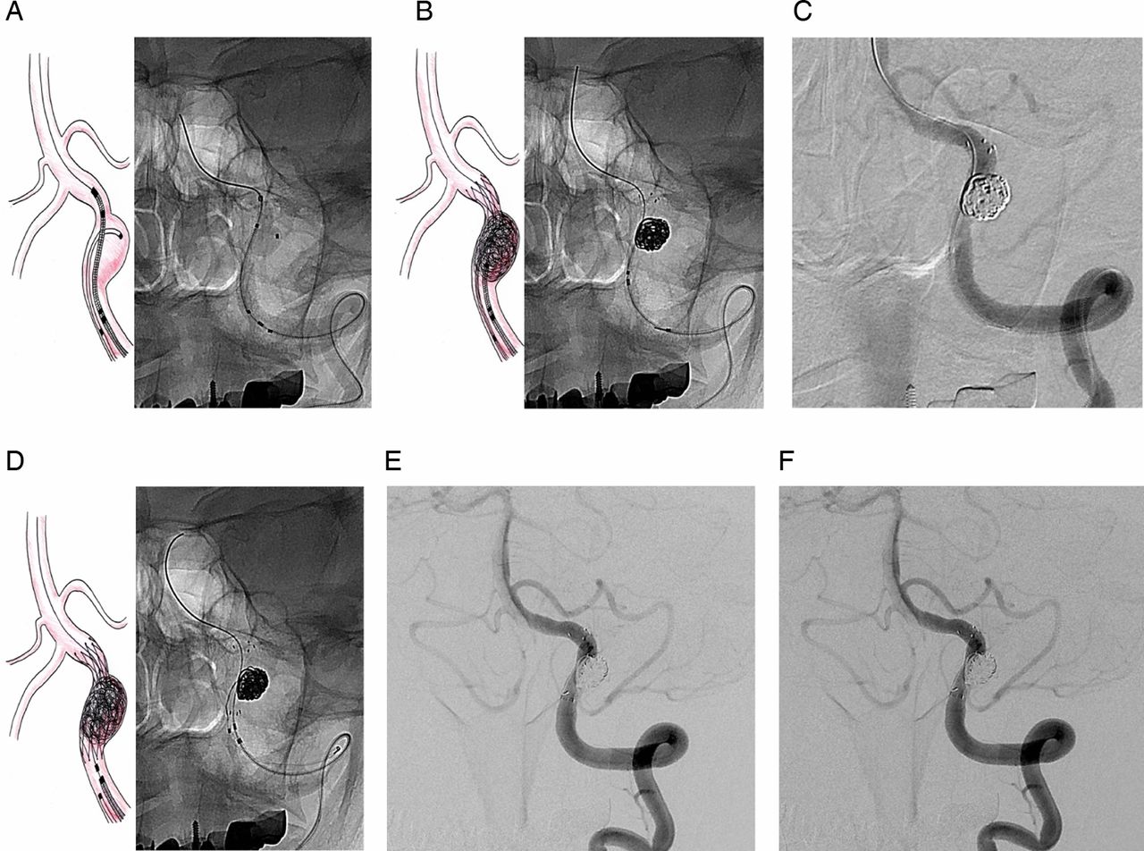
Serial schematic and case illustrations of modified stent-assisted semi-jailing technique of left vertebral artery fusiform dissecting aneurysm. (A) Neuroform stent delivery followed by microcatheter aneurysmal sac selection. (B) Partial stent deployment with circumferential and loose coil embolization. (C) Mid-term digital subtraction angiogram showing slight distal flow compromise. (D, E) Complete stent deployment and final angiogram showing complete obliteration of the aneurysmal dilation with patent parent artery. (F) Follow-up angiogram at 13 months showing no evidence of recurrence with stable complete embolization.
Clinical assessment, angiographic and clinical follow-up
Clinical assessment using the Hunt–Hess grading system was performed on the first hospital day and procedure-related complications were examined during and after the intervention. Clinical outcome was assessed using the mRS at the last clinical follow-up and defined as the final outcome. At least one follow-up DSA was performed in all 26 patients 6–12 months after treatment. The immediate angiographic result of obliteration was defined as complete occlusion (entire aneurysmal dilation obliterated) or partial occlusion (a small proximal or distal aneurysmal dilation portion opacified with contrast). Follow-up DSA results were defined as three types: (1) stable occlusion: no aneurysmal growth or no contrast opacification at aneurysmal dilation; (2) regrowth: contrast opacification at aneurysmal dilation or enlargement of dilation; or (3) parent artery occlusion.
Results
The clinical and angiographic follow-up results of the patients are summarized in tables 1 and 2. All patients except the three who died had angiographic follow-up at 6–32 months (mean 10.4 months). Fourteen patients had hemorrhagic type VAD and 12 had non-hemorrhagic type acute VAD. Data on clinical and angiographic follow-up for at least 6 months were obtained from 23 patients (88.5%). All six with dominant side ruptured VADs failed BTO and nine non-hemorrhagic VADs were treated using a reconstructive technique. Failed BTO means that ischemic symptoms developed during temporary BTO or BTO was precluded by the patient's initial poor clinical status. The immediate angiographic results of reconstructive treatment were complete occlusion in 13 patients and partial occlusion in three. Clinical outcomes were favorable in 22 patients (84.6%), one patient developed a severe disability, and three patients (11.5%) died due to poor initial clinical grade. Follow-up DSA results of nine cases of internal trapping and one case of proximal occlusion all showed stable occlusion. Among the 15 cases who had reconstructive treatment, follow-up DSAs were available in the 13 surviving patients, 10 of whom demonstrated stable occlusion (figure 2). Two patients with hemorrhagic type VAD who were treated with a reconstructive technique developed regrowth of the coiled arterial wall. Successful retreatment was achieved in one case (case 5 in table 1) and the other patient with minimal regrowth demonstrated stable occlusion at the second follow-up DSA 21 months later (case 11 in table 1). In-stent parent artery occlusion occurred in one case of hemorrhagic type VAD without symptoms (case 12 in table 1). There was no recurrent bleeding or ischemic symptoms during the clinical follow-up period of 6–67 months (mean 36.7 months).

(A) A middle-aged patient with a ruptured right distal vertebral artery fusiform dissecting aneurysm with early termination of the left vertebral artery at the posterior inferior cerebellar artery. (B) Mid-term native image of modified stent-assisted semi-jailing technique (left) and final native image of additional stent-within-a-stent technique (right). (C, D) Final and 13 month follow-up angiograms showing complete obliteration of the aneurysmal dilation and stable complete occlusion.
Discussion
Although there have been some reports on the natural course of acute VAD, it is still not well understood. The treatment strategy for acute VAD is also controversial. However, most agree that prevention of rebleeding of ruptured VAD is an important and essential goal of treatment. During the past decade many studies on the feasibility and effectiveness of internal trapping of VADs have been reported, and this technique is believed to be the treatment of choice for ruptured VAD.6–8 ,19 ,20 The current study also confirmed that internal trapping of the dissecting segment appears to be an effective and safe treatment method. However, this deconstructive treatment is not possible or may be inappropriate in some situations such as dominant VAD rupture with poor collateral flow, dominant PI type VAD rupture, or non-hemorrhagic type symptomatic fusiform VAD. Proximal occlusion of the parent artery is an alternative option, but it does not completely prevent rebleeding due to retrograde flow from the basilar artery.21–23
On the other hand, reconstructive treatment using stents or coils may be an appropriate and acceptable technique when DSA confirms inadequate collateral flow or a dissected segment involves a major branch, particularly PICA. Some studies have reported on reconstructive treatment of VADs. Reconstructive treatment includes stent only therapy, stent-assisted coil embolization, multiple stent technique, or a combination of these. Theoretically, the stent only or multiple stent techniques are ideal for arterial dissection, but they do not completely protect from rebleeding in cases of rupture.17 ,24–26 Therefore, stent-assisted coil embolization has been applied to VADs and is an attractive reconstructive option for acute VAD.11 ,13–16 ,27 In this technique the stent works as a scaffold for the coil mesh and maintains the patency of the arterial lumen. Three-dimensionally, stents provide an artificial aneurysm neck and prevent distal coil migration or coil loop protrusion into the parent artery. In addition, the stent can divert flow from the dissecting aneurysm and promote the healing process. Endovascular flow diversion has been suggested as an advanced treatment of intracranial arterial dissection. Theoretically, dissecting aneurysms have a segmental defect of the intima. Thus, stents with dense mesh and small porosity could facilitate the healing process of the dissecting aneurysm. Furthermore, flow diverters could modify aneurysmal hemodynamics and facilitate thrombosis of the dilated aneurysmal portion. The Pipeline embolization device was recently introduced as the first flow-diverting stent which aimed to decrease the shear stress of the aneurysmal wall. Several articles have reported on the efficacy of the Pipeline embolization device for intracranial VADs; however, their case sizes were small.28–31 Although the use of this device has shown promising results, it has several disadvantages. In cases of ruptured VAD, the patient is unable to take premedication of antiplatelet agents and therefore thromboembolic complications could occur frequently. In addition, the possibility of in-stent stenosis due to intimal hyperplasia and obliteration of perforating arteries is a problem.32 Despite these concerns, reconstruction using flow diverters might be an attractive option for treating VAD.
There have been some reports on the relation between rebleeding and the angioarchitecture of ruptured VADs. In a nationwide Japanese study, Ono et al33 reported a significant association of ‘pearl and string’ structure with rebleeding in ruptured VADs. This result was also supported by the findings of Yamada et al5 in 2004. In our series, all hemorrhagic VADs had an angiographically fusiform dilation portion (‘pearl and string’ sign), and this result was similar to those of previous reports. The ruptured ‘pearl and string’ or fusiform dilation aneurysm may be the result of a dissection in the entire wall of the artery that reached the adventitia, which could increase the risk of rebleeding. Therefore, we have to focus on the sharpest and most dilated portion of the VAD and attempt to obliterate that portion with coils. To date, the most commonly used stent-assisted coiling techniques have been the jailing technique (microcatheter selection first and stent deployment second) or stent deployment followed by selection of a microcatheter through the stent struts. These techniques were originally suitable for wide-necked saccular aneurysms, but there are some problems in their application to fusiform dilated VADs. The jailing technique is simple and safe when aneurysmal dilation is selected using a microcatheter. However, the possibility of a microcatheter kick-back movement when coiling could be associated with a higher risk of pseudoaneurysm rupture compared with saccular aneurysms. In addition, jailing of a microcatheter does not permit circumferential coil position in a fusiform VAD. Stent deployment followed by microcatheter selection through the stent struts permits a microcatheter but it could facilitate stent migration or stent folding. In addition, as with the jailing technique, this technique does not permit circumferential coil position in a fusiform VAD. Guaranteeing the patency of the parent artery may be difficult or impossible because coil loop protrusion through the stent interstices may occur and would not be detected due to superimposition of the coils and stent mesh. Double stents (the stent-within-a-stent technique) may be a solution to ensuring the patency of the parent artery.
Modified stent-assisted semi-jailing with or without the stent-within-a-stent technique could be a solution. As described in the Methods section, a partially deployed stent (the ‘semi-jailing technique’) has several advantages. First, due to the small size of the VA compared with the carotid artery, we could not use a large guiding catheter. We placed a 6 Fr guiding catheter in the distal V2 segment of the VA, which means that we could use only one microcatheter and one stent delivery system. Therefore, the existing stent-assisted coiling technique has been the only method for prevention of distal coil migration. However, a partially deployed stent resembling a reversal cone could protect the distal portion of fusiform dilation and prevent distal coil migration and permit the kick-back movement of the microcatheter in order to decrease the risk of rupture during dense coil packing. Second, proximal stent-free space could facilitate circumferential coil packing of fusiform dilation. After partial packing of fusiform dilation and repeat angiography showed slight distal flow compromise, we deployed the stent completely. The radial forces of the stent push the coil loops laterally, securing the parent artery. The stent-within-a-stent technique is optional and could be a means for achieving definite patency.
Among the 15 cases of reconstructive treatment in our series, follow-up DSAs were available in 13 surviving patients and 10 demonstrated stable occlusion with patent parent artery. Reconstructive treatment using stent or coils, particularly the modified semi-jailing technique, is comparable to internal trapping for prevention of rebleeding of VAD and the clinical outcome is also favorable compared with those of most previous series.6–8 ,11 ,20 Two patients with hemorrhagic type VAD treated with reconstructive technique developed regrowth of the coiled arterial wall. One patient showed regrowth of the opposite side of the coiled aneurysm wall at 6 months follow-up DSA and complete occlusion was achieved using additional stent-assisted coiling. With regard to regrowth of the opposite side, in our opinion circumferential coiling of fusiform dilation using the modified stent-assisted semi-jailing technique might achieve stable occlusion and also complete occlusion. The other patient with regrowth demonstrated stable occlusion on the second follow-up DSA at 21 months. Despite the fact that this patient had hemorrhagic VAD, we could not decide whether to treat the recurred portion at first follow-up because of PICA involvement. In-stent parent artery occlusion occurred in one patient with hemorrhagic type VAD (dominant VA, PICA distal type) treated using the reconstructive technique. Follow-up angiography showed a change of VA dominancy to the contralateral side and early termination of ipsilateral VA just distal to PICA. This angiographic change may facilitate sufficient collateral flow of the posterior circulation, and the patient has been symptom-free during 16 months of clinical follow-up. Among cases of ruptured VAD, all patients were treated within 12 h of the initial hemorrhage, and emergency treatment could have prevented rebleeding in the acute state and improved the clinical outcome.
Conclusion
The results of this study have shown that internal trapping is a stable and effective treatment for acute VAD and that reconstructive treatment using a stent and coils could also be a feasible and safe alternative treatment modality, even in hemorrhagic type VAD. However, close serial DSA follow-up is essential
References
Footnotes
Contributors JIL and KHN collected the data and drafted the article. JKK, CHC, SHC and THL provided patients and revised the draft paper.
Competing interests None.
Ethics approval Approved by Pusan National University Institutional Review Board (E-2014060).
Provenance and peer review Not commissioned; externally peer reviewed.
Data sharing statement All data in this manuscript are available to all.