Article Text

Statistics from Altmetric.com
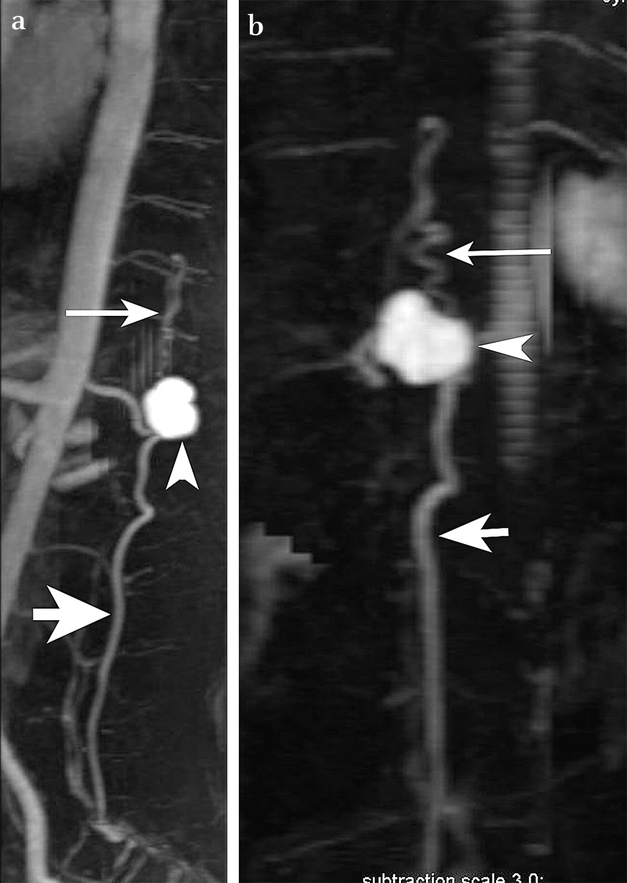
A 30-year-old woman presented with progressive paraparesis and urinary incontinence for 1 month. Physical examination revealed weakness at the plantar and dorsal flexors of the ankle and toes. Routine blood tests were normal. MRI showed spinal cord compression. The imaging sequences also showed an intraspinal paramedullary partially thrombosed aneurysm, compressing the spinal cord at T12 level, and several flow voids superior and inferior to the aneursym which indicated a spinal arteriovenous malformation (figure 1). Contrast-enhanced MRI angiography revealed a giant aneurysm originating from the anterior spinal artery and spinal arteriovenous malformation (figure 2). These MRI and MRI angiography findings were shown during the surgery, too.

(a) Sagittal, (b) multiplanar reconstruction coronal and (c) axially T2 weighted MRI images showing a dilated anterior spinal artery (arrow), giant aneurysm (double arrow), dilated draining vein (short arrow) and compressed spinal cord (arrowhead).

(a) Sagittal and (b) coronal contrast-enhanced MRI angiography showing a dilated anterior spinal artery (arrow), giant aneurysm (arrowhead) and dilated draining vein (thick arrow).
Anterior spinal artery aneurysm (ASAA) is a rare vascular pathology. These aneurysms are generally associated with vascular lesions such as spinal arteriovenous malformations, coarctation of aorta and vasculitis. Isolated aneurysms are rare. ASAA are usually a result of haemodynamic anomalies and are frequently associated with vascular lesions with high blood flow. In contrast, intracranial aneurysms are mostly of congenital origin. Therefore, these lesions may be classified as flow-directed aneurysms.1–3 These aneurysms may occur at all segments of the spinal artery, except bifurcations and generally fusiform type rather than sacculary.1
ASAA is usually small in calibre, and giant aneurysms are very rare. Most of the reported aneurysms are no larger than 3 mm in diameter.1 Patients with ASAA are usually presented by subarachnoid haemorrhagy in clinical practice. Symptoms due to giant aneurysm compressing the spinal cord are rarely seen.4 5
Spinal angiography is the best diagnostic modality for the detection of ASAA. The other diagnostic modalities such as MRI and CT angiography have a lower spatial resolution compared with angiography. AASA may be easily missed by CT or MRI angiography as ASAA are small in diameter. Only one case has been diagnosed by MRI angiography in the literature so far.1 In our case, MRI angiography demonstrated a dilated anterior spinal artery, a giant aneurysm originating from the anterior spinal artery and a dilated draining vein. As far as we know, a giant anterior spinal artery aneurysm demonstrated by MRI angiography has not been reported to date.
Spinal angiography is still the gold standard for diagnosis of ASAA, especially for small aneurysms. MRI angiography may be used to demonstrate ASAA non-invasively and can also be used as a pioneering method of invasive angiography.
Footnotes
Competing interests None.
Patient consent Obtained.
Provenance and peer review Not commissioned; not externally peer reviewed.