Article Text

Abstract
Introduction/Purpose MRI fusion is a recently developed image targeting technique that volumetrically overlays an MRI sequence onto a live fluoroscopic image updated in real time. The ability to utilize the superior soft tissue resolution of MRI for interventional lesion targeting without the practical constraints of an interventional MRI suite is potentially a powerful tool. Lesions that are well-visualized under MRI but possessing little fluoroscopic contrast are otherwise untreatable in the angio suite and ideally suited to this new technique. We present four consecutive cases involving small intraorbital masses in children where MRI-fusion volumetric overlay techniques were used to precisely target the lesions for the purposes of both percutaneous biopsy and venolymphatic malformation sclerotherapy.
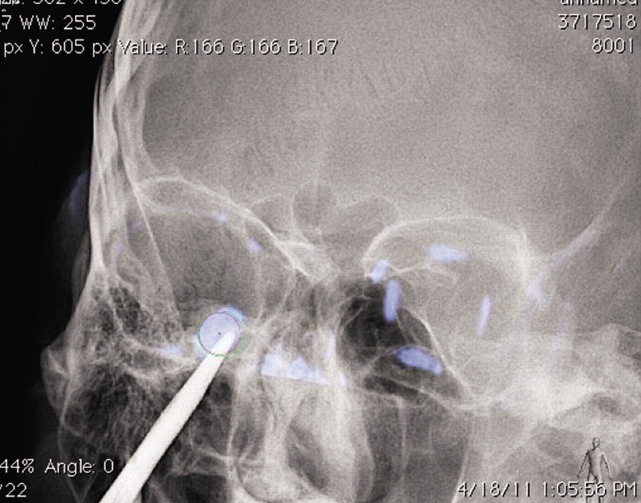
Materials/Methods Four pediatric patients were referred for percutaneous biopsy (n=1) and sclerotherapy (n=3) for small intraorbital masses. The DICOM MRI data sequences that best demonstrated the target lesions were imported to a Philips Allura fluoroscopy system. General anesthesia was administered and an XperCT scan of the head was obtained in the angiography suite. A multimodality matching overlay of the imported MRI data with the obtained XperCT data was performed to facilitate procedure planning with the MRI target superimposed over the live fluoroscopic images. Direct puncture of the target lesion was performed with a 23- or 25-gauge needle. Percutaneous sclerotherapy intralesional needle tip confirmation was assessed with aspiration and contrast injection. The targeted sclerotherapy lesions were then slowly injected manually with a mixture of Bleomycin and contrast with intermittent fluoroscopic evaluation to ensure intralesional placement. The biopsy case specimen was aspirated with a 23 gauge needle and a cell smear and core sample were submitted to Pathology.
Results The treatment/biopsy needles were successfully placed in all four lesions in the angiographic suite using this technique. The needle was placed intralesional on the first pass in all four cases without complication with needle repositioning in the treatment cases as necessary for aspiration. No contrast extravasation or venous drainage was identified in the treatment group with intralesional contrast and Bleomycin/contrast injection. The biopsy patient had a diagnostic sample obtained while the sclerotherapy lesions were all decreased in size on follow-up imaging with resolution of their clinical symptoms. No sclerotherapy patient has required additional treatment.
Conclusion MRI fusion with real-time fluoroscopic imaging allows for treatment of previously non-visualized lesions in the angiographic suite with sufficient soft tissue detail to perform precise percutaneous orbital interventions in a safe and therapeutic manner.
Competing interests None.
