Article Text

Abstract
Introduction Although coil embolization of brain aneurysms is safe and effective, aneurysm recurrence remains a limitation requiring surveillance imaging. Recently, MR angiography (MRA) has become a viable option for non-invasive follow-up. Often, a baseline MRA within one week of coiling is acquired for comparison. The coil mass produces magnetic susceptibility artifact that may obscure the evaluation of the host vasculature and the aneurysm. Although previous research into MR artifact has been done in vitro, the dynamic characteristics of a functional vascular system cannot be simulated. Our aim in this study was to evaluate the MR susceptibility artifact from coil masses over time using different MRA techniques.
Materials and Methods Four mongrel canines were used: two venous side-wall aneurysms were created on each common carotid artery for a total of four aneurysms in each animal. Aneurysms were embolized receiving only one type of coils, GDC or a new coil prototype. After coiling, the animals were transferred to the 3T MRI. MRI protocol included axial 3D TOF MRA (TE of 3.5, 5 and 6.9), phase contrast MRA, and coronal contrast-enhanced MRA. Serial MRI studies were repeated at 1, 4, 6, 8, 14, and 28 weeks. The volume of the coil mass was measured using 3D radiography. MRA sequences were analyzed using Mimics 13.1 for 3D reconstruction and volume measurements. An overestimation factor (OEF) was calculated: OEF=VSA/VCM; where the numerator represents the volume of the MR susceptibility artifact and the denominator is the true volume of the coil mass measured by 3D radiography.
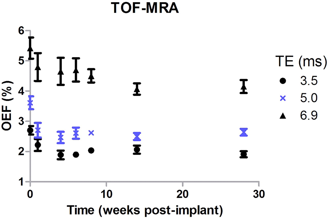
Results OEFs are largest immediately after embolization, and show a gradual decay until approximately 4 weeks (Abstract O-030 figure 1), when there is stabilization of the size of the artifact. By 6 weeks we found that there was mild coil compaction (average coil mass volume decrease by 9.4%). However, the susceptibility artifact reduced substantially during the same time period; with the OEFs decreasing by 30% after 6 weeks. The decrease of the artifact is currently under evaluation. Histopathological analysis of explanted aneurysms did not show corrosion of the metal detachment zone. The effect of compaction has also been excluded as a possible hypothesis in phantom experiments.
Conclusion MR susceptibility artifact changes over time, being maximal in the post embolization setting and decaying until 4 weeks. The clinical implications of this study indicate that baseline MRA for comparison with future imaging should be acquired after 4 weeks post procedure.
Statistics from Altmetric.com
Footnotes
Disclosures G Spilberg: None. S Carniato: Stryker Neurovascular. R King: None. R Murphy: Stryker Neurovascular. M Gounis: Stryker Neurovascular. A Wakhloo: Stryker Neurovascular.