Article Text

Abstract
Introduction Dolichoectatic dilatation in intracranial arteries may enlarge, however the evolution of this progression has not been described. We present 3 cases with intracranial vascular imaging spanning for a period of 10 years demonstrating growth of dolichoectatic dilatations to large and giant aneurysms, which were eventually treated by various modalities.
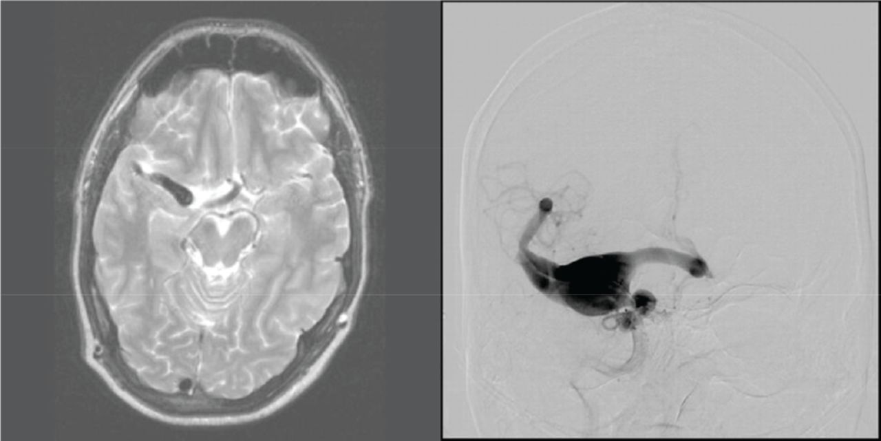
Cases Case 1: 49 year old man who had brain imaging in 2002 demonstrating dolichoectatic dilatation of the right internal carotid, anterior cerebral and middle cerebral arteries with maximal diameter of 6mm, presented in 2011 with seizures related to mass effect in the right mesial temporal lobe. Cerebral angiogram demonstrated growth of the aneurysm to a maximal diameter of 29mm, involving the right internal carotid, anterior and middle cerebral artery (figure demonstrating MRI from 2002 and angiogram from 2011). The patient was treated successfully with high flow bypass and right ICA endovascular sacrifice.
Case 2: 63 year old woman who had brain MRI in 2000 demonstrating no aneurysms, presented in 2011 with headache and subacute subarachnoid haemorrhage. The angiogram demonstrated dysplastic left MCA with fusiform dilatation and 3 irregular aneurysms. She was successfully treated with SCA/MCA bypass.
Case 3: 37 year old man with an MRI from 2000 demonstrating dolichoectatic basilar artery with maximal diameter of 6mm, presented in 2013 with brainstem compression and the angiogram showed growth into a dysplastic giant basilar aneurysm with maximal diameter of 30mm. The patient was treated surgically.
Discussion These cases describe symptomatic and imaging progression of dolichoectatic vessels to large and giant aneurysms in the anterior and posterior circulation for extended period of time. Interval and duration of follow up in this patient population has not been well described, however, progression may occur for long period of time, as demonstrated in our cases with imaging for over ten years. Frequent and long term imaging evaluation may be recommended in the follow up of these patients.
Disclosures E. Cheng-Ching: None. G. Toth: None. R. Burgess: None. M. Hussain: None. M. Bain: None.